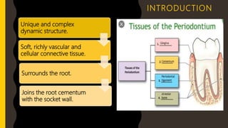
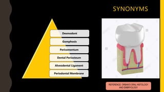
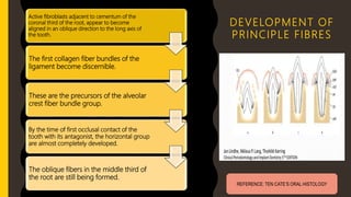
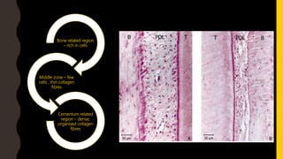
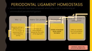
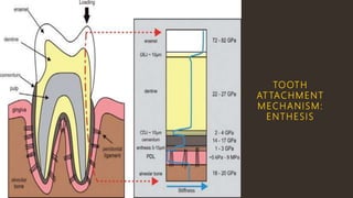
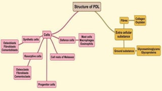
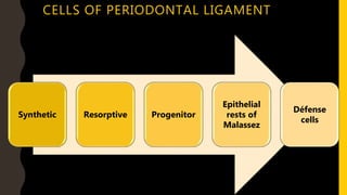
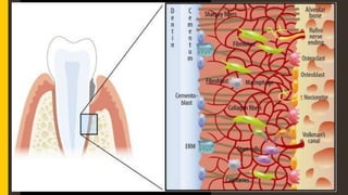
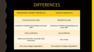
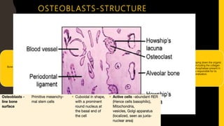
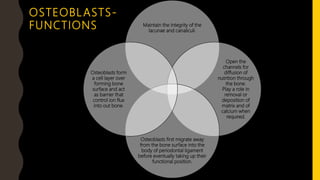
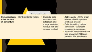
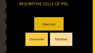
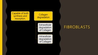
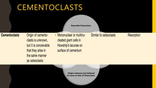
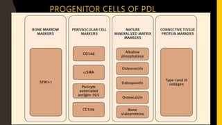
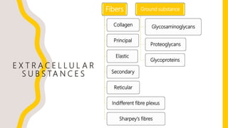
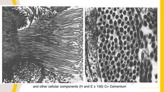
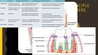
P E R I O D O N TA L L I G A M E N T
D R . T. D I N E S H K U M A R , M D S
D E P T. O F O R A L PAT H O LO G Y
S R M D E N TA L C O L L E G E , R A M A P U R A M
S R M I N S T I T U T E O F S C I E N C E & T E C H N O LO G Y
CONTENTS
INTRODUCTION
DEFINITION
SYNONYMS
EXTENT AND SHAPE
DEVELOPMENT
PERIODONTAL LIGAMENT HOMEOSTASIS
CELL BIOLOGY OF THE NORMAL PERIODONTIUM
CELLULAR EVENTS
EXTRACELLULAR SUBSTANCES
STRUCTURES OF THE CONNECTIVE TISSUE
FUNCTIONS OF THE PDL
AGE CHANGES
UNIQUE FEATURES
CLINICAL CONSIDERATIONS
INTRODUCTION
Unique and complex
dynamic structure.
Soft, richly vascular and
cellular connective tissue.
Surrounds the root.
Joins the root cementum
with the socket wall.
DEFINITIONS
• Periodontal ligament is composed of soft complex vascular and highly
cellular connective tissue that surrounds the tooth roots and connects to
the inner wall of the alveolar bone. (Mc Culloch CA, Lekic P, Mc Kee MD
Periodontal 2000 24:56,2000)
• It is that soft, specialized CT situated between the cementum covering the
root of the tooth and bone forming the socket wall. (A.R.Tencate 1971)
• The periodontal ligament occupies the periodontal space, which is located
between the cementum and the periodontal surface of alveolar bone and
extends coronally to the most apical part of the lamina propria of the
gingiva. (Orban)
SYNONYMS
Desmodont
Gomphosis
Pericementum
Dental Periosteum
Alveodental Ligament
Periodontal Membrane
REFERENCE: ORBAN’S ORAL HISTOLOGY
AND EMBRYOLOGY
EXTENT AND SHAPE
Shape : An hour glass and is
narrowest at the mid root level
Width : 0.15-0.38mm.
Radiographic appearance:
radiolucent bounded by
radiopaque cementum and lamina
dura of the alveolar bone proper
The average width of the
periodontal space is 0.1–0.25 mm
REFERENCE: ORBAN’S ORAL HISTOLOGY
AND EMBRYOLOGY
DEVELOPMENT
 All periodontal tissues originate from the dental
follicle
 Begins with root formation & prior to tooth
eruption. (Tencate et al.,1972).
 At Late bell stage - when amelogenesis and
dentinogenesis are well advanced, the inner and
the outer enamel epithelium at the cervical loop
of enamel organs form a doubled layered
epithelial root sheath which proliferates apically
to map out the shape of future root called the
HERTWIG’S EPITHELIAL ROOT SHEATH REFERENCE: ORBAN’S ORAL
HISTOLOGY AND EMBRYOLOGY; TEN
CATE’S ORAL HISTOLOGY
DEVELOPMENT
Establish continuity across the ligament space
Assembles as collagen bundles on the bone and cementum surface
Type I collagen is secreted
Actively synthesize & deposit collagen fibrils and glycoproteins in
developing PDL (Grant’s 1989; Ten Cate’s 1971)
As the root formation continues, cells in the peri follicular
gain their polarity, cellular volume & become widely separated
DEVELOPMENT OF
PRINCIPLE FIBRES
Active fibroblasts adjacent to cementum of the
coronal third of the root, appear to become
aligned in an oblique direction to the long axis of
the tooth.
The first collagen fiber bundles of the
ligament become discernible.
These are the precursors of the alveolar
crest fiber bundle group.
By the time of first occlusal contact of the
tooth with its antagonist, the horizontal group
are almost completely developed.
The oblique fibers in the middle third of
the root are still being formed.
REFERENCE: TEN CATE’S ORAL HISTOLOGY
Bone related region
– rich in cells
Middle zone – few
cells , thin collagen
fibres
Cementum related
region – dense,
organised collagen
fibres
PERIODONTAL LIGAMENT HOMEOSTASIS
• Various molecules have been proposed, which play a role in maintaining an
unmineralized periodontal ligament.
Msx-2
Osteopontin
and Bone
Sialoprotein
Matrix ‘Gla’
protein
RGD-
cementum
attachment
protein
TGF-𝜷
Prostaglandins
REFERENCE: ORBAN’S ORAL
HISTOLOGY AND EMBRYOLOGY
Msx – 2
Prevents osteogenic
differentiation of
PDL fibroblasts by
repressing Runx2
transcriptional
activity
Matrix ‘Gla’ protein
Inhibitor of
mineralisation –
preserves the width
of the ligament
Prostaglandins
Inhibits mineralised
bone nodule
formation
Bone sialoprotein vs
osteopontin
Helps to maintain
the unmineralized
ligament region
HOMEOSTASIS
↑Functional demand
Width increases by 50%
Fibre bundles increased
in thickness
↓Functional demand
Narrowing of the
ligament
↓Number & thickness of
fibre bundles
TOOTH
AT TACHMENT
MECHANISM:
ENTHESIS
S T R U C T U R E O F T H E
P E R I O D O N TA L L I G A M E N T
CELLS OF PERIODONTAL LIGAMENT
Synthetic Resorptive Progenitor
Epithelial
rests of
Malassez
Défense
cells
SYNTHETIC
CELLS OF
PDL
FIBROBLAST OSTEOBLAST
CEMENTOBLAST
Most
predominant
cell type of
periodontal
ligament.
Found between
the fibers of
the periodontal
ligament,
where they are
surrounded by
fibers and
ground
substance.
STRUCTURE:
Ovoid or
elongated cells
oriented along
the principal
fibres.
Large cells with
an extensive
cytoplasm
Associated with
protein
synthesis &
secretion (e.g.
RER, several
Golgi complex,
& many
secretary
vesicles)
FIBROBLASTS
DIFFERENCES
PERIODONTAL LIGAMENT FIBROBLASTS GINGIVAL FIBROBLASTS
Ectomesenchymal origin Mesodermal origin
Expression of alkaline phosphatase and cyclic
AMP
Expression of alkaline phosphatase and
cyclic AMP
More proliferative Less proliferative
Motile and contractile. Generate tooth
movement.
Non motile
Also cause collagen degradation Not capable of collagen degradation
Bone forming cells derived from
osteoprogenitor cells.
Found on the surface of the alveolar
bone .
Seen on surfaces of bone giving an
epithelium like appearance. Shape
varies (seen as oral, triangular, cuboidal)
etc.
Responsible for laying down the organic
matrix of bone including the collagen
fibers. Alkaline phosphatase present in
osteoblasts is responsible for its
mineralization.
OSTEOBLASTS-STRUCTURE
Maintain the integrity of the
lacunae and canaliculi.
Open the
channels for
diffusion of
nutrition through
the bone.
Play a role in
removal or
deposition of
matrix and of
calcium when
required.
Osteoblasts first migrate away
from the bone surface into the
body of periodontal ligament
before eventually taking up their
functional position.
Osteoblasts form
a cell layer over
forming bone
surface and act
as barrier that
control ion flux
into out bone.
OSTEOBLASTS-
FUNCTIONS
These are the cells
responsible for secreting
the organic matrix (mainly
collagen) of cementum.
As distinct layer of cells on
the root surface, similar to
osteoblastic layer but not
regular in arrangement.
CEMENTOBL ASTS
RESORPTIVE CELLS OF PDL
Osteoclast
Cementoclast Fibroblast
FIBROBLASTS
Capable of both
synthesis and
resorption.
Collagen
degradation
Extracellular
degradation
of Collagen
Intracellular
degradation
of Collagen
OSTEOCLASTS-STRUCTURE
Present on the
surface of bone
where it is removed.
At such locations
cells occupy pits
called Resorption
bays or lacunae of
Howship's.
Osteoclasts are
formed by fusion of
mononuclear cells
arising from bone
marrow.
They do not cover
the whole of
resorbing surface at
any one time, rather
they service a much
larger area by
demonstrating
considerable
Inhibitor: Osteo-
protegrin
CEMENTOCLASTS
Resemble Osteoclasts.
Located in
Howship's
lacunae.
Origin unknown but believed
be same as that of Osteoclasts.
Since constant
deposition of
cementum
occurs, these
cells are not
seen during
normal
functioning of
cementum.
PROGENITOR CELLS OF PDL
EPITHELIAL RESTS OF MALASSEZ
Represent the remnants
of developmental
Hertwig’s epithelial root
sheath which is involved
in differentiation of root
odontoblasts and also
secrete enamel like
proteins on the root
surface.
Form a latticework in the
periodontal ligament
appear as either isolated
clusters of cells or
interlacing strands.
Arranged in close
packing of cuboidal cells,
completely surrounded
by connective tissue
DEFENSE
CELLS
E X T R A C E L L U L A R
S U B S TA N C E S
Fibers
Collagen
Principal
Elastic
Secondary
Reticular
Indifferent fibre plexus
Sharpey's fibres
Ground substance
Glycosaminoglycans
Proteoglycans
Glycoproteins
C O L L A G E N
The collagen is gathered to form bundles approximately
5 µm in diameter. These bundles are termed PRINCIPAL
FIBERS.
Composed mainly of amino acids.
The main types of: Type I and Type III.
More than 70% of periodontal ligament collagen is type
SYNTHESIS OF COLLAGEN
Fibroblast
Tropocollagen
Microfibril
Fibril
Fiber
Bundle
PRINCIPLE
FIBERS
Alveolar
Crest
Horizontal Oblique Interradicular Apical
INTERMEDIATE PLEXUS
Earlier it was believed that
principal fibers follow a wavy
course from cementum to bone
and are joined in the mid region
of the periodontal space giving
rise to a zone of distinct
appearance i.e the Intermediate
plexus .
Research over past years suggests
that cemental fibers meet and
fuse with osseous fibers, no such
plexus remains. Secondly the
entire PDL is metabolically active ,
not just the middle or
intermediate zone (Thomas M.
The recent concept is that, fibers
cross the entire width of
periodontal space but branch en
route and join neighboring fibers
to from a complex three
dimensional network .
EL A STIC FI BRES
ELASTIC
FIBRES
IMMATURE
ELAUNIN OXYTALIN
MATURE
ELASTIN
O T H E R F I B R E S
RETICULAR FIBERS
• These are immature collagen fibers with argyrophilic staining properties and
are related to basement membrane of blood vessels and epithelial cells
which lie within the periodontal ligament.
SECONDARY FIBERS
 Represent the newly formed collagenous elements, not yet incorporated
into principal fiber bundle.
 Located between and among the principal fibers.
 These are relatively non-directional and randomly oriented.
 Appear to transverse the periodontal ligament space corono-apically and
are often associated with path of vasculature and nervous elements.
SHARPEY’S FIBERS
Collagen fibers are embedded
into cementum on one side of
the periodontal space and into
alveolar bone on the other.
Periodontal ligament - Part 1

Periodontal ligament - Part 1

  • 1.
    P E RI O D O N TA L L I G A M E N T D R . T. D I N E S H K U M A R , M D S D E P T. O F O R A L PAT H O LO G Y S R M D E N TA L C O L L E G E , R A M A P U R A M S R M I N S T I T U T E O F S C I E N C E & T E C H N O LO G Y
  • 2.
    CONTENTS INTRODUCTION DEFINITION SYNONYMS EXTENT AND SHAPE DEVELOPMENT PERIODONTALLIGAMENT HOMEOSTASIS CELL BIOLOGY OF THE NORMAL PERIODONTIUM CELLULAR EVENTS EXTRACELLULAR SUBSTANCES STRUCTURES OF THE CONNECTIVE TISSUE FUNCTIONS OF THE PDL AGE CHANGES UNIQUE FEATURES CLINICAL CONSIDERATIONS
  • 3.
    INTRODUCTION Unique and complex dynamicstructure. Soft, richly vascular and cellular connective tissue. Surrounds the root. Joins the root cementum with the socket wall.
  • 4.
    DEFINITIONS • Periodontal ligamentis composed of soft complex vascular and highly cellular connective tissue that surrounds the tooth roots and connects to the inner wall of the alveolar bone. (Mc Culloch CA, Lekic P, Mc Kee MD Periodontal 2000 24:56,2000) • It is that soft, specialized CT situated between the cementum covering the root of the tooth and bone forming the socket wall. (A.R.Tencate 1971) • The periodontal ligament occupies the periodontal space, which is located between the cementum and the periodontal surface of alveolar bone and extends coronally to the most apical part of the lamina propria of the gingiva. (Orban)
  • 5.
  • 6.
    EXTENT AND SHAPE Shape: An hour glass and is narrowest at the mid root level Width : 0.15-0.38mm. Radiographic appearance: radiolucent bounded by radiopaque cementum and lamina dura of the alveolar bone proper The average width of the periodontal space is 0.1–0.25 mm REFERENCE: ORBAN’S ORAL HISTOLOGY AND EMBRYOLOGY
  • 7.
    DEVELOPMENT  All periodontaltissues originate from the dental follicle  Begins with root formation & prior to tooth eruption. (Tencate et al.,1972).  At Late bell stage - when amelogenesis and dentinogenesis are well advanced, the inner and the outer enamel epithelium at the cervical loop of enamel organs form a doubled layered epithelial root sheath which proliferates apically to map out the shape of future root called the HERTWIG’S EPITHELIAL ROOT SHEATH REFERENCE: ORBAN’S ORAL HISTOLOGY AND EMBRYOLOGY; TEN CATE’S ORAL HISTOLOGY
  • 8.
    DEVELOPMENT Establish continuity acrossthe ligament space Assembles as collagen bundles on the bone and cementum surface Type I collagen is secreted Actively synthesize & deposit collagen fibrils and glycoproteins in developing PDL (Grant’s 1989; Ten Cate’s 1971) As the root formation continues, cells in the peri follicular gain their polarity, cellular volume & become widely separated
  • 9.
    DEVELOPMENT OF PRINCIPLE FIBRES Activefibroblasts adjacent to cementum of the coronal third of the root, appear to become aligned in an oblique direction to the long axis of the tooth. The first collagen fiber bundles of the ligament become discernible. These are the precursors of the alveolar crest fiber bundle group. By the time of first occlusal contact of the tooth with its antagonist, the horizontal group are almost completely developed. The oblique fibers in the middle third of the root are still being formed. REFERENCE: TEN CATE’S ORAL HISTOLOGY
  • 10.
    Bone related region –rich in cells Middle zone – few cells , thin collagen fibres Cementum related region – dense, organised collagen fibres
  • 11.
    PERIODONTAL LIGAMENT HOMEOSTASIS •Various molecules have been proposed, which play a role in maintaining an unmineralized periodontal ligament. Msx-2 Osteopontin and Bone Sialoprotein Matrix ‘Gla’ protein RGD- cementum attachment protein TGF-𝜷 Prostaglandins REFERENCE: ORBAN’S ORAL HISTOLOGY AND EMBRYOLOGY Msx – 2 Prevents osteogenic differentiation of PDL fibroblasts by repressing Runx2 transcriptional activity Matrix ‘Gla’ protein Inhibitor of mineralisation – preserves the width of the ligament Prostaglandins Inhibits mineralised bone nodule formation Bone sialoprotein vs osteopontin Helps to maintain the unmineralized ligament region
  • 12.
    HOMEOSTASIS ↑Functional demand Width increasesby 50% Fibre bundles increased in thickness ↓Functional demand Narrowing of the ligament ↓Number & thickness of fibre bundles
  • 13.
  • 14.
    S T RU C T U R E O F T H E P E R I O D O N TA L L I G A M E N T
  • 15.
    CELLS OF PERIODONTALLIGAMENT Synthetic Resorptive Progenitor Epithelial rests of Malassez Défense cells
  • 16.
  • 17.
    Most predominant cell type of periodontal ligament. Foundbetween the fibers of the periodontal ligament, where they are surrounded by fibers and ground substance. STRUCTURE: Ovoid or elongated cells oriented along the principal fibres. Large cells with an extensive cytoplasm Associated with protein synthesis & secretion (e.g. RER, several Golgi complex, & many secretary vesicles) FIBROBLASTS
  • 18.
    DIFFERENCES PERIODONTAL LIGAMENT FIBROBLASTSGINGIVAL FIBROBLASTS Ectomesenchymal origin Mesodermal origin Expression of alkaline phosphatase and cyclic AMP Expression of alkaline phosphatase and cyclic AMP More proliferative Less proliferative Motile and contractile. Generate tooth movement. Non motile Also cause collagen degradation Not capable of collagen degradation
  • 19.
    Bone forming cellsderived from osteoprogenitor cells. Found on the surface of the alveolar bone . Seen on surfaces of bone giving an epithelium like appearance. Shape varies (seen as oral, triangular, cuboidal) etc. Responsible for laying down the organic matrix of bone including the collagen fibers. Alkaline phosphatase present in osteoblasts is responsible for its mineralization. OSTEOBLASTS-STRUCTURE
  • 20.
    Maintain the integrityof the lacunae and canaliculi. Open the channels for diffusion of nutrition through the bone. Play a role in removal or deposition of matrix and of calcium when required. Osteoblasts first migrate away from the bone surface into the body of periodontal ligament before eventually taking up their functional position. Osteoblasts form a cell layer over forming bone surface and act as barrier that control ion flux into out bone. OSTEOBLASTS- FUNCTIONS
  • 21.
    These are thecells responsible for secreting the organic matrix (mainly collagen) of cementum. As distinct layer of cells on the root surface, similar to osteoblastic layer but not regular in arrangement. CEMENTOBL ASTS
  • 22.
    RESORPTIVE CELLS OFPDL Osteoclast Cementoclast Fibroblast
  • 23.
    FIBROBLASTS Capable of both synthesisand resorption. Collagen degradation Extracellular degradation of Collagen Intracellular degradation of Collagen
  • 24.
    OSTEOCLASTS-STRUCTURE Present on the surfaceof bone where it is removed. At such locations cells occupy pits called Resorption bays or lacunae of Howship's. Osteoclasts are formed by fusion of mononuclear cells arising from bone marrow. They do not cover the whole of resorbing surface at any one time, rather they service a much larger area by demonstrating considerable Inhibitor: Osteo- protegrin
  • 25.
    CEMENTOCLASTS Resemble Osteoclasts. Located in Howship's lacunae. Originunknown but believed be same as that of Osteoclasts. Since constant deposition of cementum occurs, these cells are not seen during normal functioning of cementum.
  • 26.
  • 27.
    EPITHELIAL RESTS OFMALASSEZ Represent the remnants of developmental Hertwig’s epithelial root sheath which is involved in differentiation of root odontoblasts and also secrete enamel like proteins on the root surface. Form a latticework in the periodontal ligament appear as either isolated clusters of cells or interlacing strands. Arranged in close packing of cuboidal cells, completely surrounded by connective tissue
  • 28.
  • 29.
    E X TR A C E L L U L A R S U B S TA N C E S Fibers Collagen Principal Elastic Secondary Reticular Indifferent fibre plexus Sharpey's fibres Ground substance Glycosaminoglycans Proteoglycans Glycoproteins
  • 30.
    C O LL A G E N The collagen is gathered to form bundles approximately 5 µm in diameter. These bundles are termed PRINCIPAL FIBERS. Composed mainly of amino acids. The main types of: Type I and Type III. More than 70% of periodontal ligament collagen is type
  • 31.
  • 32.
  • 33.
    INTERMEDIATE PLEXUS Earlier itwas believed that principal fibers follow a wavy course from cementum to bone and are joined in the mid region of the periodontal space giving rise to a zone of distinct appearance i.e the Intermediate plexus . Research over past years suggests that cemental fibers meet and fuse with osseous fibers, no such plexus remains. Secondly the entire PDL is metabolically active , not just the middle or intermediate zone (Thomas M. The recent concept is that, fibers cross the entire width of periodontal space but branch en route and join neighboring fibers to from a complex three dimensional network .
  • 34.
    EL A STICFI BRES ELASTIC FIBRES IMMATURE ELAUNIN OXYTALIN MATURE ELASTIN
  • 35.
    O T HE R F I B R E S
  • 36.
    RETICULAR FIBERS • Theseare immature collagen fibers with argyrophilic staining properties and are related to basement membrane of blood vessels and epithelial cells which lie within the periodontal ligament. SECONDARY FIBERS  Represent the newly formed collagenous elements, not yet incorporated into principal fiber bundle.  Located between and among the principal fibers.  These are relatively non-directional and randomly oriented.  Appear to transverse the periodontal ligament space corono-apically and are often associated with path of vasculature and nervous elements.
  • 37.
    SHARPEY’S FIBERS Collagen fibersare embedded into cementum on one side of the periodontal space and into alveolar bone on the other.