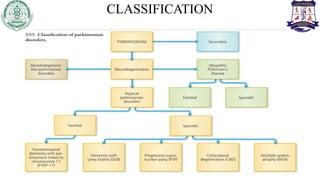
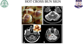
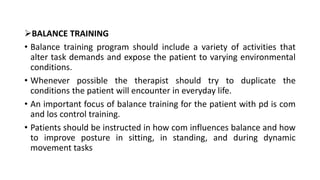
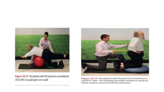
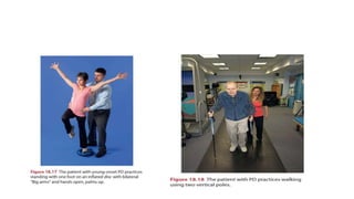
Presented By – Sapna Dhote
(MPT-1)
Neuro-Physiotherapy Department
CONTENT
• Introduction
• Epidemiology
• Classification
• Aetiology
• Relevant anatomy
• Pathophysiology
• Clinical features
• Evaluation
• Diagnosis
• Treatment
• Recent advances
INTRODUCTION
• Parkinson’s disease (PD):It is a progressive disorder of the central
nervous system (CNS) with both motor and non-motor symptoms.
• PD is a common disease that affects an estimated 1million American
and an estimated 7 to 10 million people worldwide.
• The prevalence of the disease is expected to increase substantially in
the coming years due to the aging of the population.
• The average age of onset is 50-60 years.
CLASSIFICATION
PATHOPHYSIOLOGY
• Parkinsonism is a generic term used to describe a group of disorders
with primary disturbance in the dopamine system of basal ganglia
(BG).
• BG is a network of sub cortical nuclei consisting of caudate nucleus,
putamen ,globus pallidus, and subthalamic nucleus with along with
substantia nigra.
• The BG engage in number of parallel circuit or loops ,only few of
which are motor .
DIRECT AND INDIRECT MOTOR LOOPS
PARKINSON’S PLUS SYNDROME
• A group of neurodegenerative diseases can affect the substantia
nigra and produce Parkinsonian symptoms along with other
neurological sign.
• These diseases include strionigral degeneration, shy-drager
syndrome, progressive supranuclear palsy, olivopontocerebellar
atrophy and cortical basal ganglion degeneration.
• In addition, parkinsonian symptoms can be exhibited in patients
with multi –infract vascular disease, Alzheimer’s disease,
Lewy’s body disease, normal pressure hydrocephalus, Wilson’s
disease, juvenile Huntington’s disease.
• Many of these conditions are rare and affect relatively small
numbers of individuals.
• Early in their course ,these disease may present with rigidity and
bradykinesia indistinguishable from PD .
• However other diagnostic symptoms eventually appear.
MULTIPLE SYSTEM ATROPHY
DEFINITION AND EPIDEMIOLOGY
- Multiple system atrophy (MSA) is a sporadic, adult onset,
progressive, neurodegenerative disease of unknown etiology,
characterized by autonomic dysfunction combined with
parkinsonism and/or ataxia. While clinically protean, different
manifestations of the disease are unified by common cellular
pathology featuring glial cytoplasmic inclusions.
The term was proposed as shorthand to cover many cases
described under different titles that overlapped with each other,
including:
• Sporadic olivopontocerebellar atrophy (OPCA):
• Shy–Drager syndrome (SDS)
• Striatonigral degeneration (SND)
• Etiology and pathophysiology: Complex and not well understood.
• Clinical features:Clinical diagnosis has been revised in 2008 by a group of experts via a
consensus conference. Any combination of symptoms and signs of documented autonomic
dysfunction, which, like PD, may pre-date the motor features:
• Orthostatic hypotension.
• Impotence.
• Urinary incontinence.
• Plus one of the following:
• Predominantly cerebellar signs: dysarthria, gait ataxia, oculomotor dysfunction (termed
MSA-C).
• Predominantly parkinsonism, with poor, mild, or Transient response to levodopa therapy
(termed MSA-P).
• The following sign present and are considered ‘red flags’ of supportive features for the diagnosis of
MSA:
• Pyramidal signs: brisk reflexes, Babinski sign, spastic quadriparesis (about 50% of patients).
• Stridor, inspiratory sighs, dysphonia, new or increased snoring.
• Rapidly progressive parkinsonism.
• Dysphagia within 5 years of motor onset.
• Orofacial Dystonia.
• Disproportionate antecolic.
• Camptocormia (severe anterior spine flexion) and/or
• Pisa syndrome (severe lateral spine flexion).
• Contractures of hands/feet.
• Pathologic laughter or crying.
• Myoclonic, jerky action and postural tremor
INVESTIGATIONS
• CT is used to exclude some of the differential diagnoses and to
identify atrophy of the cerebellum and brainstem, particularly the
pons, inferior olives, vermis, and cerebellar peduncles; however,
MRI has a much higher resolution for these structures.
• MRI brain:
1. Hot cross Bun sign –pons (MSA-c)
2. Putaminal rim-MSA-p
• PET scan:18F-fluorodeoxyglucose PET: decreased glucose
metabolism is found in cerebellum, thalamus, putamen, and cortex .
HOT CROSS BUN SIGN
TREATMENT
• Parkinsonism: dopaminergic agents: similar strategies
to PD with the following caveats:
• The response to levodopa is usually transient, poor, waning, or absent, due to
striatal pathology;
• The minimal responsiveness (or lack of) to levodopa is therefore a clinical clue to
the diagnosis of MSA.
• Levodopa may induce dyskinesias Of head and neck in 50% Of MSA-P patient.
• Dopamine agonists or other agents such as MAO inhibitors and amantadine are
second- and third-line therapies due to side-effect profiles and decreased efficacy.
PALLIATIVE THERAPIES
• Continuous positive airway pressure (CPAP) for prominent
stridor; tracheostomy rarely needed and may fatally exacerbate
sleep disordered Breathing.
• Botulinum toxin for drooling, dystonia, or contractures.
• Percutaneous endoscopic gastrostomy (PEG) for severe
dysphagia.
• Physical, occupational, and speech therapies.
PROGRESSIVE SUPRANUCLEAR PALSY
DEFINITION AND EPIDEMIOLOGY
• PSP is a multisystem neurodegenerative disease of the basal ganglia and
brainstem, also known as the Steele–Richardson–Olszewski syndrome,
which presents with a disturbance of balance, impaired downward gaze,
subcortical FLD, and levodopa-unresponsive parkinsonism.
• Patients suffer from progressive dysphagia and dysarthria. Death typically
occurs from complications of immobility, aspiration, or falls.
• Second most common form of parkinsonism after PD.
• Estimated prevalence: 6.4 per 100,000.
• Age of onset: 40–60 years of age, mean onset 60–65
CLINICAL FEATURES
• Typical findings include: Insidious onset of a progressive, symmetric
(but may be asymmetric) parkinsonian syndrome, unresponsive to
levodopa, characterized by:
• Staring, nonblinking, wide eyed (lid retracted) facies, described as
worried or surprised, with suggestion of the term ‘procerus sign’ to
describe the often seen characteristic forehead wrinkling.
• Axial (neck and trunk) dystonia and rigidity and symmetric
bradykinesia.
• Retrocollis or dystonic arm.
• Unsteady gait (wide-based, shuffling)
• Supranuclear ophthalmoparesis, initially involving vertical(particularly downgaze) and
subsequently horizontal eye movements.
• Mild subcortical dementia, characterized by slowness of central processing time.
• Pseudobulbar palsy.
• Dysarthria (spastic: voice has a strained, harsh quality).
• Dysphagia.
• Spasticity of lower and (less so) upper limbs, with hyper-reflexia and extensor plantar responses.
• Bladder and bowel dysfunction in end-stage disease.
• Frontal lobe signs (bradyphrenia, perseveration, primitive reflexes: forced grasping, pout and
imitation and utilization behavior).
• Stuttering speech, torticollis, and blepharospasm may occur.
• Segmental dystonia or myoclonus may occur.
• Nocturnal disturbances: prolonged latency of sleep onset, prolonged wakefulness, frequent early
morning awakenings, reduced total sleep tim
INVESTIGATIONS
• Cranial CT scan may show enlargement of the third ventricle and interpeduncular cistern due to
atrophy of the mid brain.
• MRI brain can show various signs of brainstem atrophy such as reduction in size of part of the
substantia nigra (as in PD). Reduced diameter of the midbrain on sagittal imaging has been termed
the ‘hummingbird sign’
• Treatment
• Treatment is symptomatic.
• Parkinsonism:
• Dopaminergic drugs may reduce the symptoms in some cases but a sustained response is rare.
• Amantadine may cause temporary improvement in a small subset of patients.
• Higher doses of levodopa may be needed for clinical response (up to 1 g daily).
• Levodopa-induced dyskinesias are rare, but dystonia may occur (dysarthria, apraxia of eyelid
closure).
• Visual disturbances:
• Artificial tears (for decreased blink rate).
• Palliative therapies:
• Speech and swallowing assessment and management (dietary
changes, gastrostomy tube).
• Physical and occupational therapy.
• Patient and family support: lay associations, supportive
psychotherapy
HUMMING BIRD SIGN
CORTICAL-BASAL GANGLIONIC
DEGENERATION
DEFINITION AND EPIDEMIOLOGY
• CBD is a rare, sporadic, progressive extrapyramidal degenerative disease of unknown
etiology, characterized by co-occurrence of cortical and basal ganglia signs and
symptoms.
Clinical Features
• Clinical presentation of CBD varies widely, making the disease one of the most
misdiagnosed neurodegenerative disorders. Patients may present with either dementia or
primarily motor features with relatively preserved intellect until late in the disease.
Symptoms May Include
• An asymmetric akinetic–rigid syndrome with dystonic posturing of the hand, with the
wrist flexed and the thumb flexed across the palm .This may spread to the ipsilateral foot
followed by the contralateral limbs.
• The hand often becomes functionally useless due to a severe apraxia rather than to any
weakness
• Tremor : Attempted movement of an affected limb.
• May evoke episodes of fine myoclonus in the forearm flexor muscles that can be misinterpreted as
an action tremor.
• Gait disorder/postural instability: with parkinsonian, apraxic , dystonic, or spastic features.
• Choreoathetosis.
• Focal stimulus-sensitive myoclonus and action tremor.
• Cortical sensory loss.
• Some overlap with PSP, including supranuclear gaze palsy.
• Corticospinal tract signs: Babinski signs, spasticity.
• Pseudobulbar palsy.
• Cortical sensory loss, altered visuospatial function.
• Behavioral manifestations: Frontal lobe-type behavior and language disturbances.
• Dysarthria is a relatively late sign.
INVESTIGATIONS
MRI may be normal or nonspecific, or may demonstrate:
• Asymmetric atrophy in posterior frontal and parietal regions
contralateral to the most affected side atrophy of the cerebral
peduncle.
• Hyperintensity in the subcortical white matter.
• PET/SPECT show focal/asymmetric hypoperfusion on
functional imaging, maximal in the frontoparietal cortex.
TREATMENT
• Symptomatic therapy only, as there is no cure or neuroprotective
agent.
• DEMENTIA WITH LEWY BODIES (DLB)
Definition And Epidemiology
• A dementia and motor syndrome associated with the widespread
presence of Lewy bodies, characterized clinically by fluctuating visual
hallucinations and delusions, parkinsonism (muscle rigidity and
bradykinesia), progressive dementia, and a poor tolerance of
neuroleptic drugs.
PHYSIOTHERAPY INTERVENTION
• Motor learning strategies
• High amplitude exercise training
• Relaxation Exercises
• Flexibility Exercises
• Resistance training
• Functional training
• Balance training
• Pulmonary rehabilitation
• Speech therapy
• Group and home exercises
MOTOR LEARNING
• Patients with PD typically demonstrate motor learning deficits,
including slower learning rates reduced efficiency, And increased
context-specificity of learning.
• Learning complex movement sequences and movements dependent
on internally generated cues are more difficult than those dependent
on external cues.
• In the early and middle stages of the disease, patients can improve
their performance through practice and by using additional sensory
information
• In more advanced stages and in the presence of pronounced cognitive
deficits, training will likely be less successful . the therapist needs to
structure treatment sessions to optimize motor learning.
• Use of structured instructional sets has been shown to improve
movement speed and consistency. For example, walking patterns can
be improved With focused instructions of “swing your arms,”“Walk
fast,” or “take large steps.
• External cues have been shown to be effective in triggering sequential
movements and improving movement characteristics in individuals
with mild to moderate PD.
• Visual cues include
• Stationary floor markings (E.G., Brightly colored lines on the floor
placed perpendicular to the gait path and spaced about one step
length apart) and dynamic transportable cues (E.G., Laser light
signals).
RHYTHMIC AUDITORY STIMULATION (RAS) INCLUDES
• Use of a metronome beat or a steady beat from a musical listening device.
• Multisensory cueing (use of both visual and auditory cueing) has been used
for patients with pd.
HIGH AMPLITUDE TRAINING
• Patients are guided by a physical therapist to exercise at a high intensity
(8/10 Borg’s RPE scale) for 1 hour 4 times a week for 4 weeks with large
amplitude, multiple repetitions , and whole body movements that increase
in complexity
• Examples of the exercises and patient directions include the following:
• “Reach left arm across the body to opposite side, keep hand open, palm
up, right leg fully extended, toe pushing into the floor. Repeat on other leg
and alternate
RELAXATION EXERCISES
• Gentle rocking can be used to produce generalized relaxation of
excessive muscle tension owing to rigidity.
• a rocking chair can be used to temporarily relax the patient and
enhance sit-to-stand transfers. During therapy, slow, rhythmic,
rotational movements of the extremities and trunk can precede
interventions such as ROM and stretching, and functional training.
• Another strategy to promote relaxation is emphasis on diaphragmatic
breathing during exercise
FLEXIBILITY EXERCISE
• The purpose of flexibility exercise (stretching) is to improve ROM and
physical function. A combination of static (PROM), dynamic (AROM),
and facilitated PNF exercises is used to achieve maximum ROM.
• Flexibility exercises should be performed a minimum 2 to 3 days per
week and ideally 5 to 7 days per week
• Special consideration should be given to stretching common areas of
limitation .
• Stretching can be combined with joint mobilization techniques to
reduce tightness of the joint capsule or of ligaments around a joint .
• By using selected grades of accessory movement, both improved
ROM and decreased pain can be achieved.
RESISTANCE TRAINING
• Resistance training is indicated for patients with PD who demonstrate
primary muscle weakness with impaired motor unit recruitment and
rate of force development and disuse weakness associated with
prolonged inactivity.
• Specific areas of weakness are targeted.
BALANCE TRAINING
• Balance training program should include a variety of activities that
alter task demands and expose the patient to varying environmental
conditions.
• Whenever possible the therapist should try to duplicate the
conditions the patient will encounter in everyday life.
• An important focus of balance training for the patient with pd is com
and los control training.
• Patients should be instructed in how com influences balance and how
to improve posture in sitting, in standing, and during dynamic
movement tasks
FUNCTIONAL TRAINING
• An exercise program should be based on focused practice of
functional skills.
• The overall emphasis is on improving functional mobility with specific
emphasis on improving mobility of axial structures, the head, trunk,
hips, and shoulders.
• Progression to more difficult motor activities should be gradual.
RECENT ADVANCES
• Multimodal Balance Training Supported by
Rhythmical Auditory Stimuli in Parkinson's
Disease: A Randomized Clinical Trial :- Tamine T
C Capato 1 2, Nienke M de Vries 1, Joanna IntHout 3, Egberto R
Barbosa 2, Jorik Nonnekes 4, Bastiaan R Bloem 1
REFERENCE
• Physical Rehabilitation (O'Sullivan, Physical Rehabilitation)
• Hankey's Clinical Neurology Second Edition
• Umphred's Neurological Rehabilitation Sixth Edition
THANK YOU

Parkinson Plus Seminar PPT.pptx

  • 1.
    Presented By –Sapna Dhote (MPT-1) Neuro-Physiotherapy Department
  • 2.
    CONTENT • Introduction • Epidemiology •Classification • Aetiology • Relevant anatomy • Pathophysiology • Clinical features • Evaluation • Diagnosis • Treatment • Recent advances
  • 3.
    INTRODUCTION • Parkinson’s disease(PD):It is a progressive disorder of the central nervous system (CNS) with both motor and non-motor symptoms. • PD is a common disease that affects an estimated 1million American and an estimated 7 to 10 million people worldwide. • The prevalence of the disease is expected to increase substantially in the coming years due to the aging of the population. • The average age of onset is 50-60 years.
  • 4.
  • 5.
    PATHOPHYSIOLOGY • Parkinsonism isa generic term used to describe a group of disorders with primary disturbance in the dopamine system of basal ganglia (BG). • BG is a network of sub cortical nuclei consisting of caudate nucleus, putamen ,globus pallidus, and subthalamic nucleus with along with substantia nigra. • The BG engage in number of parallel circuit or loops ,only few of which are motor .
  • 7.
  • 8.
    PARKINSON’S PLUS SYNDROME •A group of neurodegenerative diseases can affect the substantia nigra and produce Parkinsonian symptoms along with other neurological sign. • These diseases include strionigral degeneration, shy-drager syndrome, progressive supranuclear palsy, olivopontocerebellar atrophy and cortical basal ganglion degeneration. • In addition, parkinsonian symptoms can be exhibited in patients with multi –infract vascular disease, Alzheimer’s disease, Lewy’s body disease, normal pressure hydrocephalus, Wilson’s disease, juvenile Huntington’s disease.
  • 9.
    • Many ofthese conditions are rare and affect relatively small numbers of individuals. • Early in their course ,these disease may present with rigidity and bradykinesia indistinguishable from PD . • However other diagnostic symptoms eventually appear.
  • 10.
    MULTIPLE SYSTEM ATROPHY DEFINITIONAND EPIDEMIOLOGY - Multiple system atrophy (MSA) is a sporadic, adult onset, progressive, neurodegenerative disease of unknown etiology, characterized by autonomic dysfunction combined with parkinsonism and/or ataxia. While clinically protean, different manifestations of the disease are unified by common cellular pathology featuring glial cytoplasmic inclusions.
  • 11.
    The term wasproposed as shorthand to cover many cases described under different titles that overlapped with each other, including: • Sporadic olivopontocerebellar atrophy (OPCA): • Shy–Drager syndrome (SDS) • Striatonigral degeneration (SND)
  • 12.
    • Etiology andpathophysiology: Complex and not well understood. • Clinical features:Clinical diagnosis has been revised in 2008 by a group of experts via a consensus conference. Any combination of symptoms and signs of documented autonomic dysfunction, which, like PD, may pre-date the motor features: • Orthostatic hypotension. • Impotence. • Urinary incontinence. • Plus one of the following: • Predominantly cerebellar signs: dysarthria, gait ataxia, oculomotor dysfunction (termed MSA-C). • Predominantly parkinsonism, with poor, mild, or Transient response to levodopa therapy (termed MSA-P).
  • 13.
    • The followingsign present and are considered ‘red flags’ of supportive features for the diagnosis of MSA: • Pyramidal signs: brisk reflexes, Babinski sign, spastic quadriparesis (about 50% of patients). • Stridor, inspiratory sighs, dysphonia, new or increased snoring. • Rapidly progressive parkinsonism. • Dysphagia within 5 years of motor onset. • Orofacial Dystonia. • Disproportionate antecolic. • Camptocormia (severe anterior spine flexion) and/or • Pisa syndrome (severe lateral spine flexion). • Contractures of hands/feet. • Pathologic laughter or crying. • Myoclonic, jerky action and postural tremor
  • 14.
    INVESTIGATIONS • CT isused to exclude some of the differential diagnoses and to identify atrophy of the cerebellum and brainstem, particularly the pons, inferior olives, vermis, and cerebellar peduncles; however, MRI has a much higher resolution for these structures. • MRI brain: 1. Hot cross Bun sign –pons (MSA-c) 2. Putaminal rim-MSA-p • PET scan:18F-fluorodeoxyglucose PET: decreased glucose metabolism is found in cerebellum, thalamus, putamen, and cortex .
  • 15.
  • 16.
    TREATMENT • Parkinsonism: dopaminergicagents: similar strategies to PD with the following caveats: • The response to levodopa is usually transient, poor, waning, or absent, due to striatal pathology; • The minimal responsiveness (or lack of) to levodopa is therefore a clinical clue to the diagnosis of MSA. • Levodopa may induce dyskinesias Of head and neck in 50% Of MSA-P patient. • Dopamine agonists or other agents such as MAO inhibitors and amantadine are second- and third-line therapies due to side-effect profiles and decreased efficacy.
  • 17.
    PALLIATIVE THERAPIES • Continuouspositive airway pressure (CPAP) for prominent stridor; tracheostomy rarely needed and may fatally exacerbate sleep disordered Breathing. • Botulinum toxin for drooling, dystonia, or contractures. • Percutaneous endoscopic gastrostomy (PEG) for severe dysphagia. • Physical, occupational, and speech therapies.
  • 18.
    PROGRESSIVE SUPRANUCLEAR PALSY DEFINITIONAND EPIDEMIOLOGY • PSP is a multisystem neurodegenerative disease of the basal ganglia and brainstem, also known as the Steele–Richardson–Olszewski syndrome, which presents with a disturbance of balance, impaired downward gaze, subcortical FLD, and levodopa-unresponsive parkinsonism. • Patients suffer from progressive dysphagia and dysarthria. Death typically occurs from complications of immobility, aspiration, or falls. • Second most common form of parkinsonism after PD. • Estimated prevalence: 6.4 per 100,000. • Age of onset: 40–60 years of age, mean onset 60–65
  • 19.
    CLINICAL FEATURES • Typicalfindings include: Insidious onset of a progressive, symmetric (but may be asymmetric) parkinsonian syndrome, unresponsive to levodopa, characterized by: • Staring, nonblinking, wide eyed (lid retracted) facies, described as worried or surprised, with suggestion of the term ‘procerus sign’ to describe the often seen characteristic forehead wrinkling. • Axial (neck and trunk) dystonia and rigidity and symmetric bradykinesia. • Retrocollis or dystonic arm. • Unsteady gait (wide-based, shuffling)
  • 20.
    • Supranuclear ophthalmoparesis,initially involving vertical(particularly downgaze) and subsequently horizontal eye movements. • Mild subcortical dementia, characterized by slowness of central processing time. • Pseudobulbar palsy. • Dysarthria (spastic: voice has a strained, harsh quality). • Dysphagia. • Spasticity of lower and (less so) upper limbs, with hyper-reflexia and extensor plantar responses. • Bladder and bowel dysfunction in end-stage disease. • Frontal lobe signs (bradyphrenia, perseveration, primitive reflexes: forced grasping, pout and imitation and utilization behavior). • Stuttering speech, torticollis, and blepharospasm may occur. • Segmental dystonia or myoclonus may occur. • Nocturnal disturbances: prolonged latency of sleep onset, prolonged wakefulness, frequent early morning awakenings, reduced total sleep tim
  • 21.
    INVESTIGATIONS • Cranial CTscan may show enlargement of the third ventricle and interpeduncular cistern due to atrophy of the mid brain. • MRI brain can show various signs of brainstem atrophy such as reduction in size of part of the substantia nigra (as in PD). Reduced diameter of the midbrain on sagittal imaging has been termed the ‘hummingbird sign’ • Treatment • Treatment is symptomatic. • Parkinsonism: • Dopaminergic drugs may reduce the symptoms in some cases but a sustained response is rare. • Amantadine may cause temporary improvement in a small subset of patients. • Higher doses of levodopa may be needed for clinical response (up to 1 g daily). • Levodopa-induced dyskinesias are rare, but dystonia may occur (dysarthria, apraxia of eyelid closure).
  • 22.
    • Visual disturbances: •Artificial tears (for decreased blink rate). • Palliative therapies: • Speech and swallowing assessment and management (dietary changes, gastrostomy tube). • Physical and occupational therapy. • Patient and family support: lay associations, supportive psychotherapy
  • 23.
  • 24.
    CORTICAL-BASAL GANGLIONIC DEGENERATION DEFINITION ANDEPIDEMIOLOGY • CBD is a rare, sporadic, progressive extrapyramidal degenerative disease of unknown etiology, characterized by co-occurrence of cortical and basal ganglia signs and symptoms. Clinical Features • Clinical presentation of CBD varies widely, making the disease one of the most misdiagnosed neurodegenerative disorders. Patients may present with either dementia or primarily motor features with relatively preserved intellect until late in the disease. Symptoms May Include • An asymmetric akinetic–rigid syndrome with dystonic posturing of the hand, with the wrist flexed and the thumb flexed across the palm .This may spread to the ipsilateral foot followed by the contralateral limbs. • The hand often becomes functionally useless due to a severe apraxia rather than to any weakness
  • 25.
    • Tremor :Attempted movement of an affected limb. • May evoke episodes of fine myoclonus in the forearm flexor muscles that can be misinterpreted as an action tremor. • Gait disorder/postural instability: with parkinsonian, apraxic , dystonic, or spastic features. • Choreoathetosis. • Focal stimulus-sensitive myoclonus and action tremor. • Cortical sensory loss. • Some overlap with PSP, including supranuclear gaze palsy. • Corticospinal tract signs: Babinski signs, spasticity. • Pseudobulbar palsy. • Cortical sensory loss, altered visuospatial function. • Behavioral manifestations: Frontal lobe-type behavior and language disturbances. • Dysarthria is a relatively late sign.
  • 26.
    INVESTIGATIONS MRI may benormal or nonspecific, or may demonstrate: • Asymmetric atrophy in posterior frontal and parietal regions contralateral to the most affected side atrophy of the cerebral peduncle. • Hyperintensity in the subcortical white matter. • PET/SPECT show focal/asymmetric hypoperfusion on functional imaging, maximal in the frontoparietal cortex.
  • 27.
    TREATMENT • Symptomatic therapyonly, as there is no cure or neuroprotective agent. • DEMENTIA WITH LEWY BODIES (DLB) Definition And Epidemiology • A dementia and motor syndrome associated with the widespread presence of Lewy bodies, characterized clinically by fluctuating visual hallucinations and delusions, parkinsonism (muscle rigidity and bradykinesia), progressive dementia, and a poor tolerance of neuroleptic drugs.
  • 28.
    PHYSIOTHERAPY INTERVENTION • Motorlearning strategies • High amplitude exercise training • Relaxation Exercises • Flexibility Exercises • Resistance training • Functional training • Balance training • Pulmonary rehabilitation • Speech therapy • Group and home exercises
  • 29.
    MOTOR LEARNING • Patientswith PD typically demonstrate motor learning deficits, including slower learning rates reduced efficiency, And increased context-specificity of learning. • Learning complex movement sequences and movements dependent on internally generated cues are more difficult than those dependent on external cues. • In the early and middle stages of the disease, patients can improve their performance through practice and by using additional sensory information
  • 30.
    • In moreadvanced stages and in the presence of pronounced cognitive deficits, training will likely be less successful . the therapist needs to structure treatment sessions to optimize motor learning. • Use of structured instructional sets has been shown to improve movement speed and consistency. For example, walking patterns can be improved With focused instructions of “swing your arms,”“Walk fast,” or “take large steps.
  • 31.
    • External cueshave been shown to be effective in triggering sequential movements and improving movement characteristics in individuals with mild to moderate PD. • Visual cues include • Stationary floor markings (E.G., Brightly colored lines on the floor placed perpendicular to the gait path and spaced about one step length apart) and dynamic transportable cues (E.G., Laser light signals).
  • 32.
    RHYTHMIC AUDITORY STIMULATION(RAS) INCLUDES • Use of a metronome beat or a steady beat from a musical listening device. • Multisensory cueing (use of both visual and auditory cueing) has been used for patients with pd. HIGH AMPLITUDE TRAINING • Patients are guided by a physical therapist to exercise at a high intensity (8/10 Borg’s RPE scale) for 1 hour 4 times a week for 4 weeks with large amplitude, multiple repetitions , and whole body movements that increase in complexity • Examples of the exercises and patient directions include the following: • “Reach left arm across the body to opposite side, keep hand open, palm up, right leg fully extended, toe pushing into the floor. Repeat on other leg and alternate
  • 34.
    RELAXATION EXERCISES • Gentlerocking can be used to produce generalized relaxation of excessive muscle tension owing to rigidity. • a rocking chair can be used to temporarily relax the patient and enhance sit-to-stand transfers. During therapy, slow, rhythmic, rotational movements of the extremities and trunk can precede interventions such as ROM and stretching, and functional training. • Another strategy to promote relaxation is emphasis on diaphragmatic breathing during exercise
  • 35.
    FLEXIBILITY EXERCISE • Thepurpose of flexibility exercise (stretching) is to improve ROM and physical function. A combination of static (PROM), dynamic (AROM), and facilitated PNF exercises is used to achieve maximum ROM. • Flexibility exercises should be performed a minimum 2 to 3 days per week and ideally 5 to 7 days per week • Special consideration should be given to stretching common areas of limitation .
  • 36.
    • Stretching canbe combined with joint mobilization techniques to reduce tightness of the joint capsule or of ligaments around a joint . • By using selected grades of accessory movement, both improved ROM and decreased pain can be achieved.
  • 37.
    RESISTANCE TRAINING • Resistancetraining is indicated for patients with PD who demonstrate primary muscle weakness with impaired motor unit recruitment and rate of force development and disuse weakness associated with prolonged inactivity. • Specific areas of weakness are targeted.
  • 38.
    BALANCE TRAINING • Balancetraining program should include a variety of activities that alter task demands and expose the patient to varying environmental conditions. • Whenever possible the therapist should try to duplicate the conditions the patient will encounter in everyday life. • An important focus of balance training for the patient with pd is com and los control training. • Patients should be instructed in how com influences balance and how to improve posture in sitting, in standing, and during dynamic movement tasks
  • 40.
    FUNCTIONAL TRAINING • Anexercise program should be based on focused practice of functional skills. • The overall emphasis is on improving functional mobility with specific emphasis on improving mobility of axial structures, the head, trunk, hips, and shoulders. • Progression to more difficult motor activities should be gradual.
  • 42.
    RECENT ADVANCES • MultimodalBalance Training Supported by Rhythmical Auditory Stimuli in Parkinson's Disease: A Randomized Clinical Trial :- Tamine T C Capato 1 2, Nienke M de Vries 1, Joanna IntHout 3, Egberto R Barbosa 2, Jorik Nonnekes 4, Bastiaan R Bloem 1
  • 43.
    REFERENCE • Physical Rehabilitation(O'Sullivan, Physical Rehabilitation) • Hankey's Clinical Neurology Second Edition • Umphred's Neurological Rehabilitation Sixth Edition
  • 44.