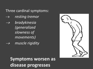
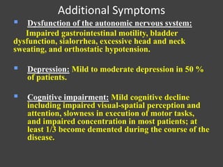
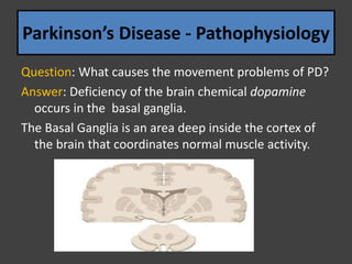
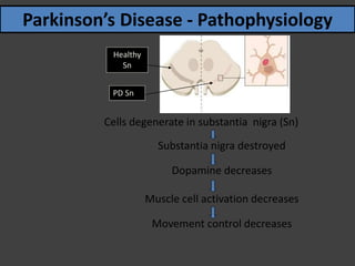
Parkinsonism
Dr. Varun SJ Doctor of Pharmacy,
INDEX
• History
• Definition
• Causes
• Risk factors
• Symptoms
• Pathophysiology
• Important questions with answers
It is a clinical syndrome characterized
by
diminished facial expression,
stooped posture,
slowness of voluntary movement,
festinating gait,
rigidity and
a “pill – rolling” tremor
 First described in 1817 by an English
physician, James Parkinson, in “An Essay
on the Shaking Palsy.”
 The famous French neurologist, Charcot,
further described the syndrome in the late
1800s
Parkinson Disease
• It is a degenerative disorder of the CNS
• The most common movement disorder
affecting 1-2 % of the general population
over the age of 65 years.
• Parkinson's disease is the most common
form of parkinsonism
What causes PD
May be combination of factors involving
genetics, environmental agents,& abnormalities
in cellular process.
Risk Factors Age - the most important risk factor
Develops around age 50
* incidence rises with age
* affects 1-2% of population > age 65
 Positive family history
 Male gender
Higher incidence in men (62%) compared to women (38%)
 Environmental exposure: Herbicide and pesticide exposure, metals
(manganese, iron), well water, farming, rural residence, wood pulp
mills; and steel alloy industries
 Life experiences (trauma, emotional stress, personality traits such as
shyness and depressiveness)
Three cardinal symptoms:
 resting tremor
 bradykinesia
(generalized
slowness of
movements)
 muscle rigidity
Symptoms worsen as
disease progresses
 Resting tremor: Most common first
symptom, usually asymmetric and most
evident in one hand with the arm at rest.
can occur in head, face, jaw, & leg
• Bradykinesia: Difficulty with daily
activities such as writing, shaving,
using a knife and fork, and opening
buttons; decreased blinking, masked
facies, slowed chewing and
swallowing.
• Rigidity: Muscle tone increased in
both flexor and extensor muscles
providing a constant resistance to
passive movements of the joints;
stooped posture, anteroflexed head,
and flexed knees and elbows.
Postural manifestations –
■ postural instability
■ rigidity
■ stooped
Additional Symptoms
 Dysfunction of the autonomic nervous system:
Impaired gastrointestinal motility, bladder
dysfunction, sialorrhea, excessive head and neck
sweating, and orthostatic hypotension.
 Depression: Mild to moderate depression in 50 %
of patients.
 Cognitive impairment: Mild cognitive decline
including impaired visual-spatial perception and
attention, slowness in execution of motor tasks,
and impaired concentration in most patients; at
least 1/3 become demented during the course of the
disease.
Parkinson’s Disease – Stages of Symptoms
Stage Symptoms
One Unilateral
Tw o Bilateral
No balanc e impairment
Three Balanc e impairment
Mild to mod erate d isease
Physic ally ind ependent
Four Severe d isability
Still able to w alk & stand unassisted
Five Wheelc hair-bound or bed rid d ened
unlessassisted
Parkinson’s Disease - Pathophysiology
Question: What causes the movement problems of PD?
Answer: Deficiency of the brain chemical dopamine
occurs in the basal ganglia.
The Basal Ganglia is an area deep inside the cortex of
the brain that coordinates normal muscle activity.
• Question: What causes the dopamine
deficiency?
• Answer: Degenerative changes in the
substantia nigra and striatum portions of the
basal ganglia reduce dopamine production.
Cross section of striatum
Substantia nigra
Cells degenerate in substantia nigra (Sn)
Substantia nigra destroyed
Dopamine decreases
Muscle cell activation decreases
Movement control decreases
Parkinson’s Disease - Pathophysiology
Healthy
Sn
PD Sn
Pathophysiology
• Imbalance of dopamine and acetylcholine
• Loss of 80 to 90% of dopaminergic production
in the substantia nigra
• Lewy Bodies
• Lewy bodies are abnormal aggregates of
protein that develop inside nerve cells in
Parkinson's disease (PD). They are
identified under the microscope when
histology is performed on the brain.
• Lewy bodies appear as spherical masses
that displace other cell components.
There are two morphological types:
classical (brain stem) Lewy bodies and
cortical Lewy bodies.
• A classical Lewy body is an eosinophilic
cytoplasmic inclusion that consists of a dense
core surrounded by a halo of 10-nm wide
radiating fibrils, the primary structural
component of which is alpha-synuclein.
• In contrast, a cortical Lewy body is less well
defined and lacks the halo. Nonetheless, it is
still made up of alpha-synuclein fibrils.
Morphology
• Macroscopic findings : substantiua nigra - pallor
• Microscopic examination:
 loss of pigmented, catecholaminergic neurons
 Lewy bodies found in remaining neurons
• Multiple system atrophy (MSA) is a progressive
neurodegenerative disorder characterized by
symptoms of autonomic nervous system failure such
as fainting spells and bladder control problems,
combined with motor control symptoms such as
tremor, rigidity, and loss of muscle coordination.
• MSA affects both men and women primarily in their
50s
Multiple System Atrophy
MSA = OPCA + SDS + SND
• Olivopontocerebellar atrophy (OPCA)
• Shy-Drager syndrome (SDS)
• Striatonigral degeneration (SND)
• The disease tends to advance rapidly
over the course of 9 to 10 years, with
progressive loss of motor skills,
eventual confinement to bed, and
death.
• There is no remission from the
disease. There is currently no cure
Multiple System Atrophy
• Neurodegenerative disorders affecting
multiple neural systems
• Characterized by the presence of glial
cytoplasmic inclusions (GCIs), typically
within the cytoplasm of
oligodentrocytes.
• α Synuclein is the major component of
the inclusion – synucleinopathy
• Unlike PD with (α synuclein inclusion) no
mutations in the gene for this synaptic protein
• (A53T, A30P and E46K) – MUTATED GENES
SPECIFIC FOR PD
• Definition of categories of MSA: The 2
categories of MSA are (1) with predominant
parkinsonism (MSA-P) and (2) with cerebellar
features (MSA-C).
• MSA-P is the category of MSA where
extrapyramidal features predominate. The term
striatonigral degeneration, parkinsonian variant
is sometimes used.
• MSA-C is the category when cerebellar ataxia
predominates. It is sometimes termed sporadic
olivopontocerebellar atrophy.
• The designation to a category MSA-P or MSA-C
depends on the dominant feature at the time of
evaluation, which can change with time.
Clinical Features
• Two principal symptoms
are parkinsonism and autonomic dysfunction,
particularly orthostatic hypotension.
• When these are present in relative isolation,
the syndromes may be referred to as
striatonigral degeneration and Shy Drager
syndrome respectively.
• Parkinsonism (akinesia and rigidity) can be
related to degree of cell loss from the
substantia nigra and striatum
• Ataxia - with changes in the circuits involving
the pons, cerebellum and inferior olive.
• autonomic symptoms - with loss from the
catecholaminergic nuclei of the medulla and
the intermediolateral cell column of the spinal
cord.
Morphology
• Macroscopic examination – atrophy of the
cerebellum, including the cerebellar peduncles,
pons, medulla, substantia nigra and striatum
• These brain regions shows neural loss as well
as cytoplasmic and nuclear inclusions
• These inclusions contains a synuclein, ubiquitin
and α B crystallin.

Parkinsonism Disease

  • 1.
    Parkinsonism Dr. Varun SJDoctor of Pharmacy,
  • 2.
    INDEX • History • Definition •Causes • Risk factors • Symptoms • Pathophysiology • Important questions with answers
  • 3.
    It is aclinical syndrome characterized by diminished facial expression, stooped posture, slowness of voluntary movement, festinating gait, rigidity and a “pill – rolling” tremor
  • 4.
     First describedin 1817 by an English physician, James Parkinson, in “An Essay on the Shaking Palsy.”  The famous French neurologist, Charcot, further described the syndrome in the late 1800s Parkinson Disease
  • 5.
    • It isa degenerative disorder of the CNS • The most common movement disorder affecting 1-2 % of the general population over the age of 65 years. • Parkinson's disease is the most common form of parkinsonism
  • 6.
    What causes PD Maybe combination of factors involving genetics, environmental agents,& abnormalities in cellular process.
  • 7.
    Risk Factors Age- the most important risk factor Develops around age 50 * incidence rises with age * affects 1-2% of population > age 65  Positive family history  Male gender Higher incidence in men (62%) compared to women (38%)  Environmental exposure: Herbicide and pesticide exposure, metals (manganese, iron), well water, farming, rural residence, wood pulp mills; and steel alloy industries  Life experiences (trauma, emotional stress, personality traits such as shyness and depressiveness)
  • 8.
    Three cardinal symptoms: resting tremor  bradykinesia (generalized slowness of movements)  muscle rigidity Symptoms worsen as disease progresses
  • 9.
     Resting tremor:Most common first symptom, usually asymmetric and most evident in one hand with the arm at rest. can occur in head, face, jaw, & leg
  • 10.
    • Bradykinesia: Difficultywith daily activities such as writing, shaving, using a knife and fork, and opening buttons; decreased blinking, masked facies, slowed chewing and swallowing.
  • 11.
    • Rigidity: Muscletone increased in both flexor and extensor muscles providing a constant resistance to passive movements of the joints; stooped posture, anteroflexed head, and flexed knees and elbows.
  • 12.
    Postural manifestations – ■postural instability ■ rigidity ■ stooped
  • 13.
    Additional Symptoms  Dysfunctionof the autonomic nervous system: Impaired gastrointestinal motility, bladder dysfunction, sialorrhea, excessive head and neck sweating, and orthostatic hypotension.  Depression: Mild to moderate depression in 50 % of patients.  Cognitive impairment: Mild cognitive decline including impaired visual-spatial perception and attention, slowness in execution of motor tasks, and impaired concentration in most patients; at least 1/3 become demented during the course of the disease.
  • 14.
    Parkinson’s Disease –Stages of Symptoms Stage Symptoms One Unilateral Tw o Bilateral No balanc e impairment Three Balanc e impairment Mild to mod erate d isease Physic ally ind ependent Four Severe d isability Still able to w alk & stand unassisted Five Wheelc hair-bound or bed rid d ened unlessassisted
  • 15.
    Parkinson’s Disease -Pathophysiology Question: What causes the movement problems of PD? Answer: Deficiency of the brain chemical dopamine occurs in the basal ganglia. The Basal Ganglia is an area deep inside the cortex of the brain that coordinates normal muscle activity.
  • 16.
    • Question: Whatcauses the dopamine deficiency? • Answer: Degenerative changes in the substantia nigra and striatum portions of the basal ganglia reduce dopamine production. Cross section of striatum Substantia nigra
  • 17.
    Cells degenerate insubstantia nigra (Sn) Substantia nigra destroyed Dopamine decreases Muscle cell activation decreases Movement control decreases Parkinson’s Disease - Pathophysiology Healthy Sn PD Sn
  • 18.
    Pathophysiology • Imbalance ofdopamine and acetylcholine • Loss of 80 to 90% of dopaminergic production in the substantia nigra • Lewy Bodies
  • 19.
    • Lewy bodiesare abnormal aggregates of protein that develop inside nerve cells in Parkinson's disease (PD). They are identified under the microscope when histology is performed on the brain. • Lewy bodies appear as spherical masses that displace other cell components. There are two morphological types: classical (brain stem) Lewy bodies and cortical Lewy bodies.
  • 20.
    • A classicalLewy body is an eosinophilic cytoplasmic inclusion that consists of a dense core surrounded by a halo of 10-nm wide radiating fibrils, the primary structural component of which is alpha-synuclein. • In contrast, a cortical Lewy body is less well defined and lacks the halo. Nonetheless, it is still made up of alpha-synuclein fibrils.
  • 21.
    Morphology • Macroscopic findings: substantiua nigra - pallor • Microscopic examination:  loss of pigmented, catecholaminergic neurons  Lewy bodies found in remaining neurons
  • 22.
    • Multiple systematrophy (MSA) is a progressive neurodegenerative disorder characterized by symptoms of autonomic nervous system failure such as fainting spells and bladder control problems, combined with motor control symptoms such as tremor, rigidity, and loss of muscle coordination. • MSA affects both men and women primarily in their 50s Multiple System Atrophy
  • 23.
    MSA = OPCA+ SDS + SND • Olivopontocerebellar atrophy (OPCA) • Shy-Drager syndrome (SDS) • Striatonigral degeneration (SND)
  • 24.
    • The diseasetends to advance rapidly over the course of 9 to 10 years, with progressive loss of motor skills, eventual confinement to bed, and death. • There is no remission from the disease. There is currently no cure
  • 25.
    Multiple System Atrophy •Neurodegenerative disorders affecting multiple neural systems • Characterized by the presence of glial cytoplasmic inclusions (GCIs), typically within the cytoplasm of oligodentrocytes. • α Synuclein is the major component of the inclusion – synucleinopathy
  • 26.
    • Unlike PDwith (α synuclein inclusion) no mutations in the gene for this synaptic protein • (A53T, A30P and E46K) – MUTATED GENES SPECIFIC FOR PD
  • 27.
    • Definition ofcategories of MSA: The 2 categories of MSA are (1) with predominant parkinsonism (MSA-P) and (2) with cerebellar features (MSA-C). • MSA-P is the category of MSA where extrapyramidal features predominate. The term striatonigral degeneration, parkinsonian variant is sometimes used. • MSA-C is the category when cerebellar ataxia predominates. It is sometimes termed sporadic olivopontocerebellar atrophy. • The designation to a category MSA-P or MSA-C depends on the dominant feature at the time of evaluation, which can change with time.
  • 28.
    Clinical Features • Twoprincipal symptoms are parkinsonism and autonomic dysfunction, particularly orthostatic hypotension. • When these are present in relative isolation, the syndromes may be referred to as striatonigral degeneration and Shy Drager syndrome respectively.
  • 29.
    • Parkinsonism (akinesiaand rigidity) can be related to degree of cell loss from the substantia nigra and striatum • Ataxia - with changes in the circuits involving the pons, cerebellum and inferior olive. • autonomic symptoms - with loss from the catecholaminergic nuclei of the medulla and the intermediolateral cell column of the spinal cord.
  • 30.
    Morphology • Macroscopic examination– atrophy of the cerebellum, including the cerebellar peduncles, pons, medulla, substantia nigra and striatum • These brain regions shows neural loss as well as cytoplasmic and nuclear inclusions • These inclusions contains a synuclein, ubiquitin and α B crystallin.