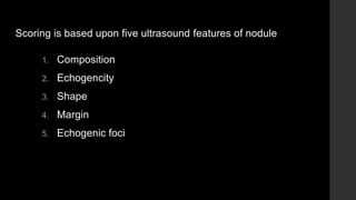
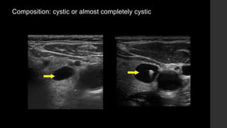
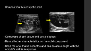
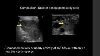
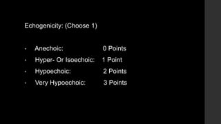
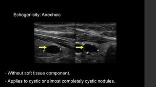
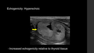
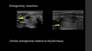
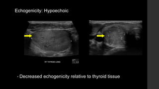
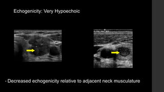
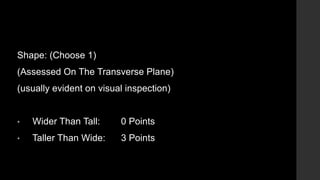
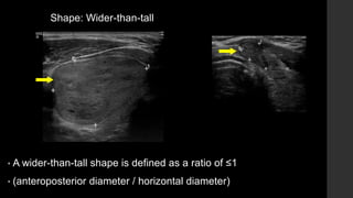
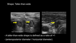
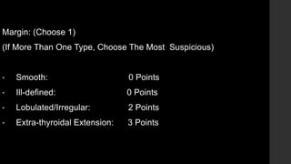
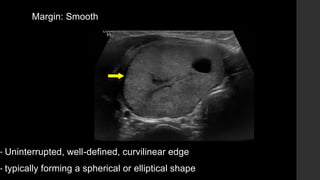
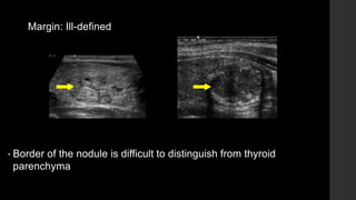
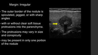
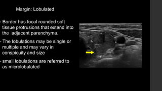
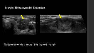
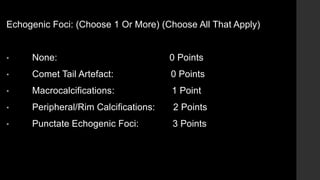
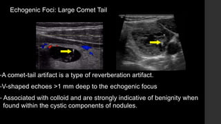
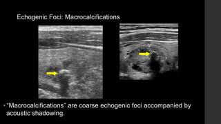
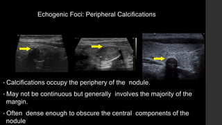
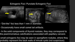
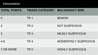
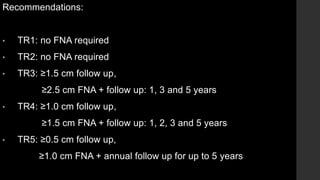
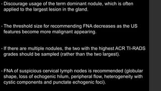
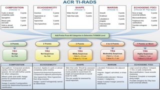
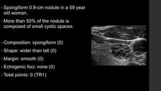
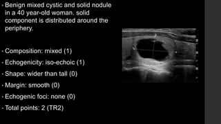
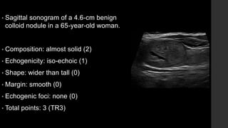
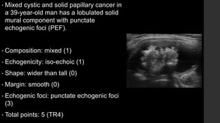
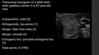
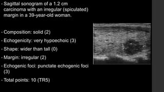
Thyroid Imaging Reporting & Data System
(ACR TI-RADS)
Dr Haseeb Manzoor
Department Of Radiology
Shalamar Hospital
• TI-RADS is a reporting system for thyroid nodules proposed by the
american college of radiology (ACR)
• This uses a standardized scoring system for reports
• Malignancy risk stratification
• Provides users recommendations for:
• When to use fine needle aspiration (FNA)
• When to suggest ultrasound follow-up
• When to leave alone nodules
Scoring is based upon five ultrasound features of nodule
1. Composition
2. Echogencity
3. Shape
4. Margin
5. Echogenic foci
Composition: (choose 1)
• Cystic or completely cystic: 0 points
• Spongiform: 0 points
• Mixed cystic and solid: 1 point
• Solid or almost completely solid: 2 points
Composition: cystic or almost completely cystic
Composition: Spongiform
• Composed predominately (>50%) of tiny cystic spaces.
• Spongiform is a benign finding.
• If there is concern for irregular margins or suspicious echogenic
foci, reconsider your choice of spongiform.
Composition: Mixed cystic solid
• Composed of soft tissue and cystic spaces.
• Base all other characteristics on the solid component.
• Solid material that is eccentric and has an acute angle with the
nodule’s wall is suspicious.
Composition: Solid or almost completely solid
• Composed entirely or nearly entirely of soft tissue, with only a
few tiny cystic spaces
Echogenicity: (Choose 1)
• Anechoic: 0 Points
• Hyper- Or Isoechoic: 1 Point
• Hypoechoic: 2 Points
• Very Hypoechoic: 3 Points
Echogenicity: Anechoic
• Without soft tissue component.
• Applies to cystic or almost completely cystic nodules.
Echogenicity: Hyperechoic
• Increased echogenicity relative to thyroid tissue
Echogenicity: Isoechoic
• Similar echogenicity relative to thyroid tissue
Echogenicity: Hypoechoic
• Decreased echogenicity relative to thyroid tissue
Echogenicity: Very Hypoechoic
• Decreased echogenicity relative to adjacent neck musculature
Shape: (Choose 1)
(Assessed On The Transverse Plane)
(usually evident on visual inspection)
• Wider Than Tall: 0 Points
• Taller Than Wide: 3 Points
Shape: Wider-than-tall
• A wider-than-tall shape is defined as a ratio of ≤1
• (anteroposterior diameter / horizontal diameter)
Shape: Taller-than-wide
• A taller-than-wide shape is defined as a ratio of >1
• (anteroposterior diameter / horizontal diameter)
Margin: (Choose 1)
(If More Than One Type, Choose The Most Suspicious)
• Smooth: 0 Points
• Ill-defined: 0 Points
• Lobulated/Irregular: 2 Points
• Extra-thyroidal Extension: 3 Points
Margin: Smooth
• Uninterrupted, well-defined, curvilinear edge
• typically forming a spherical or elliptical shape
Margin: Ill-defined
• Border of the nodule is difficult to distinguish from thyroid
parenchyma
Margin: Irregular
• The outer border of the nodule is
spiculated, jagged, or with sharp
angles
• with or without clear soft tissue
protrusions into the parenchyma.
• The protrusions may vary in size
and conspicuity
• may be present in only one portion
of the nodule
Margin: Lobulated
• Border has focal rounded soft
tissue protrusions that extend into
the adjacent parenchyma.
• The lobulations may be single or
multiple and may vary in
conspicuity and size
• small lobulations are referred to
as microlobulated
Margin: Extrathyroidal Extension
• Nodule extends through the thyroid margin
Echogenic Foci: (Choose 1 Or More) (Choose All That Apply)
• None: 0 Points
• Comet Tail Artefact: 0 Points
• Macrocalcifications: 1 Point
• Peripheral/Rim Calcifications: 2 Points
• Punctate Echogenic Foci: 3 Points
Echogenic Foci: Large Comet Tail
•A comet-tail artifact is a type of reverberation artifact.
•V-shaped echoes >1 mm deep to the echogenic focus
• Associated with colloid and are strongly indicative of benignity when
found within the cystic components of nodules.
Echogenic Foci: Macrocalcifications
• “Macrocalcifications” are coarse echogenic foci accompanied by
acoustic shadowing.
Echogenic Foci: Peripheral Calcifications
• Calcifications occupy the periphery of the nodule.
• May not be continuous but generally involves the majority of the
margin.
• Often dense enough to obscure the central components of the
nodule
Echogenic Foci: Punctate Echogenic Foci
• “Dot-like” foci less than 1 mm in diameter.
• Occasionally have small comet tail artifacts.
• In the solid components of thyroid nodules, they may correspond to
the psammomatous calcifications associated with papillary cancers
•small echogenic foci may be seen in spongiform nodules, where they
probably represent the back walls of minute cysts (not suspicious).
Assumptions (ACR)
• If margin cannot be determined, choose “ill-defined margin” (0
points).
• If echogenicity cannot be determined, choose “isoechoic” (1 point).
• If composition cannot be determined, choose “solid” (2 points).
Interpretation
TOTAL POINTS TIRADS CATEGORY MALIGNANCY RISK
0 TR 1 BENIGN
2 TR 2 NOT SUSPICIOUS
3 TR 3 MILDLY SUSPICIOUS
4-6 TR 4 MODERATELY SUSPICIOUS
7 OR MORE TR 5 HIGHLY SUSPICIOUS
Recommendations:
• TR1: no FNA required
• TR2: no FNA required
• TR3: ≥1.5 cm follow up,
≥2.5 cm FNA + follow up: 1, 3 and 5 years
• TR4: ≥1.0 cm follow up,
≥1.5 cm FNA + follow up: 1, 2, 3 and 5 years
• TR5: ≥0.5 cm follow up,
≥1.0 cm FNA + annual follow up for up to 5 years
• Discourage usage of the term dominant nodule, which is often
applied to the largest lesion in the gland.
• The threshold size for recommending FNA decreases as the US
features become more malignant appearing.
• If there are multiple nodules, the two with the highest ACR TI-RADS
grades should be sampled (rather than the two largest).
• FNA of suspicious cervical lymph nodes is recommended (globular
shape, loss of echogenic hilum, peripheral flow, heterogeneity with
cystic components and punctate echogenic foci).
EXAMPLE CASES
• Spongiform 0.9-cm nodule in a 59 year
old woman.
• More than 50% of the nodule is
composed of small cystic spaces.
• Composition: spongiform (0)
• Shape: wider than tall (0)
• Margin: smooth (0)
• Echogenic foci: none (0)
• Total points: 0 (TR1)
• Benign mixed cystic and solid nodule
in a 40 year-old woman. solid
component is distributed around the
periphery.
• Composition: mixed (1)
• Echogenicity: iso-echoic (1)
• Shape: wider than tall (0)
• Margin: smooth (0)
• Echogenic foci: none (0)
• Total points: 2 (TR2)
• Sagittal sonogram of a 4.6-cm benign
colloid nodule in a 65-year-old woman.
• Composition: almost solid (2)
• Echogenicity: iso-echoic (1)
• Shape: wider than tall (0)
• Margin: smooth (0)
• Echogenic foci: none (0)
• Total points: 3 (TR3)
• Mixed cystic and solid papillary cancer in
a 39-year-old man has a lobulated solid
mural component with punctate
echogenic foci (PEF).
• Composition: mixed (1)
• Echogenicity: iso-echoic (1)
• Shape: wider than tall (0)
• Margin: smooth (0)
• Echogenic foci: punctate echogenic foci
(3)
• Total points: 5 (TR4)
• Transverse sonogram of a taller-than
wide papillary cancer in a 47-year-old
man.
• Composition: solid (2)
• Echogenicity: iso-echoic (1)
• Shape: taller than wide (3)
• Margin: smooth (0)
• Echogenic foci: punctate echogenic foci
(3)
• Total points: 9 (TR5)
• Sagittal sonogram of a 1.2 cm
carcinoma with an irregular (spiculated)
margin in a 39-year-old woman.
• Composition: solid (2)
• Echogenicity: very hypoechoic (3)
• Shape: wider than tall (0)
• Margin: irregular (2)
• Echogenic foci: punctate echogenic foci
(3)
• Total points: 10 (TR5)
References
• ACR Thyroid Imaging, Reporting and Data System Lexicon Directory
• ACR Thyroid Imaging, Reporting and Data System (TI-RADS): White Paper of the ACR TI-RADS
Committee
• Thyroid Imaging Reporting and Data System (TI-RADS): A User’s Guide

thyroid imaging reporting & data system

  • 1.
    Thyroid Imaging Reporting& Data System (ACR TI-RADS) Dr Haseeb Manzoor Department Of Radiology Shalamar Hospital
  • 2.
    • TI-RADS isa reporting system for thyroid nodules proposed by the american college of radiology (ACR) • This uses a standardized scoring system for reports • Malignancy risk stratification • Provides users recommendations for: • When to use fine needle aspiration (FNA) • When to suggest ultrasound follow-up • When to leave alone nodules
  • 3.
    Scoring is basedupon five ultrasound features of nodule 1. Composition 2. Echogencity 3. Shape 4. Margin 5. Echogenic foci
  • 4.
    Composition: (choose 1) •Cystic or completely cystic: 0 points • Spongiform: 0 points • Mixed cystic and solid: 1 point • Solid or almost completely solid: 2 points
  • 5.
    Composition: cystic oralmost completely cystic
  • 6.
    Composition: Spongiform • Composedpredominately (>50%) of tiny cystic spaces. • Spongiform is a benign finding. • If there is concern for irregular margins or suspicious echogenic foci, reconsider your choice of spongiform.
  • 7.
    Composition: Mixed cysticsolid • Composed of soft tissue and cystic spaces. • Base all other characteristics on the solid component. • Solid material that is eccentric and has an acute angle with the nodule’s wall is suspicious.
  • 8.
    Composition: Solid oralmost completely solid • Composed entirely or nearly entirely of soft tissue, with only a few tiny cystic spaces
  • 9.
    Echogenicity: (Choose 1) •Anechoic: 0 Points • Hyper- Or Isoechoic: 1 Point • Hypoechoic: 2 Points • Very Hypoechoic: 3 Points
  • 10.
    Echogenicity: Anechoic • Withoutsoft tissue component. • Applies to cystic or almost completely cystic nodules.
  • 11.
    Echogenicity: Hyperechoic • Increasedechogenicity relative to thyroid tissue
  • 12.
    Echogenicity: Isoechoic • Similarechogenicity relative to thyroid tissue
  • 13.
    Echogenicity: Hypoechoic • Decreasedechogenicity relative to thyroid tissue
  • 14.
    Echogenicity: Very Hypoechoic •Decreased echogenicity relative to adjacent neck musculature
  • 15.
    Shape: (Choose 1) (AssessedOn The Transverse Plane) (usually evident on visual inspection) • Wider Than Tall: 0 Points • Taller Than Wide: 3 Points
  • 16.
    Shape: Wider-than-tall • Awider-than-tall shape is defined as a ratio of ≤1 • (anteroposterior diameter / horizontal diameter)
  • 17.
    Shape: Taller-than-wide • Ataller-than-wide shape is defined as a ratio of >1 • (anteroposterior diameter / horizontal diameter)
  • 18.
    Margin: (Choose 1) (IfMore Than One Type, Choose The Most Suspicious) • Smooth: 0 Points • Ill-defined: 0 Points • Lobulated/Irregular: 2 Points • Extra-thyroidal Extension: 3 Points
  • 19.
    Margin: Smooth • Uninterrupted,well-defined, curvilinear edge • typically forming a spherical or elliptical shape
  • 20.
    Margin: Ill-defined • Borderof the nodule is difficult to distinguish from thyroid parenchyma
  • 21.
    Margin: Irregular • Theouter border of the nodule is spiculated, jagged, or with sharp angles • with or without clear soft tissue protrusions into the parenchyma. • The protrusions may vary in size and conspicuity • may be present in only one portion of the nodule
  • 22.
    Margin: Lobulated • Borderhas focal rounded soft tissue protrusions that extend into the adjacent parenchyma. • The lobulations may be single or multiple and may vary in conspicuity and size • small lobulations are referred to as microlobulated
  • 23.
    Margin: Extrathyroidal Extension •Nodule extends through the thyroid margin
  • 24.
    Echogenic Foci: (Choose1 Or More) (Choose All That Apply) • None: 0 Points • Comet Tail Artefact: 0 Points • Macrocalcifications: 1 Point • Peripheral/Rim Calcifications: 2 Points • Punctate Echogenic Foci: 3 Points
  • 25.
    Echogenic Foci: LargeComet Tail •A comet-tail artifact is a type of reverberation artifact. •V-shaped echoes >1 mm deep to the echogenic focus • Associated with colloid and are strongly indicative of benignity when found within the cystic components of nodules.
  • 26.
    Echogenic Foci: Macrocalcifications •“Macrocalcifications” are coarse echogenic foci accompanied by acoustic shadowing.
  • 27.
    Echogenic Foci: PeripheralCalcifications • Calcifications occupy the periphery of the nodule. • May not be continuous but generally involves the majority of the margin. • Often dense enough to obscure the central components of the nodule
  • 28.
    Echogenic Foci: PunctateEchogenic Foci • “Dot-like” foci less than 1 mm in diameter. • Occasionally have small comet tail artifacts. • In the solid components of thyroid nodules, they may correspond to the psammomatous calcifications associated with papillary cancers •small echogenic foci may be seen in spongiform nodules, where they probably represent the back walls of minute cysts (not suspicious).
  • 29.
    Assumptions (ACR) • Ifmargin cannot be determined, choose “ill-defined margin” (0 points). • If echogenicity cannot be determined, choose “isoechoic” (1 point). • If composition cannot be determined, choose “solid” (2 points).
  • 30.
    Interpretation TOTAL POINTS TIRADSCATEGORY MALIGNANCY RISK 0 TR 1 BENIGN 2 TR 2 NOT SUSPICIOUS 3 TR 3 MILDLY SUSPICIOUS 4-6 TR 4 MODERATELY SUSPICIOUS 7 OR MORE TR 5 HIGHLY SUSPICIOUS
  • 31.
    Recommendations: • TR1: noFNA required • TR2: no FNA required • TR3: ≥1.5 cm follow up, ≥2.5 cm FNA + follow up: 1, 3 and 5 years • TR4: ≥1.0 cm follow up, ≥1.5 cm FNA + follow up: 1, 2, 3 and 5 years • TR5: ≥0.5 cm follow up, ≥1.0 cm FNA + annual follow up for up to 5 years
  • 32.
    • Discourage usageof the term dominant nodule, which is often applied to the largest lesion in the gland. • The threshold size for recommending FNA decreases as the US features become more malignant appearing. • If there are multiple nodules, the two with the highest ACR TI-RADS grades should be sampled (rather than the two largest). • FNA of suspicious cervical lymph nodes is recommended (globular shape, loss of echogenic hilum, peripheral flow, heterogeneity with cystic components and punctate echogenic foci).
  • 34.
  • 35.
    • Spongiform 0.9-cmnodule in a 59 year old woman. • More than 50% of the nodule is composed of small cystic spaces. • Composition: spongiform (0) • Shape: wider than tall (0) • Margin: smooth (0) • Echogenic foci: none (0) • Total points: 0 (TR1)
  • 36.
    • Benign mixedcystic and solid nodule in a 40 year-old woman. solid component is distributed around the periphery. • Composition: mixed (1) • Echogenicity: iso-echoic (1) • Shape: wider than tall (0) • Margin: smooth (0) • Echogenic foci: none (0) • Total points: 2 (TR2)
  • 37.
    • Sagittal sonogramof a 4.6-cm benign colloid nodule in a 65-year-old woman. • Composition: almost solid (2) • Echogenicity: iso-echoic (1) • Shape: wider than tall (0) • Margin: smooth (0) • Echogenic foci: none (0) • Total points: 3 (TR3)
  • 38.
    • Mixed cysticand solid papillary cancer in a 39-year-old man has a lobulated solid mural component with punctate echogenic foci (PEF). • Composition: mixed (1) • Echogenicity: iso-echoic (1) • Shape: wider than tall (0) • Margin: smooth (0) • Echogenic foci: punctate echogenic foci (3) • Total points: 5 (TR4)
  • 39.
    • Transverse sonogramof a taller-than wide papillary cancer in a 47-year-old man. • Composition: solid (2) • Echogenicity: iso-echoic (1) • Shape: taller than wide (3) • Margin: smooth (0) • Echogenic foci: punctate echogenic foci (3) • Total points: 9 (TR5)
  • 40.
    • Sagittal sonogramof a 1.2 cm carcinoma with an irregular (spiculated) margin in a 39-year-old woman. • Composition: solid (2) • Echogenicity: very hypoechoic (3) • Shape: wider than tall (0) • Margin: irregular (2) • Echogenic foci: punctate echogenic foci (3) • Total points: 10 (TR5)
  • 41.
    References • ACR ThyroidImaging, Reporting and Data System Lexicon Directory • ACR Thyroid Imaging, Reporting and Data System (TI-RADS): White Paper of the ACR TI-RADS Committee • Thyroid Imaging Reporting and Data System (TI-RADS): A User’s Guide