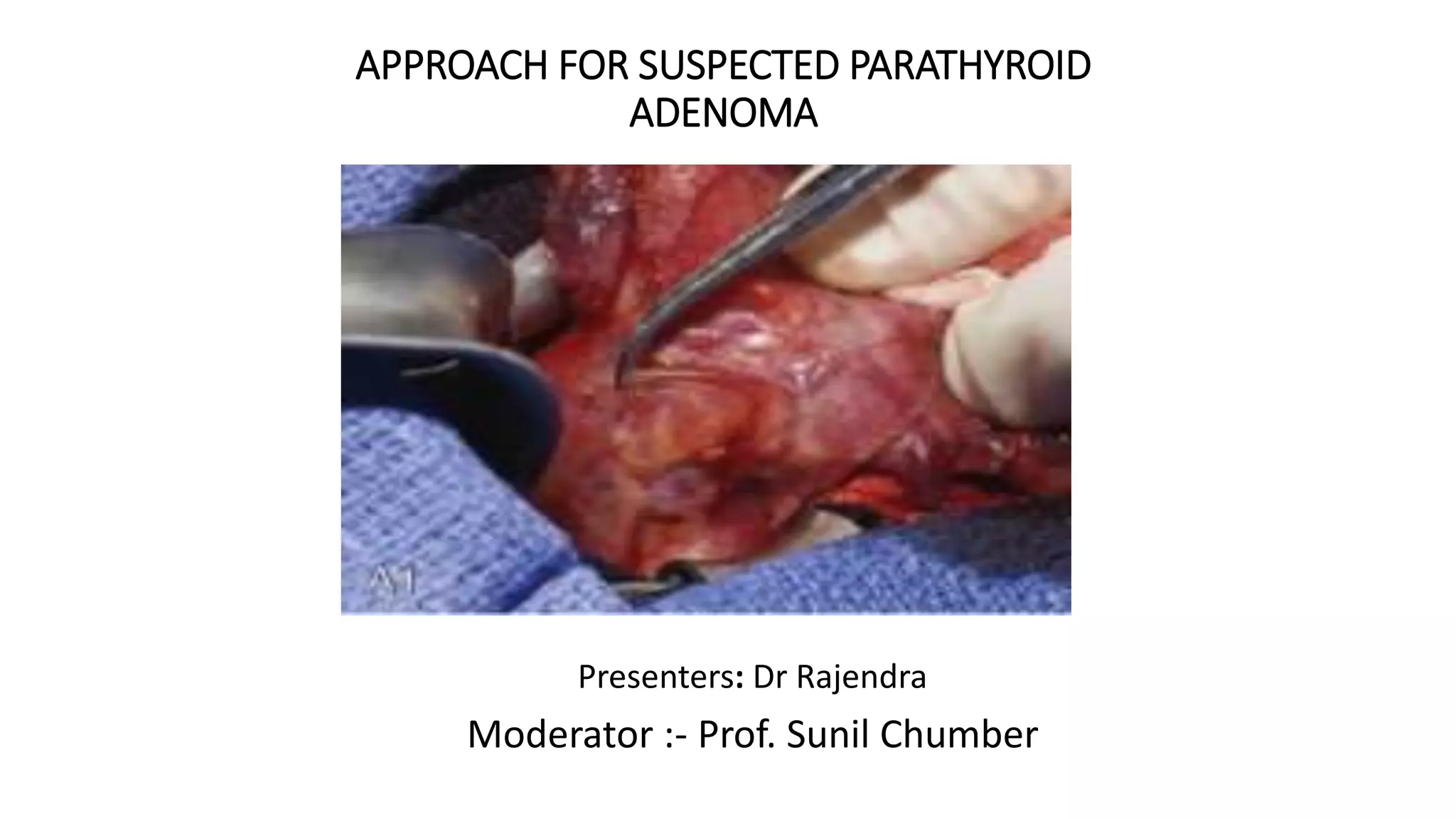
APPROACH FOR SUSPECTED PARATHYROID
ADENOMA
Presenters: Dr Rajendra
Moderator :- Prof. Sunil Chumber
Historical note
• Sir Richard Owen 1852: parathyroid
gland in a Indian rhinoceros.
• Vassale and Generali 1896: Introduced
the term "parathyroid“
• M. Askanazy 1903: Relationship
between parathyroid tumors and
osteitis fibrosa cystica
• Erdheim 1906: Relationship between
parathyroid glands and calcium
metabolism
• Mandl 1925: Removed a cervical
parathyroid adenoma
Introduction
• Embryological development and anatomy of the parathyroid glands:
prerequisites for successful surgical strategy.
Embryology of Parathyroid
• Parathyroid glands first
appear in amphibia.
• Branchial arches and
Pharyngeal pouches form in
the 4th week
• Superior Parathyroids - the 4th
Pharyngeal pouch with
thyroid
• Inferior Parathyroids – 3rd
Pharyngeal pouch with
thymus
5
Parathyroid Embryology and Anatomy
Anatomy of Parathyroid
• The considerable variability in size, shape, number, location and color
of these glands creates unique surgical challenges.
1. Autopsy studies
2. Surgical anatomy
Parathyroid Embryology and Anatomy 6
Autopsy studies
Results
• 84%- 4 glands
• 13%->4 glands: supernumerary
• Majority within the thymus or fat near the normal parathyroid gland
• More commonly found in MEN1 and Sec. hyperparathyroidism
1. Rudimentary 2%
2. Divided 6%
3. True supernumerary: >5 mg, at some distance 5%
• 3%-<4 glands
Parathyroid Embryology and Anatomy 7
•Akerstrom G etal. Surgery 1984;95:14.
Parathyroid characteristics
• Most humans : 4 glands
• 3 glands: one is missing rather absent
• Size : avg 5x3x1 mm
• Weight: 38-59 mg
• Increases till 5th decade
• Lower glands more than upper
• Increased in mal-absorption and vit D def
Parathyroid Embryology and Anatomy 8
Parathyroid characteristics
• Color :
• depends upon the age, fat content decreases
• Newborn: grey , semitransparent
• Children: light pink
• Adults: yellow
• Elders: darker
• Relation with the thyroid
• Intracapsular: enlarged glands remains in place
• Extracapsular: tends to migrate in least resistance
• Intrathyroidal: always maintan a plane of cleavage
Parathyroid Embryology and Anatomy 9
Vascular anatomy
• Terminal arteries
• Arterial supply is solitary 66%
• Sup parathyroid:
-80% ITA
-15% STA: post branch
-5% Anastomotic branches
• Inf parthyroid:
-90% ITA
-10% STA (ITA agenesis)
or anastomotic branches
• Mediastenal gland:
• Int mammary
10
Parathyroid Embryology and Anatomy
•Filament JB, et al. Anat Clin1982;3:279.
Relation with recurrent laryngeal nerve
• 93% of parathyroid glands:
predictable relation to RLNs
• Superior glands lying
posterior and superior to the
nerve
• Inferior glands lying anterior
to the nerve
Parathyroid Embryology and Anatomy 11
•Pyrtek LJ, Painter RL. Surg Gynecol Obstet 1964;119:509.
Disease spectrum of parathyroid
Hyperparathyroidism
• Primary( adenoma, hyperplasia, carcinoma)
• Secondary
• Tertiary
Hypopararthyroidism
World J Gastroenterol. 2015 May 28
Hyperparathyroidism
• Primary
Parathyroid adenoma( 80-85%,
most common )
Parathyroid hyperplasia
Parathyroid carcinoma
• Secondary
Parathyroid hyperplasia
• Tertiary
Autonomous nodule on top of
hyperplasia
• Overall prevalence of PHPT : 1 in
1000
• Higher incidence in patients > 60
years
• Female : Male - 2 to 3 : 1
• Up to 80% - asymptomatic*
A novel nomenclature to classify parathyroid
adenomas
A. Adherent to the posterior thyroid capsule-
normal superior gland location
B. Tracheo-esophageal groove
C. Tracheo-esophageal groove inferior to the
inferior thyroid pole
D. Directly over the recurrent laryngeal nerve
E. Close to the inferior thyroid pole-normal
inferior gland location
F. Within the thyro-thymic ligament,
G. Intrathyroidal
World J Surg (2009) 33:412-416
DOI 10.1007/s 00268-008-9894-0
CLINICAL FEATURES
• The pentad of symptoms—
• painful bones
• kidney stones
• abdominal groans
• psychic moans
• fatigue overtones.
• CLASSICAL FEATURES :
• osteitis fibrosa cystica,
• nephrolithiasis,
• nephrocalcinosis,
• peptic ulcer disease,
• gout, pseudogout.
CLINICAL FEATURES
Syndrome associated with
• MEN type I (MEN 1 gene)
•Parathyroid adenoma,
•(earliest & m.c manifestation is PHPT &
develops in 80-100% by 40 yrs )
•Pituitary adenoma
•Pancreatic islet cell tumor
• MEN type II
•Parathyroid adenoma,
•Thyroid medullary carcinoma &
•Pheochromocytoma
On examination
• General examination :
• Ocular : band keratopathy
• Fibro-osseous tumours- in favour of
parathyroid carcinoma.
• Neck examination-usually normal.
• Parathyroids are seldom palpable except
in patients with profound hypercalcemia
(≥14 mg/dL).
By Thomas J. Stokkermans, OD, PhD, Pankaj C. Gupta, MD,
MSc, and Rony R. Sayegh, MD
Investigations
• S. Calcium- total and ionised
• S. PTH
• 24hr urinary calcium
• S. Phosphate
• S. ALP
• 25-OH vitamin D3
• Magnesium
• X-ray :
• Hand and forearm
• skull
• Localization techniques
• Perioperative
• Intra-operative
X-ray
Trabecular bone resorption
SALT PEPPER APPEARANCE
Subperiosteal bone resorption
Phalangeal tufts.
Brown tumor
(osteitis fibrosa cystica)
Localization techniques
Localization studies in
Primary Hyperparathyroidism
Preoperative
localization
Noninvasive
imaging
Invasive
localization
Intraoperative
localization
Ultrasonography
• First modality after biochemical confirmation
• With the availability of high resolution USG, detection is easier of small adenomas
• Enlarged parathyroid glands appear as hypoechoic structures, in sharp contrast to the hyperechoic
thyroid tissue.
• Doppler is performed to test the vascularization of the area and define the arterial branches
involved.
• Concomitant thyroid pathology
A meta-analysis of preoperative localization techniques for patients with primary hyperparathyroidism.
Cheung K, Wang TS, Farrokhyar F, Roman SA, Sosa JA.
Ann Surg Oncol. 2012 Feb;19(2):577-83
Ultrasonography
• Sensitivity - 72%to 89% , PPV – 93%
• Facilitates focussed
parathyroidectomy
• Limitations :
1. Small size (<5mm)
2. Ectopic location (especially
behind trachea or clavicle)
3. Reoperative localisation
Parathyroid Scintigraphy
• Sestamibi (methoxy-isobutylisonitrile),a lipophilic compound, is
radiolabeled with 99mTc-pertechnetate .
• Retained by the mitochondria -rich oxyphil cells in parathyroid longer
than in thyroid tissue.
• Normal functioning parathyroid glands are not visualized.
Two protocols :
the single isotope-dual phase protocol and the subtraction protocol.
A. Dual phase
B. B. Subtraction Protocol:
SPECT
• Sestamibi-single photon emission computed
tomography is a 3D sestamibi scan that
provides higher-resolution imaging.
• Illustrate depth of the parathyroid gland or
glands in relation to the thyroid and improve
detection of ectopic glands
• Improves the sensitivity for identifying
abnormal parathyroid glands to 92 to 98 %.
• SPECT-CT helps to discriminate parathyroid
adenomas from other anatomic landmarks
• Specially in retrotracheal parathyroids
4D-CT
• 4D-CT gives multiplanar images and allows the visualization of differences in the perfusion characteristics
of hyperfunctioning parathyroid glands compared with normal glands and other structures
• Rapid contrast uptake and washout is characteristic of parathyroid adenomas
• Superior to SPECT-CT
• Combines both anatomical and functional
• Sensitivity=88%
• Disadvantage of 4D-CT - radiation exposure
PET
• Methionine PET scanning has high
sensitivity (about 85–90%)
• Disadvantage - half-life of 11C is
very short (20 min) and requires an
on-site cyclotron.
• Useful option in patients with
recurrent or persistent PHPT
Met-PET / CT scan located a parathyroid adenoma behind the left
thyroid lobe
18F- Fluorocholine PET
• Principle: Choline is part of the phospholipid
layer in the cell membrane
• It is hypothesized that hyperfunctioning
parathyroid cells have an increased activity
of the phospholipid/Ca2+-dependent
protein kinase ( move from cytosol to
membrane)
• Choline gets converted into
phosphatidylcholine by the enzyme choline
kinase and subsequent incorporation into
the cell membrane
MRI
• On MRI parathyroid adenoma has
intermediate to low signal intensity
on T1 imaging and high intensity on
T2 imaging. They may enhance with
gadolinium.
• Usually only as an investigation prior
to a re-exploration after failed
surgery.
• Drawbacks-size of detection is
limited to adenomas >5 mm,
localization of the superior glands is
difficult as they lie posterior to the
thyroid
Invasive Localisation
A. Selective Venous Sampling : For PTH
measurement( 1.5 to 2 times)
• It is a very sensitive test which depends
on gland function rather than size.
• Sampling is done from larger veins such
as the jugular vein, innominate vein,
superior cava and smaller veins, such as
superior, middle, and inferior thyroid
veins
• Sensitivity and specificity of the SVS -
63 to 94.7% and 86 to 100%,
respectively
Management of hypercalcemia
• Goals of therapy are aimed at
 lowering calcium levels.
 correcting dehydration and thereby increasing renal calcium excretion and
 decreasing osteoclast mediated bone resorption.
• Management of the underlying cause of hypercalcemia directs treatment strategy .
• In the majority of patients, definitive curative therapy requires surgical parathyroidectomy.
• To avoid factors which exacerbate hypercalcemia.
Management of hypercalcemia
• Hydration
• Loop diuretics ( avoid thiazides)
• calcitonin
• Biphosphonate and other medications
• Glucocorticoids
• Dialysis
• Surgery
Management of hypercalcemia
• Hydration :-
• NS 2 to 4 L IV daily for 1 to 3 days.
• Mode of action :- Enhances filtration and excretion of Ca2+
• Indication : - Severe ↑Ca++ > 14 mg per dL ,
• Cautions :- May exacerbate heart failure in elderly pt, CKD ,lowers Ca++ by 1 to
3 mg per dL
• Furosemide (Lasix)
• 10 to 20 mg IV as necessary upto 100mg/hr
• mode of action :- Inhibits calcium resorption in the distal renal tubule
• indication :- Following aggressive rehydration
• Caution:-↓K+, dehydration if used before intravascular volume is restored
• Bisphosphonates
• Pamidronate , 60 to 90 mg over 4 hours
• Zolendronic acid 4 mg IV over 15 minutes
• mode of action :- Inhibits osteoclast action and bone resorption
• indication :- Hypercalcemia of malignancy
• cautions:- Nephrotoxicity, ↓Ca++, ↓PO4, rebound ↑Ca++ in
hyperparathyroidism Maximal effects at 72 4 mg IV over 15 minutes
Management of hypercalcemia
• Calcitonin
• Dose 4 to 8 IU per kg IM or SQ every 6 hours
for 24 hours
• Mode of action :- Inhibits bone resorption,
augments Ca++ excretion
• Indication :- Initial treatment (after
rehydration) in severe ↑Ca++
• Caution:- Rebound ↑Ca++ after 24 hours,
vomiting, cramps, flushing
Rapid ↑Ca++ within 2 to 6 hours
• Glucocorticoids
• dose :- Hydrocortisone, 200 mg IV daily for 3 days
• mode of action - Inhibits vitamin D conversion to calcitriol
• Indications :- Vitamin D intoxication, hematologic malignancies,
granulomatous disease
• Caution Immune suppression, myopathy
•
Management of hypercalcemia
• Gallium nitrate :
• dose 100 to 200 mg per m2 IV over 24 hours for 5 days
• Mode of action :- Inhibits osteoclast action ↑Ca++
• Indication :- rarely used in severe ca++
• Caution :- Renal and marrow toxicity
• Dialysis :-
• Indication :- Ca++ >16 mg/dl , CHF , Renal failure.
• Hypercalcemic crisis or refractory hypercalcemia
hemodialysis against a low-calcium dialysate is more effective than
peritoneal dialysis in lowering serum calcium levels.
Surgical indication
1. Symptomatic primary HPT :
• Polydipsia and polyuria
• Nephrolithiasis
• Hypercalciuria (24hr urine calcium
>400mg/dl)
• Impaired renal function (GFR
<60cc/min)
• Osteoporosis (BMD<-2.5),fragility
fracture or vertebral compression
• Pancreatitis , PUD or GERD
• Neurocognitive dysfunction
2. Asymptomatic PHPT :
Increased parathormone levels without
symptoms
A. Age < 50 yrs
B. Serum calcium >1mg/dl above
normal/ T score < 2.5.
C. Silent nephrolithiasis/ Cr Cl < 60/
Ur Ca > 400 mg/d
D. Unable or Unwilling to comply
with surveillance
E. Patient with cardiovascular
disease
Surgical technique:
• Open standard approach
• Minimally invasive parathyroidectomy
Open minimally invasive
Minimally invasive radioguided
Video assisted
Purely endoscopic
Standard Bilateral exploration-Conventional
approach
• If parathyroid localization studies or IOPTH arent avaialable
• If localization studies fail to identify any abnormal glands
• If localization studies identify multiple abnormal glands
• If concomitant thyroid disorder requires B/L exploration
Minimally invasive video assisted
parathyroidectomy( MIVAP)
• First described by Miccoli
• Indications: <3cm size
• Previous surgery is relative contraindication
• BNE can also be performed
• With 1.5 cm incision in midline
• Miccoli reported a cure rate of 98.3%
Endoscopic parathyroidectomy
• First described by Gagner
• With 1.5 cm incision in side of target gland, anterior to the SCM
• Two 2.5mm incision along it to help CO2 and dissection
• Specially for posteriorly located parathyroids
• Help of IOPTH
Endoscopic parathyroidectomy
VATS
• For mediastinal parathyroid
• first described over 20 years ago
• Procedure of choice with early recovery and less postop pain
• Avoiding sternotomy
• Helps early recover
IOPTH
• Short half life= 3-5 min
• Miami criteria
• > 50% fall below prexcision
value demonstrates
successful
parathyroidectomy
Intraop adjuncts
• Methylene blue
• Autofluorescence imaging of parathyroid
• Near-infrared/ indocyanine green (NIR/ICG)
• Intraop radioguided surgery
• Intraop USG
Post op complications
• Hematoma
• Postoperative hypocalcemia ( transient / hungry bone syndrome )
• Permanent hypoparathyroidism
• Recurrent laryngeal nerve injury(1%)
• Persistent hyperparathyroidism—5% (PTH never touches baseline -6
mo)
• Recurrent hyperparathyroidism—initially PTH decreases but
hypercalcaemia recurs 6 months after first parathyroid surgery
Parathyroid Embryology and Anatomy 48
Surgical studies
Shen W etal. Arch Surg. 1996 ;131(8):861-7.
Reoperation for persistent or recurrent primary hyperparathyroidism
To analyze the causes and outcomes of reoperation for persistent or
recurrent primary hyperparathyroidism.
Medical records of 102 patients
Reasons for failed parathyroid operations
1. tumor in ectopic position (53%)
2. incomplete resection of multiple abnormal glands (37%)
3. adenoma in normal position missed during previous surgery (7%)
4. regrowth of previously resected tumor (3%). .
Post parathyroidectomy follow up
• Measure calcium and PTH immediate postoperatively
• Then at three month,6 month and then yearly
• Correct vitamin D deficiency if present
• No restriction on daily calcium
A total of 196 patients met inclusion criteria with an overall median follow-up time of
9.2 years IQR (interquartile range) [5.4-10.9 years].
SUMMARY
• High index of suspicion is required to diagnose hyper
parathyroidism
• Always send calcium level if patient present with recurrent
renal stones
• Low threshold for parathyroidectomy
• Reversal of most of the symptoms
• Surgery remains the treatment of choice in hypercalcemic
crisis.
• Medical therapies can be useful in many situations, but should
not be viewed as equivalent to surgery or as a potential
replacement for surgery.
“The only localization needed
for parathyroid is
good endocrine surgeon”
Thank you
Dr John Leo Doppman, M D
Interventional Radiologist

parathyroid adenoma

  • 1.
    APPROACH FOR SUSPECTEDPARATHYROID ADENOMA Presenters: Dr Rajendra Moderator :- Prof. Sunil Chumber
  • 2.
    Historical note • SirRichard Owen 1852: parathyroid gland in a Indian rhinoceros. • Vassale and Generali 1896: Introduced the term "parathyroid“ • M. Askanazy 1903: Relationship between parathyroid tumors and osteitis fibrosa cystica • Erdheim 1906: Relationship between parathyroid glands and calcium metabolism • Mandl 1925: Removed a cervical parathyroid adenoma
  • 4.
    Introduction • Embryological developmentand anatomy of the parathyroid glands: prerequisites for successful surgical strategy.
  • 5.
    Embryology of Parathyroid •Parathyroid glands first appear in amphibia. • Branchial arches and Pharyngeal pouches form in the 4th week • Superior Parathyroids - the 4th Pharyngeal pouch with thyroid • Inferior Parathyroids – 3rd Pharyngeal pouch with thymus 5 Parathyroid Embryology and Anatomy
  • 6.
    Anatomy of Parathyroid •The considerable variability in size, shape, number, location and color of these glands creates unique surgical challenges. 1. Autopsy studies 2. Surgical anatomy Parathyroid Embryology and Anatomy 6
  • 7.
    Autopsy studies Results • 84%-4 glands • 13%->4 glands: supernumerary • Majority within the thymus or fat near the normal parathyroid gland • More commonly found in MEN1 and Sec. hyperparathyroidism 1. Rudimentary 2% 2. Divided 6% 3. True supernumerary: >5 mg, at some distance 5% • 3%-<4 glands Parathyroid Embryology and Anatomy 7 •Akerstrom G etal. Surgery 1984;95:14.
  • 8.
    Parathyroid characteristics • Mosthumans : 4 glands • 3 glands: one is missing rather absent • Size : avg 5x3x1 mm • Weight: 38-59 mg • Increases till 5th decade • Lower glands more than upper • Increased in mal-absorption and vit D def Parathyroid Embryology and Anatomy 8
  • 9.
    Parathyroid characteristics • Color: • depends upon the age, fat content decreases • Newborn: grey , semitransparent • Children: light pink • Adults: yellow • Elders: darker • Relation with the thyroid • Intracapsular: enlarged glands remains in place • Extracapsular: tends to migrate in least resistance • Intrathyroidal: always maintan a plane of cleavage Parathyroid Embryology and Anatomy 9
  • 10.
    Vascular anatomy • Terminalarteries • Arterial supply is solitary 66% • Sup parathyroid: -80% ITA -15% STA: post branch -5% Anastomotic branches • Inf parthyroid: -90% ITA -10% STA (ITA agenesis) or anastomotic branches • Mediastenal gland: • Int mammary 10 Parathyroid Embryology and Anatomy •Filament JB, et al. Anat Clin1982;3:279.
  • 11.
    Relation with recurrentlaryngeal nerve • 93% of parathyroid glands: predictable relation to RLNs • Superior glands lying posterior and superior to the nerve • Inferior glands lying anterior to the nerve Parathyroid Embryology and Anatomy 11 •Pyrtek LJ, Painter RL. Surg Gynecol Obstet 1964;119:509.
  • 12.
    Disease spectrum ofparathyroid Hyperparathyroidism • Primary( adenoma, hyperplasia, carcinoma) • Secondary • Tertiary Hypopararthyroidism World J Gastroenterol. 2015 May 28
  • 13.
    Hyperparathyroidism • Primary Parathyroid adenoma(80-85%, most common ) Parathyroid hyperplasia Parathyroid carcinoma • Secondary Parathyroid hyperplasia • Tertiary Autonomous nodule on top of hyperplasia • Overall prevalence of PHPT : 1 in 1000 • Higher incidence in patients > 60 years • Female : Male - 2 to 3 : 1 • Up to 80% - asymptomatic*
  • 14.
    A novel nomenclatureto classify parathyroid adenomas A. Adherent to the posterior thyroid capsule- normal superior gland location B. Tracheo-esophageal groove C. Tracheo-esophageal groove inferior to the inferior thyroid pole D. Directly over the recurrent laryngeal nerve E. Close to the inferior thyroid pole-normal inferior gland location F. Within the thyro-thymic ligament, G. Intrathyroidal World J Surg (2009) 33:412-416 DOI 10.1007/s 00268-008-9894-0
  • 15.
    CLINICAL FEATURES • Thepentad of symptoms— • painful bones • kidney stones • abdominal groans • psychic moans • fatigue overtones. • CLASSICAL FEATURES : • osteitis fibrosa cystica, • nephrolithiasis, • nephrocalcinosis, • peptic ulcer disease, • gout, pseudogout.
  • 16.
  • 17.
    Syndrome associated with •MEN type I (MEN 1 gene) •Parathyroid adenoma, •(earliest & m.c manifestation is PHPT & develops in 80-100% by 40 yrs ) •Pituitary adenoma •Pancreatic islet cell tumor • MEN type II •Parathyroid adenoma, •Thyroid medullary carcinoma & •Pheochromocytoma
  • 18.
    On examination • Generalexamination : • Ocular : band keratopathy • Fibro-osseous tumours- in favour of parathyroid carcinoma. • Neck examination-usually normal. • Parathyroids are seldom palpable except in patients with profound hypercalcemia (≥14 mg/dL). By Thomas J. Stokkermans, OD, PhD, Pankaj C. Gupta, MD, MSc, and Rony R. Sayegh, MD
  • 19.
    Investigations • S. Calcium-total and ionised • S. PTH • 24hr urinary calcium • S. Phosphate • S. ALP • 25-OH vitamin D3 • Magnesium • X-ray : • Hand and forearm • skull • Localization techniques • Perioperative • Intra-operative
  • 20.
    X-ray Trabecular bone resorption SALTPEPPER APPEARANCE Subperiosteal bone resorption Phalangeal tufts. Brown tumor (osteitis fibrosa cystica)
  • 21.
    Localization techniques Localization studiesin Primary Hyperparathyroidism Preoperative localization Noninvasive imaging Invasive localization Intraoperative localization
  • 22.
    Ultrasonography • First modalityafter biochemical confirmation • With the availability of high resolution USG, detection is easier of small adenomas • Enlarged parathyroid glands appear as hypoechoic structures, in sharp contrast to the hyperechoic thyroid tissue. • Doppler is performed to test the vascularization of the area and define the arterial branches involved. • Concomitant thyroid pathology A meta-analysis of preoperative localization techniques for patients with primary hyperparathyroidism. Cheung K, Wang TS, Farrokhyar F, Roman SA, Sosa JA. Ann Surg Oncol. 2012 Feb;19(2):577-83
  • 23.
    Ultrasonography • Sensitivity -72%to 89% , PPV – 93% • Facilitates focussed parathyroidectomy • Limitations : 1. Small size (<5mm) 2. Ectopic location (especially behind trachea or clavicle) 3. Reoperative localisation
  • 24.
    Parathyroid Scintigraphy • Sestamibi(methoxy-isobutylisonitrile),a lipophilic compound, is radiolabeled with 99mTc-pertechnetate . • Retained by the mitochondria -rich oxyphil cells in parathyroid longer than in thyroid tissue. • Normal functioning parathyroid glands are not visualized. Two protocols : the single isotope-dual phase protocol and the subtraction protocol. A. Dual phase B. B. Subtraction Protocol:
  • 25.
    SPECT • Sestamibi-single photonemission computed tomography is a 3D sestamibi scan that provides higher-resolution imaging. • Illustrate depth of the parathyroid gland or glands in relation to the thyroid and improve detection of ectopic glands • Improves the sensitivity for identifying abnormal parathyroid glands to 92 to 98 %. • SPECT-CT helps to discriminate parathyroid adenomas from other anatomic landmarks • Specially in retrotracheal parathyroids
  • 26.
    4D-CT • 4D-CT givesmultiplanar images and allows the visualization of differences in the perfusion characteristics of hyperfunctioning parathyroid glands compared with normal glands and other structures • Rapid contrast uptake and washout is characteristic of parathyroid adenomas • Superior to SPECT-CT • Combines both anatomical and functional • Sensitivity=88% • Disadvantage of 4D-CT - radiation exposure
  • 28.
    PET • Methionine PETscanning has high sensitivity (about 85–90%) • Disadvantage - half-life of 11C is very short (20 min) and requires an on-site cyclotron. • Useful option in patients with recurrent or persistent PHPT Met-PET / CT scan located a parathyroid adenoma behind the left thyroid lobe
  • 29.
    18F- Fluorocholine PET •Principle: Choline is part of the phospholipid layer in the cell membrane • It is hypothesized that hyperfunctioning parathyroid cells have an increased activity of the phospholipid/Ca2+-dependent protein kinase ( move from cytosol to membrane) • Choline gets converted into phosphatidylcholine by the enzyme choline kinase and subsequent incorporation into the cell membrane
  • 30.
    MRI • On MRIparathyroid adenoma has intermediate to low signal intensity on T1 imaging and high intensity on T2 imaging. They may enhance with gadolinium. • Usually only as an investigation prior to a re-exploration after failed surgery. • Drawbacks-size of detection is limited to adenomas >5 mm, localization of the superior glands is difficult as they lie posterior to the thyroid
  • 32.
    Invasive Localisation A. SelectiveVenous Sampling : For PTH measurement( 1.5 to 2 times) • It is a very sensitive test which depends on gland function rather than size. • Sampling is done from larger veins such as the jugular vein, innominate vein, superior cava and smaller veins, such as superior, middle, and inferior thyroid veins • Sensitivity and specificity of the SVS - 63 to 94.7% and 86 to 100%, respectively
  • 33.
    Management of hypercalcemia •Goals of therapy are aimed at  lowering calcium levels.  correcting dehydration and thereby increasing renal calcium excretion and  decreasing osteoclast mediated bone resorption. • Management of the underlying cause of hypercalcemia directs treatment strategy . • In the majority of patients, definitive curative therapy requires surgical parathyroidectomy. • To avoid factors which exacerbate hypercalcemia.
  • 34.
    Management of hypercalcemia •Hydration • Loop diuretics ( avoid thiazides) • calcitonin • Biphosphonate and other medications • Glucocorticoids • Dialysis • Surgery
  • 35.
    Management of hypercalcemia •Hydration :- • NS 2 to 4 L IV daily for 1 to 3 days. • Mode of action :- Enhances filtration and excretion of Ca2+ • Indication : - Severe ↑Ca++ > 14 mg per dL , • Cautions :- May exacerbate heart failure in elderly pt, CKD ,lowers Ca++ by 1 to 3 mg per dL • Furosemide (Lasix) • 10 to 20 mg IV as necessary upto 100mg/hr • mode of action :- Inhibits calcium resorption in the distal renal tubule • indication :- Following aggressive rehydration • Caution:-↓K+, dehydration if used before intravascular volume is restored • Bisphosphonates • Pamidronate , 60 to 90 mg over 4 hours • Zolendronic acid 4 mg IV over 15 minutes • mode of action :- Inhibits osteoclast action and bone resorption • indication :- Hypercalcemia of malignancy • cautions:- Nephrotoxicity, ↓Ca++, ↓PO4, rebound ↑Ca++ in hyperparathyroidism Maximal effects at 72 4 mg IV over 15 minutes
  • 36.
    Management of hypercalcemia •Calcitonin • Dose 4 to 8 IU per kg IM or SQ every 6 hours for 24 hours • Mode of action :- Inhibits bone resorption, augments Ca++ excretion • Indication :- Initial treatment (after rehydration) in severe ↑Ca++ • Caution:- Rebound ↑Ca++ after 24 hours, vomiting, cramps, flushing Rapid ↑Ca++ within 2 to 6 hours • Glucocorticoids • dose :- Hydrocortisone, 200 mg IV daily for 3 days • mode of action - Inhibits vitamin D conversion to calcitriol • Indications :- Vitamin D intoxication, hematologic malignancies, granulomatous disease • Caution Immune suppression, myopathy •
  • 37.
    Management of hypercalcemia •Gallium nitrate : • dose 100 to 200 mg per m2 IV over 24 hours for 5 days • Mode of action :- Inhibits osteoclast action ↑Ca++ • Indication :- rarely used in severe ca++ • Caution :- Renal and marrow toxicity • Dialysis :- • Indication :- Ca++ >16 mg/dl , CHF , Renal failure. • Hypercalcemic crisis or refractory hypercalcemia hemodialysis against a low-calcium dialysate is more effective than peritoneal dialysis in lowering serum calcium levels.
  • 38.
    Surgical indication 1. Symptomaticprimary HPT : • Polydipsia and polyuria • Nephrolithiasis • Hypercalciuria (24hr urine calcium >400mg/dl) • Impaired renal function (GFR <60cc/min) • Osteoporosis (BMD<-2.5),fragility fracture or vertebral compression • Pancreatitis , PUD or GERD • Neurocognitive dysfunction 2. Asymptomatic PHPT : Increased parathormone levels without symptoms A. Age < 50 yrs B. Serum calcium >1mg/dl above normal/ T score < 2.5. C. Silent nephrolithiasis/ Cr Cl < 60/ Ur Ca > 400 mg/d D. Unable or Unwilling to comply with surveillance E. Patient with cardiovascular disease
  • 39.
    Surgical technique: • Openstandard approach • Minimally invasive parathyroidectomy Open minimally invasive Minimally invasive radioguided Video assisted Purely endoscopic
  • 40.
    Standard Bilateral exploration-Conventional approach •If parathyroid localization studies or IOPTH arent avaialable • If localization studies fail to identify any abnormal glands • If localization studies identify multiple abnormal glands • If concomitant thyroid disorder requires B/L exploration
  • 41.
    Minimally invasive videoassisted parathyroidectomy( MIVAP) • First described by Miccoli • Indications: <3cm size • Previous surgery is relative contraindication • BNE can also be performed • With 1.5 cm incision in midline • Miccoli reported a cure rate of 98.3%
  • 42.
    Endoscopic parathyroidectomy • Firstdescribed by Gagner • With 1.5 cm incision in side of target gland, anterior to the SCM • Two 2.5mm incision along it to help CO2 and dissection • Specially for posteriorly located parathyroids • Help of IOPTH
  • 43.
  • 44.
    VATS • For mediastinalparathyroid • first described over 20 years ago • Procedure of choice with early recovery and less postop pain • Avoiding sternotomy • Helps early recover
  • 45.
    IOPTH • Short halflife= 3-5 min • Miami criteria • > 50% fall below prexcision value demonstrates successful parathyroidectomy
  • 46.
    Intraop adjuncts • Methyleneblue • Autofluorescence imaging of parathyroid • Near-infrared/ indocyanine green (NIR/ICG) • Intraop radioguided surgery • Intraop USG
  • 47.
    Post op complications •Hematoma • Postoperative hypocalcemia ( transient / hungry bone syndrome ) • Permanent hypoparathyroidism • Recurrent laryngeal nerve injury(1%) • Persistent hyperparathyroidism—5% (PTH never touches baseline -6 mo) • Recurrent hyperparathyroidism—initially PTH decreases but hypercalcaemia recurs 6 months after first parathyroid surgery
  • 48.
    Parathyroid Embryology andAnatomy 48 Surgical studies Shen W etal. Arch Surg. 1996 ;131(8):861-7. Reoperation for persistent or recurrent primary hyperparathyroidism To analyze the causes and outcomes of reoperation for persistent or recurrent primary hyperparathyroidism. Medical records of 102 patients Reasons for failed parathyroid operations 1. tumor in ectopic position (53%) 2. incomplete resection of multiple abnormal glands (37%) 3. adenoma in normal position missed during previous surgery (7%) 4. regrowth of previously resected tumor (3%). .
  • 49.
    Post parathyroidectomy followup • Measure calcium and PTH immediate postoperatively • Then at three month,6 month and then yearly • Correct vitamin D deficiency if present • No restriction on daily calcium
  • 50.
    A total of196 patients met inclusion criteria with an overall median follow-up time of 9.2 years IQR (interquartile range) [5.4-10.9 years].
  • 51.
    SUMMARY • High indexof suspicion is required to diagnose hyper parathyroidism • Always send calcium level if patient present with recurrent renal stones • Low threshold for parathyroidectomy • Reversal of most of the symptoms • Surgery remains the treatment of choice in hypercalcemic crisis. • Medical therapies can be useful in many situations, but should not be viewed as equivalent to surgery or as a potential replacement for surgery.
  • 52.
    “The only localizationneeded for parathyroid is good endocrine surgeon” Thank you Dr John Leo Doppman, M D Interventional Radiologist