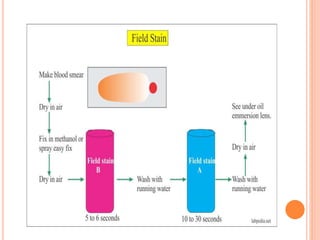
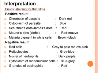
FOOD AND
MEDICAL
MICROBIOLOGY
STAINING TECHNIQUES
CALCOFLUOR STAIN (MICROSPORIDIA)
Introduction :
 Calcofluor stain binds to the chitin in the endospore
layer of the spore wall of microsporidia and
fluoresce a brilliant blue-white. This staining
technique is used for the demonstration of
microsporidia in faeces.
Method :
 Prepare a very thin smear and air dry •
 Fix the smear in methanol for 5min •
 Stain the smear with 1-2 drops of Calcofluor
solution (0.5% w/v) and leave for 2- 3min •
 Rinse under slow running water •
 Counterstain with Evans blue solution (0.1%)
for 1min •
 rinse under slow running water •
 Air dry •
 Add 1 or 2 drops of mounting fluid (Cytoseal
60) to the slide and mount with a cover slip •
 Examine microscopically under a fluorescence
(395-415nm) microscope.
Interpretation :
Positive result :
 Spores of microsporidia are typically ovoid or
piriform and fluoresce brilliant blue white. Dimension
of spores vary by species and range from 1-20µm67.
Note: Yeast cells also display a turquoise
fluorescent ring but, unlike microsporidia, will counter
stain orange in the cytoplasm.
Negative result :
 No fluorescence observed.
MICROSPORIDIA
Quality control organisms :
Positive control :
 Microsporidia species .
Negative control :
 A proven negative smear may be used as the
negative control.
Technical information :
 Fungal spores may contain chitin, and some
experience is required to differentiate spores of
microsporidia from those of fungi.
FIELD’S STAIN (PLASMODIUM SPECIES)
Introduction :
 This technique is used for the demonstration of
Plasmodium species in thick and thin blood films.
Method :
Rapid field’s staining for thin films :
This is a modification of the original Field’s stain to
enable rapid staining of fixed thin films. This method is
suitable for malaria parasites.
 Prepare a thin film and air dry .
 Fix in methanol for 1min .
 Flood the slide with 1mL of diluted Field’s stain B (1 in
4 in buffered water pH 7.2) .
 Immediately add an equal volume of undiluted field’s
stain A, mix well and leave to stain for 1min .
 Rinse the slide with clean water and drain dry.
Field’s staining for thick films:
Caution: Thick blood films are not fixed and the stains
do not kill the parasites, viruses or other pathogens
which may be present in the blood.
 Prepare a blood smear and allow to air dry. Failure to
do so will result in the blood washing off the slide.
 Hold the slide with the dried thick film facing
downwards .
 Dip the slide in the undiluted Field’s stain A for 3sec.
 Drain the excess stain by touching a corner of the slide
against the side of the container.
 Wash gently for about 3sec in clean water and agitate
gently .
 Drain off the excess water .
 Dip the slide in undiluted Field’s stain B for 3sec and
drain off the excess stain.
 Wash gently in clean water.
 Wipe the back of slide clean and place it upright in a
draining rack for the film to air dry.
 Examine with the X100 oil immersion lens. When
searching for malarial parasites, 200 microscopic fields
should be examined on the slide for at least 15min before
declaring the slide negative.
Note: If after staining, the whole film appears yellow-
brown (a sign that too much blood has been used), too
blue or too pink, do not attempt to examine it. Re-stain it
by dipping the slide in the Field’s stain A for 1sec,
followed by a gentle wash in clean water, dip in Field’s
stain B for 1sec and finally wash gently in clean water.
Interpretation :
Fields’ staining for thin films
Positive result:
 Chromatin of parasite : Dark red
 Cytoplasm of parasite : Blue
 Schüffner’s dots/James’s dots : Red
 Maurer’s dots (clefts) : Red-mauve
 Malaria pigment in white cells : Brown-black
Negative result:
 Red cells : Grey to pale mauve-pink
 Reticulocytes : Grey-blue
 Nuclei of neutrophils : Dark purple
 Cytoplasm of mononuclear cells : Blue-grey
 Granules of eosinophils : Red
Fields’ staining for thick films
Positive result :
 Chromatin of parasite : Dark red
 Cytoplasm of parasite : Blue-mauve
 Schüffner’s dots : Pale red
 Background : Pale grey/blue
Note: White cells, platelets and malaria pigment can also be
seen on thick films. Malaria pigment Yellow-brown or
yellow-black.
Negative result:
 Nuclei of small lymphocytes : Dark purple
 Nuclei of neutrophils : Dark purple
 Granules of eosinophils : Red
 Cytoplasm of mononuclear cells : Blue-grey
 Reticulum of reticulocytes : Blue-grey
TROPHOZOITE OF PLASMODIUM OVALE
QUALITY CONTROL ORGANISMS :
Positive control :
 Plasmodium species
Negative control :
 A proven negative smear may be used as the
negative control
Technical information :
 The Rapid Field’s stain is a useful method for rapid
presumptive species identification of malaria
parasites. This method shows adequate staining of
all stages including stippling. However, staining with
Giemsa is always the method of choice for
definitive species differentiation.
 With thick preparations, the end of the smear closest
to the edge of the slide that was draining should be
examined. The edges of the film will also be better
than the centre where the film may be too thick or
cracked.
LUGOL’S IODINE (PARASITES)
Introduction :
 1% Lugol’s iodine, when diluted, is used to stain
ova and protozoan cysts in wet mounts. This
method enhances their internal structures.
Method:
 Place one drop of physiological saline (0.85%) on
one end of a clean glass slide and at the other end
of the slide, place a drop of diluted Lugol’s iodine
solution .
 Use an applicator stick to place a small portion of
faeces in the saline and mix until the suspension
becomes homogenous and then make an even thin
spread .
 Use the same applicator stick to emulsify an equal
amount of faeces in the iodine strap.
 Overlay each suspension with a cover slip, being
careful to avoid producing any air bubbles.
 Examine under low power objective.
Interpretation:
Positive result:
 Protozoan nuclei take up the iodine and stain pale
brown while cytoplasm remains colourless.
Note: Trophozoites can only be detected in fresh wet
mounts before concentration.
Negative result:
 N/A
(a) Trophozoite of the parasites Giardia lamblia;
and
(b) Entymoeba histolytica as seen from the stool
of patients stained with Lugol's iodine (100×).
Quality control organisms:
Positive control:
 A proven positive smear may be used as the positive
control.
Negative control :
 A proven negative smear may be used as the
negative control.
Technical information:
 Some workers prefer to make saline and iodine
mounts on separate slides. There is less chance of
getting fluids on the microscope stage if separate
slides are used. For this method to work effectively
the 1% Lugol’s iodine solution should be a fresh
preparation (10-14 days).
 The microscope light should be reduced for low
power observations since most organisms will be
overlooked by bright light. Illumination should be
regulated so that some of the cellular elements in
the faeces show refraction. Most protozoan cysts
will refract light under these conditions.

PARASITES STAINING METHODS

  • 1.
  • 2.
  • 3.
    CALCOFLUOR STAIN (MICROSPORIDIA) Introduction:  Calcofluor stain binds to the chitin in the endospore layer of the spore wall of microsporidia and fluoresce a brilliant blue-white. This staining technique is used for the demonstration of microsporidia in faeces.
  • 4.
    Method :  Preparea very thin smear and air dry •  Fix the smear in methanol for 5min •  Stain the smear with 1-2 drops of Calcofluor solution (0.5% w/v) and leave for 2- 3min •  Rinse under slow running water •  Counterstain with Evans blue solution (0.1%) for 1min •  rinse under slow running water •  Air dry •  Add 1 or 2 drops of mounting fluid (Cytoseal 60) to the slide and mount with a cover slip •  Examine microscopically under a fluorescence (395-415nm) microscope.
  • 5.
    Interpretation : Positive result:  Spores of microsporidia are typically ovoid or piriform and fluoresce brilliant blue white. Dimension of spores vary by species and range from 1-20µm67. Note: Yeast cells also display a turquoise fluorescent ring but, unlike microsporidia, will counter stain orange in the cytoplasm. Negative result :  No fluorescence observed.
  • 6.
  • 8.
    Quality control organisms: Positive control :  Microsporidia species . Negative control :  A proven negative smear may be used as the negative control. Technical information :  Fungal spores may contain chitin, and some experience is required to differentiate spores of microsporidia from those of fungi.
  • 9.
    FIELD’S STAIN (PLASMODIUMSPECIES) Introduction :  This technique is used for the demonstration of Plasmodium species in thick and thin blood films.
  • 10.
    Method : Rapid field’sstaining for thin films : This is a modification of the original Field’s stain to enable rapid staining of fixed thin films. This method is suitable for malaria parasites.  Prepare a thin film and air dry .  Fix in methanol for 1min .  Flood the slide with 1mL of diluted Field’s stain B (1 in 4 in buffered water pH 7.2) .  Immediately add an equal volume of undiluted field’s stain A, mix well and leave to stain for 1min .  Rinse the slide with clean water and drain dry.
  • 11.
    Field’s staining forthick films: Caution: Thick blood films are not fixed and the stains do not kill the parasites, viruses or other pathogens which may be present in the blood.  Prepare a blood smear and allow to air dry. Failure to do so will result in the blood washing off the slide.  Hold the slide with the dried thick film facing downwards .  Dip the slide in the undiluted Field’s stain A for 3sec.  Drain the excess stain by touching a corner of the slide against the side of the container.  Wash gently for about 3sec in clean water and agitate gently .  Drain off the excess water .
  • 12.
     Dip theslide in undiluted Field’s stain B for 3sec and drain off the excess stain.  Wash gently in clean water.  Wipe the back of slide clean and place it upright in a draining rack for the film to air dry.  Examine with the X100 oil immersion lens. When searching for malarial parasites, 200 microscopic fields should be examined on the slide for at least 15min before declaring the slide negative. Note: If after staining, the whole film appears yellow- brown (a sign that too much blood has been used), too blue or too pink, do not attempt to examine it. Re-stain it by dipping the slide in the Field’s stain A for 1sec, followed by a gentle wash in clean water, dip in Field’s stain B for 1sec and finally wash gently in clean water.
  • 14.
    Interpretation : Fields’ stainingfor thin films Positive result:  Chromatin of parasite : Dark red  Cytoplasm of parasite : Blue  Schüffner’s dots/James’s dots : Red  Maurer’s dots (clefts) : Red-mauve  Malaria pigment in white cells : Brown-black Negative result:  Red cells : Grey to pale mauve-pink  Reticulocytes : Grey-blue  Nuclei of neutrophils : Dark purple  Cytoplasm of mononuclear cells : Blue-grey  Granules of eosinophils : Red
  • 15.
    Fields’ staining forthick films Positive result :  Chromatin of parasite : Dark red  Cytoplasm of parasite : Blue-mauve  Schüffner’s dots : Pale red  Background : Pale grey/blue Note: White cells, platelets and malaria pigment can also be seen on thick films. Malaria pigment Yellow-brown or yellow-black. Negative result:  Nuclei of small lymphocytes : Dark purple  Nuclei of neutrophils : Dark purple  Granules of eosinophils : Red  Cytoplasm of mononuclear cells : Blue-grey  Reticulum of reticulocytes : Blue-grey
  • 16.
  • 17.
    QUALITY CONTROL ORGANISMS: Positive control :  Plasmodium species Negative control :  A proven negative smear may be used as the negative control Technical information :  The Rapid Field’s stain is a useful method for rapid presumptive species identification of malaria parasites. This method shows adequate staining of all stages including stippling. However, staining with Giemsa is always the method of choice for definitive species differentiation.
  • 18.
     With thickpreparations, the end of the smear closest to the edge of the slide that was draining should be examined. The edges of the film will also be better than the centre where the film may be too thick or cracked.
  • 19.
    LUGOL’S IODINE (PARASITES) Introduction:  1% Lugol’s iodine, when diluted, is used to stain ova and protozoan cysts in wet mounts. This method enhances their internal structures. Method:  Place one drop of physiological saline (0.85%) on one end of a clean glass slide and at the other end of the slide, place a drop of diluted Lugol’s iodine solution .  Use an applicator stick to place a small portion of faeces in the saline and mix until the suspension becomes homogenous and then make an even thin spread .
  • 20.
     Use thesame applicator stick to emulsify an equal amount of faeces in the iodine strap.  Overlay each suspension with a cover slip, being careful to avoid producing any air bubbles.  Examine under low power objective. Interpretation: Positive result:  Protozoan nuclei take up the iodine and stain pale brown while cytoplasm remains colourless. Note: Trophozoites can only be detected in fresh wet mounts before concentration. Negative result:  N/A
  • 21.
    (a) Trophozoite ofthe parasites Giardia lamblia; and (b) Entymoeba histolytica as seen from the stool of patients stained with Lugol's iodine (100×).
  • 22.
    Quality control organisms: Positivecontrol:  A proven positive smear may be used as the positive control. Negative control :  A proven negative smear may be used as the negative control. Technical information:  Some workers prefer to make saline and iodine mounts on separate slides. There is less chance of getting fluids on the microscope stage if separate slides are used. For this method to work effectively the 1% Lugol’s iodine solution should be a fresh preparation (10-14 days).
  • 23.
     The microscopelight should be reduced for low power observations since most organisms will be overlooked by bright light. Illumination should be regulated so that some of the cellular elements in the faeces show refraction. Most protozoan cysts will refract light under these conditions.