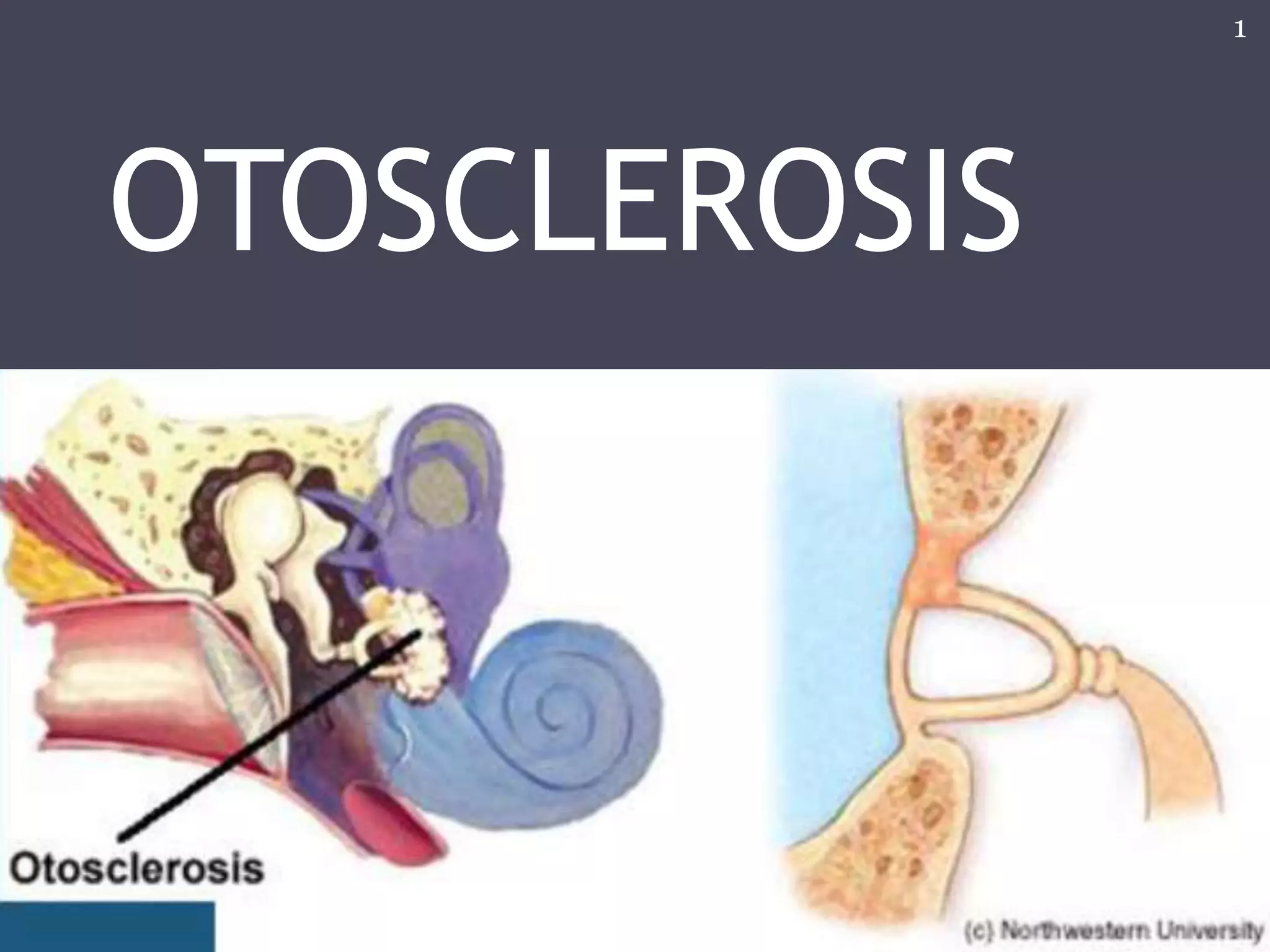
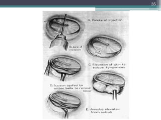
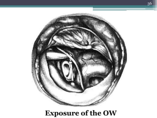
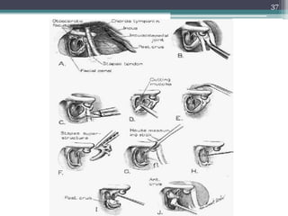
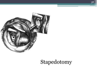
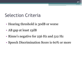
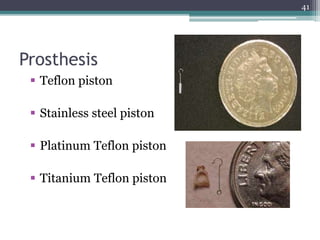
OTOSCLEROSIS
1
• Otic capsule
▫ The embryonic cartilage capsule that surrounds
the inner ear mechanism and develops into bony
tissue.
• Otic labyrinth
▫ The endolymphatic labyrinth. Consists of utricle,
saccule, cochlea, Semicircular canals,
endolymphatic duct and sac.
• Periotic labyrinth
▫ The perilymphatic labyrinth. Consists of vestibule,
scala vestibula, scala tympani and perilymphatic
space around semicircular canals.
2
3
4
Membranous Labyrinth
Otosclerosis
A primary disease of the bony otic capsule
characterized by abnormal removal of
mature bone by osteoclasts and replacement
with woven bone of greater thickness,
cellularity and vascularity.
5
• Autosomal-dominant hereditary disease
• Leads to progressive conducting hearing loss
• Most common cause of CHL in adults
• Bilateral hearing loss in 70% cases
• Male:Female is 1:2
• Most commonly effected age group is 15-45 years
old with hearing loss manifesting typically in the
third decade
• Cochlear otosclerosis manifests with sensorineural
hearing loss
6
Etiology
• The exact cause is unknown but many factors
have been proposed such as:
▫ Hereditary
▫ Endocrine (pregnancy)
▫ Trauma
▫ Infectious (eg: measles)
▫ Vascular
▫ Autoimmune
7
8
• Divided in two phases:
▫ Early spongiotic phase (otospongiosis)
 Osteocytes, histiocytes, osteoclasts
 Active reabsorption of bone
 Stains blue (blue mantles) on using H&E stain
 Dilated vessels (Schwartze’s sign)
▫ Late or Sclerotic phase
 Formation of new bone in resorption areas
 New bone is dense and sclerotic
 Stains red on using H&E stain
 Starts in endochondral bone then involves endosteal
& periosteal layers and membranous labyrinth as
disease progress
9
Histopathology
10
Histologic otosclerosis with small focus in
the anterior oval window (arrow).
Clinical otosclerosis. The lesion has
spread across the annular ligament and
fixed the stapes.
Types of Stapedial fixation
• Anterior Focus
▫ Most common, at fissula ante fenestram
• Posterior Focus
▫ Lesion spreading from posterior oval window to annular ligament
• Circumferential
▫ Lesion flows across the ligament totally obliterating the annular
ligament
• Biscuit type
▫ Lesion replacing entire footplate, but no involvement of annular
ligament leading to a solid footplate
• Obliterative
▫ Completely obliterates the oval window
11
•
12
• Other areas are:
▫ Round window, the apex of the cochlea, the
cochlear aqueduct, the semicircular canals, and
the stapes footplate itself
13
Hearing Loss
• Conducting Hearing Loss (CHL)
▫ Stapedial fixation
• Sensorineural Hearing Loss (SNHL)
▫ Cochlear otosclerosis
• Mixed
14
15
History
• Gradual onset of hearing loss progressing slowly
• In 70% cases hearing loss is bilateral
• Usually becomes apparent around the age of 30
• Loss noticeable when it reaches 25 to 30 dB
• Paracusis of Willis (characteristic of CHL)
• Unilateral loss noticed even later, problem with
localization of sound
• Tinnitus
• Positive family history
16
Physical Examination
• Otoscopy
Schwartze sign; red blush occasionally seen over
promontory or anterior to oval window
• Pneumo-otoscopy used to rule out other causes of
CHL such as middle ear serous fluid or small perforation
• Tuning Fork Test
▫ Weber Test
▫ Rinne Test
17
• Schwartze
sign
18
• Weber test
▫ Performed with 512 Hz tuning fork
▫ Lateralizes to ear with conductive or greater
conductive loss
▫ Lateralizes with 5 dB of conductive hearing loss
19
• Rinne test
▫ Performed with 512 or 1024 Hz tuning forks
▫ Compares patient perception of loudness of air
conduction versus bone conduction
▫ If BC > AC on 512 Hz fork
 Loss is 15 – 20 dB
▫ If BC > AC on 1024 Hz fork
 Loss is at least 30 db
20
Audiometry
• Pure Tone Audiometry
▫ Loss of air conduction at
lower frequencies
▫ Bone conduction normal,
sometimes shows a dip at
2000 Hz (Cahart’s notch)
which disappears after
successful surgery.
• Speech Audiometry
▫ Normal except in those with
cochlear involvement.
21
• Impedance Audiometry
▫ Tympanometry : As type curve
22
• Acoustic reflex
▫ Measure of movement of stapes at stimulus
▫ Reflex is absent in otosclerosis
23
Progressive changes in the
configuration of the acoustic
reflex with stapedial fixation.
A, healthy reflex with a
sustained change in compliance
as long as stimulus is on.
B, diphasic reflex with on-off
pattern. Seen in cases of early
otosclerotic fixation.
C, Absent acoustic reflex
Radiological Investigations
• High resolution CT scan
▫ Shows subtle areas of demineralisation
▫ In case of cochlear involvement it shows “double
ring sign”
24
Differential diagnosis
• Ossicular chain discontinuity
• Mass effect on TM or ossicular chain
• Conginital stapedial footplate fixation
• Malleus head fixtion : cause infection ,
tympanosclerosis
• Paget Dz (osteitis deformans ) :begin involve
periosteal layer & involve endochondral bone
last
• Osteogenesis imperfecta: autosomal dominant
25
26
Treatment Options
• Hearing aid
• Surgical management
• Observation
• Medical management
27
Medical management
• Aim is to Stabilize the disease by
 reduction of the osteoclastic bone resorption increase
osteoblastic bone formation
 Inhibits proteolytic enzymes that are cytotoxic to
cochlea.
• Slows the progression of sensorineural hearing
loss
• Not commonly used
28
• Sodium Fluoride therapy
 Contraindicated in patients with
 Chronic nephritis
 Chronic rheumatoid arthritis
 Pregnant and lactating women
 Children
• Bisphosphonates
29
Hearing Aid
• Very effective in early stage of disease
• But can be used in advance stage, if:
 Surgery is contraindicated
 Patient refuses the surgery
 In far-advance cases it is required, even after
stapedotomy
30
Surgical Management
• Poorer ear always chosen for surgery
• Done preferably under local anesthesia so
patient can notify surgeon if vertigo occurs
during procedure
• Options are:
 Stapedotomy
 Stapedectomy
• Lesser complications due to use of Lasers now.
31
Surgical management
• Stapedectomy :
indicated the stapes fix (A-B gap at least 30
dB) negative Rinne test at 256 , 512 Hz (
Shambaugh)
• Successful stapedectomy :
1. correct CHL
2. remove Carhart’s notch
3. closure pre-op A-B gap
32
Contraindication( Shambaugh)
1. Poor speech discrimination & Hx of vertigo in
recent month (because possibility of
endrolymohatic hydrop labyrinth open)
2. Pt with only hearing ear should be avoid
(relative)
3. Pt with ME infection or effusion (absolute)
33
Summary of small fenestra stapedotomy
• Anethesia injected
• Incisions at lateral process of malleus and inferiorly
• Tympanomeatal flap elevated
• Adequate exposure (facial nerve superiorly, pyramidal process inferiorly)
• Distance from incus to footplate measured (usu. 4.5mm)
• Microdrill used to create fenestra
• Prosthesis placed on incus and crimped firmly in place.
• Incudostapedial joint separated and stapedial tendon sectioned
• Stapes superstructure fractured and removed
• Prosthesis checked
• Tympanomeatal flap returned to normal position
34
35
36
Exposure of the OW
37
38
Stapedotomy
39
Selection Criteria
• Hearing threshold is 30dB or worse
• AB gap at least 15dB
• Rinne’s negative for 256 Hz and 512 Hz
• Speech Discrimination Score is 60% or more
40
Prosthesis
 Teflon piston
 Stainless steel piston
 Platinum Teflon piston
 Titanium Teflon piston
41
Stapedotomy v/s Stapedectomy
• Stapedotomy
oSafer
oLower rate of high frequency sensorineural
hearing loss post op.
• Stapedectomy
oLess chances of recurrence
42
Postoperative Care
• Patient’s head elevated to 30o to reduce
perilymph pressure in vestibule
• Bed rest for at least 1 hour
• If no vertigo on getting up then patient can go
home
• Patients can resume air travel after 5 days
• Follow up with audiogram in 3 weeks
43
Contraindications
• Only hearing ear
• Meniere’s disease
• Occupation
 Experience frequent change in pressure
 Works in noisy surrounding
• Otitis externa
• Perforated TM
• Young children
• Poor state of health
44
45
Intra-Operative Complications
46
• Exposed, overhanging Facial Nerve (9%)
• Chorda tympani nerve damage (30%)
• Solid or Obliterated Footplate
• Floating Footplate
• Persistent Stapedial Artery
• Perilymph Gusher
 Managed by placing tissue graft over oval window
• Tympanic Membrane perforation
• Intraoperative vertigo due to long prosthesis
• Fixed Malleus
Post-Operative Complications
• SNHL
• Vertigo
• Facial paralysis
• Tinnitus
• Taste disturbance
• Perilymph fistula
• Dead ear (1%)
• Iatrogenic tympanic membrane perforation
47
48

Otosclerosis

  • 1.
  • 2.
    • Otic capsule ▫The embryonic cartilage capsule that surrounds the inner ear mechanism and develops into bony tissue. • Otic labyrinth ▫ The endolymphatic labyrinth. Consists of utricle, saccule, cochlea, Semicircular canals, endolymphatic duct and sac. • Periotic labyrinth ▫ The perilymphatic labyrinth. Consists of vestibule, scala vestibula, scala tympani and perilymphatic space around semicircular canals. 2
  • 3.
  • 4.
  • 5.
    Otosclerosis A primary diseaseof the bony otic capsule characterized by abnormal removal of mature bone by osteoclasts and replacement with woven bone of greater thickness, cellularity and vascularity. 5
  • 6.
    • Autosomal-dominant hereditarydisease • Leads to progressive conducting hearing loss • Most common cause of CHL in adults • Bilateral hearing loss in 70% cases • Male:Female is 1:2 • Most commonly effected age group is 15-45 years old with hearing loss manifesting typically in the third decade • Cochlear otosclerosis manifests with sensorineural hearing loss 6
  • 7.
    Etiology • The exactcause is unknown but many factors have been proposed such as: ▫ Hereditary ▫ Endocrine (pregnancy) ▫ Trauma ▫ Infectious (eg: measles) ▫ Vascular ▫ Autoimmune 7
  • 8.
  • 9.
    • Divided intwo phases: ▫ Early spongiotic phase (otospongiosis)  Osteocytes, histiocytes, osteoclasts  Active reabsorption of bone  Stains blue (blue mantles) on using H&E stain  Dilated vessels (Schwartze’s sign) ▫ Late or Sclerotic phase  Formation of new bone in resorption areas  New bone is dense and sclerotic  Stains red on using H&E stain  Starts in endochondral bone then involves endosteal & periosteal layers and membranous labyrinth as disease progress 9
  • 10.
    Histopathology 10 Histologic otosclerosis withsmall focus in the anterior oval window (arrow). Clinical otosclerosis. The lesion has spread across the annular ligament and fixed the stapes.
  • 11.
    Types of Stapedialfixation • Anterior Focus ▫ Most common, at fissula ante fenestram • Posterior Focus ▫ Lesion spreading from posterior oval window to annular ligament • Circumferential ▫ Lesion flows across the ligament totally obliterating the annular ligament • Biscuit type ▫ Lesion replacing entire footplate, but no involvement of annular ligament leading to a solid footplate • Obliterative ▫ Completely obliterates the oval window 11
  • 12.
  • 13.
    • Other areasare: ▫ Round window, the apex of the cochlea, the cochlear aqueduct, the semicircular canals, and the stapes footplate itself 13
  • 14.
    Hearing Loss • ConductingHearing Loss (CHL) ▫ Stapedial fixation • Sensorineural Hearing Loss (SNHL) ▫ Cochlear otosclerosis • Mixed 14
  • 15.
  • 16.
    History • Gradual onsetof hearing loss progressing slowly • In 70% cases hearing loss is bilateral • Usually becomes apparent around the age of 30 • Loss noticeable when it reaches 25 to 30 dB • Paracusis of Willis (characteristic of CHL) • Unilateral loss noticed even later, problem with localization of sound • Tinnitus • Positive family history 16
  • 17.
    Physical Examination • Otoscopy Schwartzesign; red blush occasionally seen over promontory or anterior to oval window • Pneumo-otoscopy used to rule out other causes of CHL such as middle ear serous fluid or small perforation • Tuning Fork Test ▫ Weber Test ▫ Rinne Test 17
  • 18.
  • 19.
    • Weber test ▫Performed with 512 Hz tuning fork ▫ Lateralizes to ear with conductive or greater conductive loss ▫ Lateralizes with 5 dB of conductive hearing loss 19
  • 20.
    • Rinne test ▫Performed with 512 or 1024 Hz tuning forks ▫ Compares patient perception of loudness of air conduction versus bone conduction ▫ If BC > AC on 512 Hz fork  Loss is 15 – 20 dB ▫ If BC > AC on 1024 Hz fork  Loss is at least 30 db 20
  • 21.
    Audiometry • Pure ToneAudiometry ▫ Loss of air conduction at lower frequencies ▫ Bone conduction normal, sometimes shows a dip at 2000 Hz (Cahart’s notch) which disappears after successful surgery. • Speech Audiometry ▫ Normal except in those with cochlear involvement. 21
  • 22.
    • Impedance Audiometry ▫Tympanometry : As type curve 22
  • 23.
    • Acoustic reflex ▫Measure of movement of stapes at stimulus ▫ Reflex is absent in otosclerosis 23 Progressive changes in the configuration of the acoustic reflex with stapedial fixation. A, healthy reflex with a sustained change in compliance as long as stimulus is on. B, diphasic reflex with on-off pattern. Seen in cases of early otosclerotic fixation. C, Absent acoustic reflex
  • 24.
    Radiological Investigations • Highresolution CT scan ▫ Shows subtle areas of demineralisation ▫ In case of cochlear involvement it shows “double ring sign” 24
  • 25.
    Differential diagnosis • Ossicularchain discontinuity • Mass effect on TM or ossicular chain • Conginital stapedial footplate fixation • Malleus head fixtion : cause infection , tympanosclerosis • Paget Dz (osteitis deformans ) :begin involve periosteal layer & involve endochondral bone last • Osteogenesis imperfecta: autosomal dominant 25
  • 26.
  • 27.
    Treatment Options • Hearingaid • Surgical management • Observation • Medical management 27
  • 28.
    Medical management • Aimis to Stabilize the disease by  reduction of the osteoclastic bone resorption increase osteoblastic bone formation  Inhibits proteolytic enzymes that are cytotoxic to cochlea. • Slows the progression of sensorineural hearing loss • Not commonly used 28
  • 29.
    • Sodium Fluoridetherapy  Contraindicated in patients with  Chronic nephritis  Chronic rheumatoid arthritis  Pregnant and lactating women  Children • Bisphosphonates 29
  • 30.
    Hearing Aid • Veryeffective in early stage of disease • But can be used in advance stage, if:  Surgery is contraindicated  Patient refuses the surgery  In far-advance cases it is required, even after stapedotomy 30
  • 31.
    Surgical Management • Poorerear always chosen for surgery • Done preferably under local anesthesia so patient can notify surgeon if vertigo occurs during procedure • Options are:  Stapedotomy  Stapedectomy • Lesser complications due to use of Lasers now. 31
  • 32.
    Surgical management • Stapedectomy: indicated the stapes fix (A-B gap at least 30 dB) negative Rinne test at 256 , 512 Hz ( Shambaugh) • Successful stapedectomy : 1. correct CHL 2. remove Carhart’s notch 3. closure pre-op A-B gap 32
  • 33.
    Contraindication( Shambaugh) 1. Poorspeech discrimination & Hx of vertigo in recent month (because possibility of endrolymohatic hydrop labyrinth open) 2. Pt with only hearing ear should be avoid (relative) 3. Pt with ME infection or effusion (absolute) 33
  • 34.
    Summary of smallfenestra stapedotomy • Anethesia injected • Incisions at lateral process of malleus and inferiorly • Tympanomeatal flap elevated • Adequate exposure (facial nerve superiorly, pyramidal process inferiorly) • Distance from incus to footplate measured (usu. 4.5mm) • Microdrill used to create fenestra • Prosthesis placed on incus and crimped firmly in place. • Incudostapedial joint separated and stapedial tendon sectioned • Stapes superstructure fractured and removed • Prosthesis checked • Tympanomeatal flap returned to normal position 34
  • 35.
  • 36.
  • 37.
  • 38.
  • 39.
  • 40.
    Selection Criteria • Hearingthreshold is 30dB or worse • AB gap at least 15dB • Rinne’s negative for 256 Hz and 512 Hz • Speech Discrimination Score is 60% or more 40
  • 41.
    Prosthesis  Teflon piston Stainless steel piston  Platinum Teflon piston  Titanium Teflon piston 41
  • 42.
    Stapedotomy v/s Stapedectomy •Stapedotomy oSafer oLower rate of high frequency sensorineural hearing loss post op. • Stapedectomy oLess chances of recurrence 42
  • 43.
    Postoperative Care • Patient’shead elevated to 30o to reduce perilymph pressure in vestibule • Bed rest for at least 1 hour • If no vertigo on getting up then patient can go home • Patients can resume air travel after 5 days • Follow up with audiogram in 3 weeks 43
  • 44.
    Contraindications • Only hearingear • Meniere’s disease • Occupation  Experience frequent change in pressure  Works in noisy surrounding • Otitis externa • Perforated TM • Young children • Poor state of health 44
  • 45.
  • 46.
    Intra-Operative Complications 46 • Exposed,overhanging Facial Nerve (9%) • Chorda tympani nerve damage (30%) • Solid or Obliterated Footplate • Floating Footplate • Persistent Stapedial Artery • Perilymph Gusher  Managed by placing tissue graft over oval window • Tympanic Membrane perforation • Intraoperative vertigo due to long prosthesis • Fixed Malleus
  • 47.
    Post-Operative Complications • SNHL •Vertigo • Facial paralysis • Tinnitus • Taste disturbance • Perilymph fistula • Dead ear (1%) • Iatrogenic tympanic membrane perforation 47
  • 48.

Editor's Notes

  • #8 Heriditary being strongest.
  • #10 Schwartze’s sign not usually visible at otoscopy. TM will be normal in most cases 90%+
  • #17 Tinnitus indicates SNHL Patients are soft speakers as they hear themselves thru bone conduction
  • #21 1024 not clinically used as it only picks loss at 30 dB and by that time loss is apparent to the patient anyway
  • #22 Caharts notch a mechanical phenomenon. 2000 Hz is frequency at which ossicles resonate in humans.
  • #25 Only for complex presentations when we need to exclude differentials.
  • #28 Obervation: is an option. Only during early stage of disease when the CHL is minimum
  • #32 Laser stapedotomy
  • #35 Put pictures along with steps from Cummings volume 2 chap 144
  • #41 1--- when pt feels socially handicapped
  • #45 1---absolute Pressure change---diver, athelete, frequent air-traveller (experience vertigo)