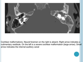
COCHLEAR
IMPLANT
DR SATINDER
COCHLEAR IMPLANT BENIFITS
 They make spoken language a viable
communication option for those who B/L
severe to profound hearing losses.
 Improve speech perception
 Speech production skills
 Contribute to improved reading outcome for
school aged students.
 Systematic rehabilitation and education
programming are necessary for CIs
recipients.
CONTRAINDICATIONS
Absolute C/I deafness due to
lesion in 8th nerve / brainstem and
cochlear aplasia.
Chronic infections middle
ear/mastoid cavity and tympanic
perforations.
If there is total/near total, b/l
cochlear ossicfication.
INDICATIONS FOR COCHLEAR IMPLANTS
Bilateral severe to profound H.L. that
is not adequately treated with std.
hearing aids.
Congenital H.L./ prelingual deafness
Acquired H.L./ postlingual deafness
Severe hearing loss that can be aided
and that deteriorates to profound loss
in childhood, adolescence, or
adulthood (prelingual).
PREOPERATIVE CONSIDERATIONS
 Collaborative effort involving patients, families,
schools, audiologists, speech/hearing therapists,
and surgeons.
 Candidates must undergo audiologic testing and
speech evaluation.
 Candidates are recommended to undergo a hearing
aid trial to test whether hearing aids are sufficient or
an implant may be more beneficial.
 Infants with hearing impairment should also
undergo a trial with hearing aids to assess potential
benefits of amplification.
•MRI and/or CT scanning are needed to
assess anatomy and to evaluate for cochlear,
vestibular, and facial nerve locations, as well
as to confirm the presence of the auditory
nerve (CN VIII).
• Implants for children aged 12 months or
older with profound b/l H.L. and for adults with
similar H.L. who demonstrate limited benefit
from hearing aid use as demonstrated by
speech perception test scores.
AIM OF PRE-OP IMAGING
Cochleo-vestiublar anomalies
Evidence of luminal
obstruction
Additional finding that can
complicate surgical/ post-op
recovery.
IMAGING
 Detect inner ear
abnormality.
 Tracing course of facial
nerve in complex cong.
malformed cases.
 In case of narrow IAM
absence of the bony
cochlear nerve canal at
the modiolus.
 Detection of high riding
jugular bulb cases.
 Presence of round or oval
window
 Detect labyrinthine
ossification
 Early fibrotic
obstructions.
 Identification of
cochlear N.
 Potency of cochlear
duct
 Large vestibular
aqueduct syndrome
 CNS abnormalities
HRCT MRI
Complete labyrinthine ossification.
Axial HRCT showing no visualized
cochlear lumen.
Dehiscent jugular bulb. Note the absence
of a bony plate between the anterior wall of
the jugular bulb and the tympanic cavity on
axial HRCT.
Labyrinthitis ossificans. Cochlea on the left is obliterated by bone after meningitis. Scala
tympani of the cochlea on the right was patent, and the patient underwent successful
implantation with complete electrode insertion
Cochlear malformations. Neural foramen on the right is absent. Right arrow indicates a
rudimentary vestibule. On the left is a severe cochlear malformation (large arrow). Small
arrow indicates the internal auditory canal
APPROACHES
TRANSMASTOID APPROACH
MIDDLE CRANIAL FOSSA
APPROACH
MINIMALLY INVASIVE COCHLEAR
IMPLANT SURGERY
FOLLOW-UP/MONITORING
Candidate attend weekly speech
therapy sessions, language
expression, social skills, lip reading
and hearing tactics
Meet with the surgeon in the follow-
up period to address any wound-
healing issues
Prognosis for hearing improvement
and improved quality of life
COMPLICATIONS
Flap dehiscence,
Seroma formation,
Implant migration,
Facial nerve stimulation,
Perilymphatic or CSF gusher,
Device failure.
Mennigitis

Cochlear implant

  • 1.
  • 2.
    COCHLEAR IMPLANT BENIFITS They make spoken language a viable communication option for those who B/L severe to profound hearing losses.  Improve speech perception  Speech production skills  Contribute to improved reading outcome for school aged students.  Systematic rehabilitation and education programming are necessary for CIs recipients.
  • 3.
    CONTRAINDICATIONS Absolute C/I deafnessdue to lesion in 8th nerve / brainstem and cochlear aplasia. Chronic infections middle ear/mastoid cavity and tympanic perforations. If there is total/near total, b/l cochlear ossicfication.
  • 4.
    INDICATIONS FOR COCHLEARIMPLANTS Bilateral severe to profound H.L. that is not adequately treated with std. hearing aids. Congenital H.L./ prelingual deafness Acquired H.L./ postlingual deafness Severe hearing loss that can be aided and that deteriorates to profound loss in childhood, adolescence, or adulthood (prelingual).
  • 5.
    PREOPERATIVE CONSIDERATIONS  Collaborativeeffort involving patients, families, schools, audiologists, speech/hearing therapists, and surgeons.  Candidates must undergo audiologic testing and speech evaluation.  Candidates are recommended to undergo a hearing aid trial to test whether hearing aids are sufficient or an implant may be more beneficial.  Infants with hearing impairment should also undergo a trial with hearing aids to assess potential benefits of amplification.
  • 6.
    •MRI and/or CTscanning are needed to assess anatomy and to evaluate for cochlear, vestibular, and facial nerve locations, as well as to confirm the presence of the auditory nerve (CN VIII). • Implants for children aged 12 months or older with profound b/l H.L. and for adults with similar H.L. who demonstrate limited benefit from hearing aid use as demonstrated by speech perception test scores.
  • 7.
    AIM OF PRE-OPIMAGING Cochleo-vestiublar anomalies Evidence of luminal obstruction Additional finding that can complicate surgical/ post-op recovery.
  • 8.
    IMAGING  Detect innerear abnormality.  Tracing course of facial nerve in complex cong. malformed cases.  In case of narrow IAM absence of the bony cochlear nerve canal at the modiolus.  Detection of high riding jugular bulb cases.  Presence of round or oval window  Detect labyrinthine ossification  Early fibrotic obstructions.  Identification of cochlear N.  Potency of cochlear duct  Large vestibular aqueduct syndrome  CNS abnormalities HRCT MRI
  • 9.
    Complete labyrinthine ossification. AxialHRCT showing no visualized cochlear lumen. Dehiscent jugular bulb. Note the absence of a bony plate between the anterior wall of the jugular bulb and the tympanic cavity on axial HRCT.
  • 10.
    Labyrinthitis ossificans. Cochleaon the left is obliterated by bone after meningitis. Scala tympani of the cochlea on the right was patent, and the patient underwent successful implantation with complete electrode insertion
  • 11.
    Cochlear malformations. Neuralforamen on the right is absent. Right arrow indicates a rudimentary vestibule. On the left is a severe cochlear malformation (large arrow). Small arrow indicates the internal auditory canal
  • 12.
    APPROACHES TRANSMASTOID APPROACH MIDDLE CRANIALFOSSA APPROACH MINIMALLY INVASIVE COCHLEAR IMPLANT SURGERY
  • 13.
    FOLLOW-UP/MONITORING Candidate attend weeklyspeech therapy sessions, language expression, social skills, lip reading and hearing tactics Meet with the surgeon in the follow- up period to address any wound- healing issues Prognosis for hearing improvement and improved quality of life
  • 14.
    COMPLICATIONS Flap dehiscence, Seroma formation, Implantmigration, Facial nerve stimulation, Perilymphatic or CSF gusher, Device failure. Mennigitis