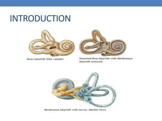
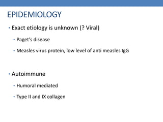
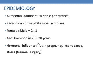
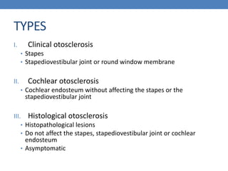
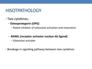
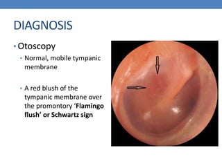
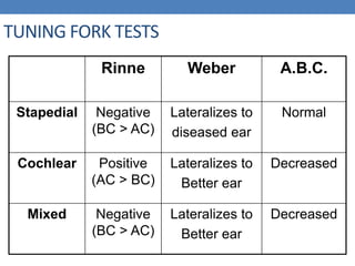
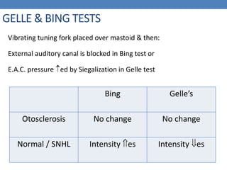
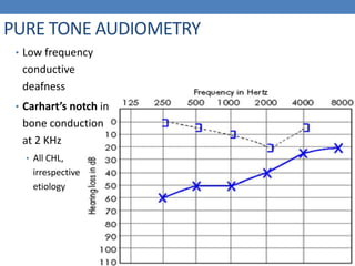
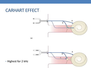
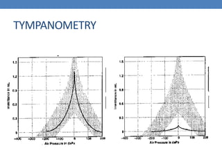
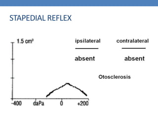
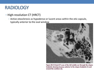
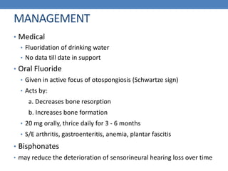
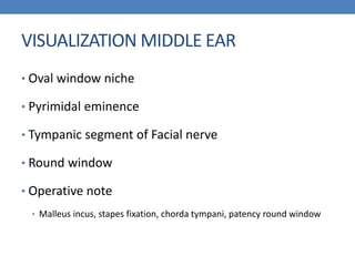
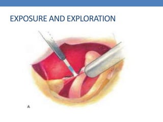
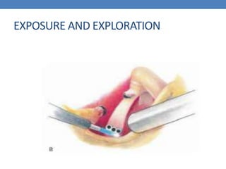
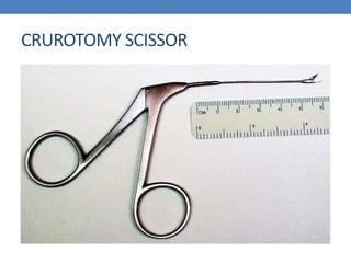
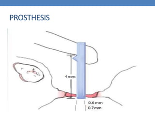
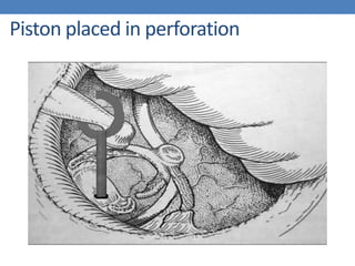
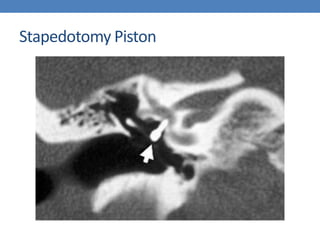
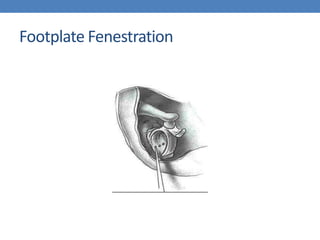
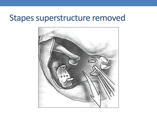
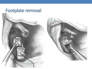
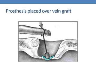
OTOSCLEROSIS
Dr. Rajesh Maharjan
ENT 1st year Resident
GMC
INTRODUCTION
• Localized hereditary disorder of bone metabolism of
otic capsule enchondral bone that is characterized by
disordered resorption and deposition of bone
• Bone resorption by osteoclasts and new bone
formation by osteoblasts, accompanied by vascular
proliferation and tissue stroma
• New vascular spongy bone formation causes ankylosis
Antonio Valsalva
Italian Anatomist
First described
ankylosis of stapes
in 1741
Joseph Toynbee
• First described the
condition causing a
hearing loss by fixation of
the stapes in 1860
Anton von Tröltsch
• 1869
• Final inactive
sclerotic stage of the
disease, "sclerosis”
Adam Politzer
1893
Coined the term
Otosclerosis
in
Friedrich Siebenmann
Swiss otolaryngologist
Coined the term
Otospongiosis
in 1912
INTRODUCTION
EPIDEMIOLOGY
• Exact etiology is unknown (? Viral)
• Paget’s disease
• Measles virus protein, low level of anti measles IgG
• Autoimmune
• Humoral mediated
• Type II and IX collagen
EPIDEMIOLOGY
• Autosomal dominant: variable penetrance
• Race: common in white races & Indians
• Female : Male = 2 : 1
• Age: Common in 20 - 30 years
• Hormonal influence: es in pregnancy, menopause,
stress (trauma, surgery)
TYPES
I. Clinical otosclerosis
• Stapes
• Stapediovestibular joint or round window membrane
II. Cochlear otosclerosis
• Cochlear endosteum without affecting the stapes or the
stapediovestibular joint
III. Histological otosclerosis
• Histopathological lesions
• Do not affect the stapes, stapediovestibular joint or cochlear
endosteum
• Asymptomatic
HISOTPATHOLOGY
• Two cytokines,
• Osteoprotegerin (OPG)
• Potent inhibitor of osteoclast activation and maturation
• RANKL (receptor activator nuclear-kb ligand)
• Osteoclast activator
• Breakage in signaling pathway between two cytokines
LIGHT AND ELECTRON MICROSCOPY
• Earliest indication
• Resorption of enchondral bone around blood vessels
• Enlargement of perivascular spaces followed by deposition of immature
(woven) bone
• Active otosclerotic foci
• Increased vascularity and increased bone turnover
• Inactive (sclerotic) foci
• Dense mineralized bone
• The connective tissue stroma in the foci consists
• Fibroblasts and osteocytes
• Complete absence of acute inflammatory cells
ORIGIN AND SITES
• Wall anterior to the oval window > round window
niche > cochlear apex
• Less frequent sites
• Posterior to the oval window
• The internal auditory canal
• Around the cochlear duct and the semicircular canals
• Middle ear ossicles
• Labyrinthine spaces and the vestibular aqueduct
CAVITATING OTOSCLEROSIS
• Large cavities may form within otosclerotic foci
• If contact with endosteum of scala tympani
• 3rd window
• Persistence of air bone gap following stapedectomy
• Communicate with the CSF space in the internal
auditory canal
• Result in gushers during cochlear implantation
TYPES OF STAPEDIAL OTOSCLEROSIS
1. Anterior focus (commonest): 2 mm anterior to oval Window
2. Posterior focus: 2 mm behind oval window
3. Circumferential: involves footplate margin only
Types of Stapedial Otosclerosis
4. Biscuit type: footplate involved, margin is free
5. Obliterative: obliterates oval window completely
Symptoms of Otosclerosis
• Hearing loss: Bilateral, slowly progressive
• Conductive: stapedial otosclerosis
• Sensori-neural: cochlear otosclerosis  hyalinization and atrophy of spiral
ligament
• Mixed: stapedial + cochlear otosclerosis
• Vestibular symptoms
• Vestibular ganglion degeneration
• Asso. Meniere’s disease
• Vestibular aqueduct filled with active otosclerotic foci
Symptoms of Otosclerosis
3. Paracusis Willis: Better hearing in noisy surroundings (people
increase their voice intensity & pt’s speech discrimination
becomes better).
DIAGNOSIS
• Otoscopy
• Normal, mobile tympanic
membrane
• A red blush of the
tympanic membrane over
the promontory ‘Flamingo
flush’ or Schwartz sign
TUNING FORK TESTS
Rinne Weber A.B.C.
Stapedial Negative
(BC > AC)
Lateralizes to
diseased ear
Normal
Cochlear Positive
(AC > BC)
Lateralizes to
Better ear
Decreased
Mixed Negative
(BC > AC)
Lateralizes to
Better ear
Decreased
GELLE & BING TESTS
Vibrating tuning fork placed over mastoid & then:
External auditory canal is blocked in Bing test or
E.A.C. pressure ed by Siegalization in Gelle test
Bing Gelle’s
Otosclerosis No change No change
Normal / SNHL Intensity es Intensity es
PURE TONE AUDIOMETRY
• Low frequency
conductive
deafness
• Carhart’s notch in
bone conduction
at 2 KHz
• All CHL,
irrespective
etiology
CARHART EFFECT
• Highest for 2 kHz
TYMPANOMETRY
STAPEDIAL REFLEX
RADIOLOGY
• High-resolution CT (HRCT)
• Active otosclerosis as hypodense or lucent areas within the otic capsule,
typically anterior to the oval window
SURGICAL DIAGNOSIS
• Middle ear anatomy normal
• Bone around oval window whiter than normal but no
clear junction
• Footplate thicker
HISTOLOGICAL DIAGNOSIS
• Only if stapedectomy performed
• Stapedotomy
• Stapes superstructure
• Non otosclerotic bone
DIFFERENTIAL DIAGNOSIS
• Otitis Media with Effusion: type B tympanogram
• Adhesive Otitis Media: absence of T.M. mobility
• Tympanosclerosis: white patch on T.M.
• Ossicular discontinuity: type Ad tympanogram
• Congenital ossicular chain fixation: tympanotomy
• Malleus head fixation: tympanotomy
MANAGEMENT
• Medical
• Fluoridation of drinking water
• No data till date in support
• Oral Fluoride
• Given in active focus of otospongiosis (Schwartze sign)
• Acts by:
a. Decreases bone resorption
b. Increases bone formation
• 20 mg orally, thrice daily for 3 - 6 months
• S/E arthritis, gastroenteritis, anemia, plantar fascitis
• Bisphonates
• may reduce the deterioration of sensorineural hearing loss over time
HEARING AIDS
• Unfit for surgery
• Disadvantage
• Aesthetic concern as patient usually
of young age
• BAHA (Bone Anchored Hearing
Aid)
• An only hearing ear with otosclerosis
• Difficulty using a conventional aid
• Post-fenestration cavity
FAR-ADVANCED OTOSCLEROSIS
• Average air-conduction of > 85 db
• Bone conduction immeasurable due to limits of
audiometric equipment
• Distinguish from Pure SNHL
• Timescale of the hearing impairment
• Family history, previous audiograms showing an air–bone gap
• No CT evidence of cochlear otosclerosis
• Managed in 1 of 3 ways
• Stapedectomy and subsequent conventional hearing aids
• Cochlear implantation
• The new direct acoustic cochlear stimulation (DACS) device
DIRECT ACOUSTIC COCHLEAR STIMULATION
(DACS)
• The device consists of an active middle ear implant
attached to a stapes prosthesis
• Mixed hearing loss where a successful stapedotomy
alone would not allow the patient to manage without
a hearing aid
• Indications
• Minimum average bone-conduction of over 30 db and with
an additional air–bone gap of over 30 db
COCHLEAR IMPLANT
• Patients with FAO or failed stapedotomy may be candidates for
cochlear implantation
• There may be problems in electrode placement due to
otosclerotic ossification of the round window or basal turn of
the cochlear which may necessitate extra drilling
SURGERY
SURGERY
• 1878
• Transtympanic mobilization and
removal of the stapes
• 1899
• 6th International Otologic
Congress in London
Johannes Kessel
SURGERY
• 1956
• Binocular microscope
• Stapedotomy
• Ossicular
reconstruction/teflon piston
• Vein graft
John Shea
INDICATIONS
• Hearing threshold for air conduction 30 dB or more
• Average air bone gap at least 15 dB or more at
frequencies of 250 Hz to 1 kHz
• Negative Rinne at 512 Hz
INDICATIONS
• Bilateral involvement
• worse hearing ear/ interval 1 yr
• Symmetric loss
• Patients preference
• Concomitant sensorineural loss is not a
contraindication for surgery
CONTRAINDICATIONS
• Only hearing ear
• Infection of external or middle ear
• Pregnancy
• Tympanic membrane perforation
FACTORS TO CONSIDER
• Age
• Children
• CT temporal bone
• Congenital Middle/inner ear malformation
• No upper age limit
• Occupations
• Scuba diving, commercial air travellers, parachuting
• Barotrauma
• Tympanometric pressure of 400 mm of H2O, if no nystagmus
• Work in noisy environment
• Ménière’s disease
• Distended saccule close to oval window
INFORMED CONSENT
• Description of the procedure and discussion of all
potential risks
• Occupational dependence on taste
OPERATIVE NOTE
• Shape and mobility of the incus and malleus
• Presence of otosclerosis
• Fixation of the stapes
• Patency of the round window
• Location of and the bone covering the facial nerve
• Status of the chorda tympani
ANESTHESIA
• Local anesthesia
• Time saving
• Intraoperative report of vestibular symptoms
• Prevention of excessive inner ear irritation
• General
• Painless
• Motionless operative field
SURGICAL TECHNIQUE
POSITIONING
• Supine head tilted 10-15 down
• Head turned to contralateral shoulder
• Tympanic membrane almost horizontal plane
EXPOSURE AND EXPLORATION
• Speculum holder attached to the bed or the headrest
• Size
• Too small
• Move against EAC
• Restrict vision and instrumentation
• Too large
• Push soft tissue of EAC medially
• Obstruct view
• Should wedge into lateral aspect of bony canal
EXPOSURE AND EXPLORATION
• Local anesthesia 1% lidocaine and 1:100,000
epinephrine
• Minimizes bleeding
• 27 gauge needle 4 quadrants of cartilage EAC
• Lateral to junction
• Bevelled 30-gauge needle into subperiosteal plane at
6 and 12 o’ clock
EXPOSURE AND EXPLORATION
• Permeatal incision
Tympanomeatal flap raised
Posterosuperior annulus
CHORDA TYMPANI SEPARATED
VISUALIZATION MIDDLE EAR
• Oval window niche
• Pyrimidal eminence
• Tympanic segment of Facial nerve
• Round window
• Operative note
• Malleus incus, stapes fixation, chorda tympani, patency round window
EXPOSURE AND EXPLORATION
CRUROTOMY SCISSOR
EXPOSURE AND EXPLORATION
JOINT KNIFE
EXPOSURE AND EXPLORATION
FENESTRATION
FENESTRATION
• Site
• Middle and inferior 3rd
• Greatest distance from footplate to membranous labyrinth
• Argon vs Carbon dioxide
• Visible light range
• Laser vs Microdrill
• Cut and coagulate
• No pressure and movement
• Less irritation and trauma perilymph
PROSTHESIS
Piston placed in perforation
ALLIGATOR FORCEPS
• Grasped by its loop
• Pace to fenestra and
incus in one movement
• Site
• Narrowest area of incus
CRIMPING
• Differential movement
• Delayed failure
POSITION
• Perpendicular to footplate
and long axis incus
• Fitting fenestra snugly but
freely moving
Tympanomeatal flap put back
Stapedotomy Piston
STAPEDECTOMY
STAPEDECTOY
• Free floating footplate
• Communited fracture footplate
• Inadvertently removed during superstrucure removal
• Revision surgery
• Instrument for fenestration not availabe
STAPEDOTOMY VS STAPEDECTOMY
• Hearing difference not significant
• Vestibular symptoms
• Occurrence, duration, and severity
Footplate Fenestration
Stapes superstructure removed
Footplate removal
Prosthesis placed over vein graft
COMPLICATIONS OF
STAPES SURGERY
INTRAOPERATIVE
• Tear tympanomeatal flap
• Perforation TM
• Subluxation Incus
• Facial nerve overhanging
• Obliterative otosclerosis of the oval window
• Otosclerosis involving round window
• Malleus ankylosis
• Perilymph gusher
• Floating and depressed footplate
POST OPERATIVE
• Facial palsy
• Chorda tympani dysfunction
• Otitis media
• Vertigo
• Sensorineuronal hearing loss
• Conductive hearing loss
• Malfunctioning prosthesis Malleus fixation OM, DSSC, round window
obliteration
REFERENCE
• Scott-Brow’s Otorhinolaryngology, Head and Neck
Surgery, 8th edition
• Glasscock-Shambaugh, Surgery of the ear, 6th edition
Otosclerosis

Otosclerosis

  • 1.
  • 2.
    INTRODUCTION • Localized hereditarydisorder of bone metabolism of otic capsule enchondral bone that is characterized by disordered resorption and deposition of bone • Bone resorption by osteoclasts and new bone formation by osteoblasts, accompanied by vascular proliferation and tissue stroma • New vascular spongy bone formation causes ankylosis
  • 3.
    Antonio Valsalva Italian Anatomist Firstdescribed ankylosis of stapes in 1741
  • 4.
    Joseph Toynbee • Firstdescribed the condition causing a hearing loss by fixation of the stapes in 1860
  • 5.
    Anton von Tröltsch •1869 • Final inactive sclerotic stage of the disease, "sclerosis”
  • 6.
    Adam Politzer 1893 Coined theterm Otosclerosis in
  • 7.
  • 8.
  • 9.
    EPIDEMIOLOGY • Exact etiologyis unknown (? Viral) • Paget’s disease • Measles virus protein, low level of anti measles IgG • Autoimmune • Humoral mediated • Type II and IX collagen
  • 10.
    EPIDEMIOLOGY • Autosomal dominant:variable penetrance • Race: common in white races & Indians • Female : Male = 2 : 1 • Age: Common in 20 - 30 years • Hormonal influence: es in pregnancy, menopause, stress (trauma, surgery)
  • 11.
    TYPES I. Clinical otosclerosis •Stapes • Stapediovestibular joint or round window membrane II. Cochlear otosclerosis • Cochlear endosteum without affecting the stapes or the stapediovestibular joint III. Histological otosclerosis • Histopathological lesions • Do not affect the stapes, stapediovestibular joint or cochlear endosteum • Asymptomatic
  • 12.
    HISOTPATHOLOGY • Two cytokines, •Osteoprotegerin (OPG) • Potent inhibitor of osteoclast activation and maturation • RANKL (receptor activator nuclear-kb ligand) • Osteoclast activator • Breakage in signaling pathway between two cytokines
  • 13.
    LIGHT AND ELECTRONMICROSCOPY • Earliest indication • Resorption of enchondral bone around blood vessels • Enlargement of perivascular spaces followed by deposition of immature (woven) bone • Active otosclerotic foci • Increased vascularity and increased bone turnover • Inactive (sclerotic) foci • Dense mineralized bone • The connective tissue stroma in the foci consists • Fibroblasts and osteocytes • Complete absence of acute inflammatory cells
  • 14.
    ORIGIN AND SITES •Wall anterior to the oval window > round window niche > cochlear apex • Less frequent sites • Posterior to the oval window • The internal auditory canal • Around the cochlear duct and the semicircular canals • Middle ear ossicles • Labyrinthine spaces and the vestibular aqueduct
  • 15.
    CAVITATING OTOSCLEROSIS • Largecavities may form within otosclerotic foci • If contact with endosteum of scala tympani • 3rd window • Persistence of air bone gap following stapedectomy • Communicate with the CSF space in the internal auditory canal • Result in gushers during cochlear implantation
  • 16.
    TYPES OF STAPEDIALOTOSCLEROSIS 1. Anterior focus (commonest): 2 mm anterior to oval Window 2. Posterior focus: 2 mm behind oval window 3. Circumferential: involves footplate margin only
  • 17.
    Types of StapedialOtosclerosis 4. Biscuit type: footplate involved, margin is free 5. Obliterative: obliterates oval window completely
  • 18.
    Symptoms of Otosclerosis •Hearing loss: Bilateral, slowly progressive • Conductive: stapedial otosclerosis • Sensori-neural: cochlear otosclerosis  hyalinization and atrophy of spiral ligament • Mixed: stapedial + cochlear otosclerosis • Vestibular symptoms • Vestibular ganglion degeneration • Asso. Meniere’s disease • Vestibular aqueduct filled with active otosclerotic foci
  • 19.
    Symptoms of Otosclerosis 3.Paracusis Willis: Better hearing in noisy surroundings (people increase their voice intensity & pt’s speech discrimination becomes better).
  • 21.
    DIAGNOSIS • Otoscopy • Normal,mobile tympanic membrane • A red blush of the tympanic membrane over the promontory ‘Flamingo flush’ or Schwartz sign
  • 22.
    TUNING FORK TESTS RinneWeber A.B.C. Stapedial Negative (BC > AC) Lateralizes to diseased ear Normal Cochlear Positive (AC > BC) Lateralizes to Better ear Decreased Mixed Negative (BC > AC) Lateralizes to Better ear Decreased
  • 23.
    GELLE & BINGTESTS Vibrating tuning fork placed over mastoid & then: External auditory canal is blocked in Bing test or E.A.C. pressure ed by Siegalization in Gelle test Bing Gelle’s Otosclerosis No change No change Normal / SNHL Intensity es Intensity es
  • 24.
    PURE TONE AUDIOMETRY •Low frequency conductive deafness • Carhart’s notch in bone conduction at 2 KHz • All CHL, irrespective etiology
  • 25.
  • 26.
  • 27.
  • 28.
    RADIOLOGY • High-resolution CT(HRCT) • Active otosclerosis as hypodense or lucent areas within the otic capsule, typically anterior to the oval window
  • 29.
    SURGICAL DIAGNOSIS • Middleear anatomy normal • Bone around oval window whiter than normal but no clear junction • Footplate thicker
  • 30.
    HISTOLOGICAL DIAGNOSIS • Onlyif stapedectomy performed • Stapedotomy • Stapes superstructure • Non otosclerotic bone
  • 31.
    DIFFERENTIAL DIAGNOSIS • OtitisMedia with Effusion: type B tympanogram • Adhesive Otitis Media: absence of T.M. mobility • Tympanosclerosis: white patch on T.M. • Ossicular discontinuity: type Ad tympanogram • Congenital ossicular chain fixation: tympanotomy • Malleus head fixation: tympanotomy
  • 32.
    MANAGEMENT • Medical • Fluoridationof drinking water • No data till date in support • Oral Fluoride • Given in active focus of otospongiosis (Schwartze sign) • Acts by: a. Decreases bone resorption b. Increases bone formation • 20 mg orally, thrice daily for 3 - 6 months • S/E arthritis, gastroenteritis, anemia, plantar fascitis • Bisphonates • may reduce the deterioration of sensorineural hearing loss over time
  • 33.
    HEARING AIDS • Unfitfor surgery • Disadvantage • Aesthetic concern as patient usually of young age • BAHA (Bone Anchored Hearing Aid) • An only hearing ear with otosclerosis • Difficulty using a conventional aid • Post-fenestration cavity
  • 34.
    FAR-ADVANCED OTOSCLEROSIS • Averageair-conduction of > 85 db • Bone conduction immeasurable due to limits of audiometric equipment • Distinguish from Pure SNHL • Timescale of the hearing impairment • Family history, previous audiograms showing an air–bone gap • No CT evidence of cochlear otosclerosis • Managed in 1 of 3 ways • Stapedectomy and subsequent conventional hearing aids • Cochlear implantation • The new direct acoustic cochlear stimulation (DACS) device
  • 35.
    DIRECT ACOUSTIC COCHLEARSTIMULATION (DACS) • The device consists of an active middle ear implant attached to a stapes prosthesis • Mixed hearing loss where a successful stapedotomy alone would not allow the patient to manage without a hearing aid • Indications • Minimum average bone-conduction of over 30 db and with an additional air–bone gap of over 30 db
  • 36.
    COCHLEAR IMPLANT • Patientswith FAO or failed stapedotomy may be candidates for cochlear implantation • There may be problems in electrode placement due to otosclerotic ossification of the round window or basal turn of the cochlear which may necessitate extra drilling
  • 37.
  • 38.
    SURGERY • 1878 • Transtympanicmobilization and removal of the stapes • 1899 • 6th International Otologic Congress in London Johannes Kessel
  • 39.
    SURGERY • 1956 • Binocularmicroscope • Stapedotomy • Ossicular reconstruction/teflon piston • Vein graft John Shea
  • 40.
    INDICATIONS • Hearing thresholdfor air conduction 30 dB or more • Average air bone gap at least 15 dB or more at frequencies of 250 Hz to 1 kHz • Negative Rinne at 512 Hz
  • 41.
    INDICATIONS • Bilateral involvement •worse hearing ear/ interval 1 yr • Symmetric loss • Patients preference • Concomitant sensorineural loss is not a contraindication for surgery
  • 42.
    CONTRAINDICATIONS • Only hearingear • Infection of external or middle ear • Pregnancy • Tympanic membrane perforation
  • 43.
    FACTORS TO CONSIDER •Age • Children • CT temporal bone • Congenital Middle/inner ear malformation • No upper age limit • Occupations • Scuba diving, commercial air travellers, parachuting • Barotrauma • Tympanometric pressure of 400 mm of H2O, if no nystagmus • Work in noisy environment • Ménière’s disease • Distended saccule close to oval window
  • 44.
    INFORMED CONSENT • Descriptionof the procedure and discussion of all potential risks • Occupational dependence on taste
  • 45.
    OPERATIVE NOTE • Shapeand mobility of the incus and malleus • Presence of otosclerosis • Fixation of the stapes • Patency of the round window • Location of and the bone covering the facial nerve • Status of the chorda tympani
  • 46.
    ANESTHESIA • Local anesthesia •Time saving • Intraoperative report of vestibular symptoms • Prevention of excessive inner ear irritation • General • Painless • Motionless operative field
  • 47.
  • 48.
    POSITIONING • Supine headtilted 10-15 down • Head turned to contralateral shoulder • Tympanic membrane almost horizontal plane
  • 49.
    EXPOSURE AND EXPLORATION •Speculum holder attached to the bed or the headrest • Size • Too small • Move against EAC • Restrict vision and instrumentation • Too large • Push soft tissue of EAC medially • Obstruct view • Should wedge into lateral aspect of bony canal
  • 50.
    EXPOSURE AND EXPLORATION •Local anesthesia 1% lidocaine and 1:100,000 epinephrine • Minimizes bleeding • 27 gauge needle 4 quadrants of cartilage EAC • Lateral to junction • Bevelled 30-gauge needle into subperiosteal plane at 6 and 12 o’ clock
  • 51.
    EXPOSURE AND EXPLORATION •Permeatal incision
  • 52.
  • 53.
  • 54.
  • 55.
    VISUALIZATION MIDDLE EAR •Oval window niche • Pyrimidal eminence • Tympanic segment of Facial nerve • Round window • Operative note • Malleus incus, stapes fixation, chorda tympani, patency round window
  • 56.
  • 57.
  • 58.
  • 59.
  • 60.
  • 61.
  • 62.
  • 63.
    FENESTRATION • Site • Middleand inferior 3rd • Greatest distance from footplate to membranous labyrinth • Argon vs Carbon dioxide • Visible light range • Laser vs Microdrill • Cut and coagulate • No pressure and movement • Less irritation and trauma perilymph
  • 64.
  • 65.
    Piston placed inperforation
  • 66.
    ALLIGATOR FORCEPS • Graspedby its loop • Pace to fenestra and incus in one movement • Site • Narrowest area of incus
  • 67.
  • 68.
    POSITION • Perpendicular tofootplate and long axis incus • Fitting fenestra snugly but freely moving
  • 69.
  • 70.
  • 71.
  • 72.
    STAPEDECTOY • Free floatingfootplate • Communited fracture footplate • Inadvertently removed during superstrucure removal • Revision surgery • Instrument for fenestration not availabe
  • 73.
    STAPEDOTOMY VS STAPEDECTOMY •Hearing difference not significant • Vestibular symptoms • Occurrence, duration, and severity
  • 74.
  • 75.
  • 76.
  • 77.
  • 78.
  • 79.
    INTRAOPERATIVE • Tear tympanomeatalflap • Perforation TM • Subluxation Incus • Facial nerve overhanging • Obliterative otosclerosis of the oval window • Otosclerosis involving round window • Malleus ankylosis • Perilymph gusher • Floating and depressed footplate
  • 80.
    POST OPERATIVE • Facialpalsy • Chorda tympani dysfunction • Otitis media • Vertigo • Sensorineuronal hearing loss • Conductive hearing loss • Malfunctioning prosthesis Malleus fixation OM, DSSC, round window obliteration
  • 81.
    REFERENCE • Scott-Brow’s Otorhinolaryngology,Head and Neck Surgery, 8th edition • Glasscock-Shambaugh, Surgery of the ear, 6th edition

Editor's Notes

  • #3 The normal temporal bone has embryonic cartilage rests called "globuli interossei." These rests are associated with sites of predilection in otosclerosis
  • #26 0.5,1,2,4 air bone gap is greatest Magnitude of air bone gap 10 irrespective of what frequencies involved