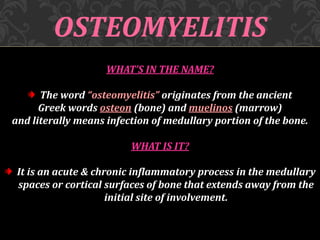
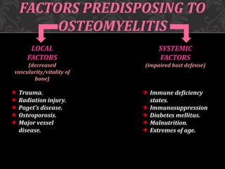
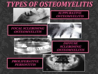
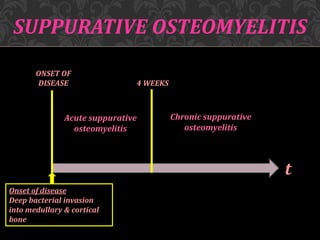
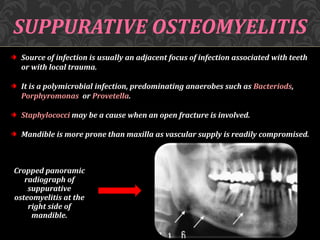
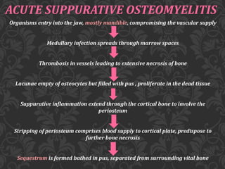
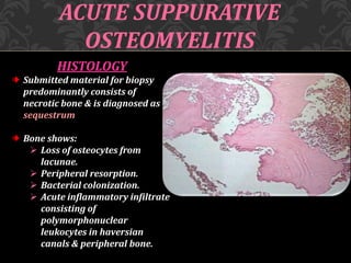
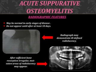
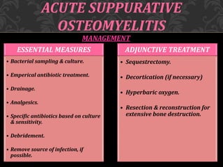
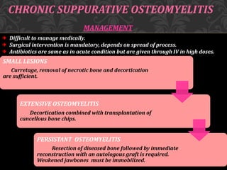
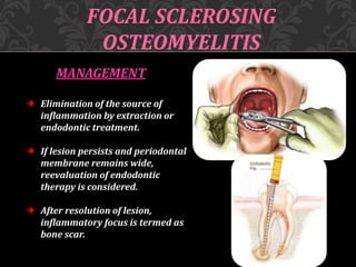
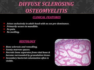
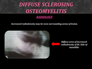
PRESENTED BY:
SAPEEDEH AFZAL
ROLL # 10
(PATHOGENESIS & MANAGEMENT)
R
O
A
D
M
A
P
WHAT IS OSTEOMYELITIS
FACTORS PREDISPOSING TO OSTEOMYELITIS
PATHOGENESIS OF OSTEOMYELITIS
TYPES OF OSTEOMYELITIS
 PATHOGENESIS
 CLINICAL FEATURES
 HISTOLOGY
 RADIOLOGY
 MANAGEMENT
OSTEOMYELITIS
WHAT’S IN THE NAME?
The word “osteomyelitis” originates from the ancient
Greek words osteon (bone) and muelinos (marrow)
and literally means infection of medullary portion of the bone.
WHAT IS IT?
It is an acute & chronic inflammatory process in the medullary
spaces or cortical surfaces of bone that extends away from the
initial site of involvement.
FACTORS PREDISPOSING TO
OSTEOMYELITIS
LOCAL
FACTORS
(decreased
vascularity/vitality of
bone)
Trauma.
Radiation injury.
Paget’s disease.
Osteoporosis.
Major vessel
disease.
SYSTEMIC
FACTORS
(impaired host defense)
Immune deficiency
states.
Immunosuppression
Diabetes mellitus.
Malnutrition.
Extremes of age.
PATHOGENESIS OF
OSTEOMYELITIS
Inflammatory process of entire bone including cortex &
periosteum, not just confined to endosteum
Inflammatory condition beginning in medullary cavity &
havarsian system & extending to involve periosteum of
affected area
Local factors decreases the vitality of bone
Systemic conditions comprises the defense system of the host
TYPES OF OSTEOMYELITIS
SUPPURATIVE
OSTEOMYELITIS
FOCAL SCLEROSING
OSTEOMYELITIS
DIFFUSE
SCLEROSING
OSTEOMYELITIS
PROLIFERATIVE
PERIOSTITIS
ONSET OF
DISEASE 4 WEEKS
Acute suppurative
osteomyelitis
Chronic suppurative
osteomyelitis
t
Onset of disease
Deep bacterial invasion
into medullary & cortical
bone
SUPPURATIVE OSTEOMYELITIS
Source of infection is usually an adjacent focus of infection associated with teeth
or with local trauma.
It is a polymicrobial infection, predominating anaerobes such as Bacteriods,
Porphyromonas or Provetella.
Staphylococci may be a cause when an open fracture is involved.
Mandible is more prone than maxilla as vascular supply is readily compromised.
Cropped panoramic
radiograph of
suppurative
osteomyelitis at the
right side of
mandible.
ACUTE SUPPURATIVE OSTEOMYELITIS
Organisms entry into the jaw, mostly mandible, compromising the vascular supply
Medullary infection spreads through marrow spaces
Thrombosis in vessels leading to extensive necrosis of bone
Lacunae empty of osteocytes but filled with pus , proliferate in the dead tissue
Suppurative inflammation extend through the cortical bone to involve the
periosteum
Stripping of periosteum comprises blood supply to cortical plate, predispose to
further bone necrosis
Sequestrum is formed bathed in pus, separated from surrounding vital bone
ACUTE SUPPURATIVE
OSTEOMYELITIS
CLINICAL FEATURES
EARLY :
 Severe throbbing, deep- seated pain.
 Swelling due to inflammatory
edema.
 Gingiva appears red, swollen &
tender.
LATE :
 Distension of periosteum with pus.
FINAL:
 Subperiosteal bone formation cause
swelling to become firm.
HISTOLOGY
Submitted material for biopsy
predominantly consists of
necrotic bone & is diagnosed as
sequestrum
Bone shows:
 Loss of osteocytes from
lacunae.
 Peripheral resorption.
 Bacterial colonization.
 Acute inflammatory infiltrate
consisting of
polymorphonuclear
leukocytes in haversian
canals & peripheral bone.
ACUTE SUPPURATIVE
OSTEOMYELITIS
ACUTE SUPPURATIVE
OSTEOMYELITIS
RADIOGRAPHIC FEATURES
May be normal in early stages of disease .
Do not appear until after at least 10 days.
After sufficient bone
resorption irregular, mot-
eaten areas of radiolucency
may appear.
Radiograph may
demonstrate ill-defined
radiolucency.
MANAGEMENT
ACUTE SUPPURATIVE
OSTEOMYELITIS
ESSENTIAL MEASURES
• Bacterial sampling & culture.
• Emperical antibiotic treatment.
• Drainage.
• Analgesics.
• Specific antibiotics based on culture
& sensitivity.
• Debridement.
• Remove source of infection, if
possible.
ADJUNCTIVE TREATMENT
• Sequestrectomy.
• Decortication (if necessary)
• Hyperbaric oxygen.
• Resection & reconstruction for
extensive bone destruction.
ACUTE SUPPURATIVE
OSTEOMYELITIS
COMPLICATIONS
Rare but include:
 Pathological fracture Extensive bone destruction.
 Chronic osteomyelitis Inadequate treatment.
 Cellulitis Spread of virulent bacteria.
 Septicemia Immuno-compromised patient.
CHRONIC SUPPURATIVE OSTEOMYELITIS
• Inadequate treatment of acute osteomyelitis
• Periodontal diseases
• Pulpal infections
• Extraction wounds
• Infected fractures
Infection in the medulllary spaces spread and form granulation
tissue
Granulation tissue forms dense scar to wall off the infected area
Encircled dead space acts as a reserviour for bacteria &
antibiotics have great difficulty reaching the site
CHRONIC SUPPURATIVE
OSTEOMYELITIS
CLINICAL FEATURES
Swelling
Pain
Sinus formation
Purulent discharge
Sequestrum formation
Tooth loss
Pathologic fracture
CHRONIC SUPPURATIVE
OSTEOMYELITIS
HISTOLOGY
Inflammed connective tissue filling inter-trabecular areas of
bone.
Scattered sequestra.
Pockets of abscess.
CHRONIC SUPPURATIVE
OSTEOMYELITIS
RADIOLOGY
Patchy, ragged & ill defined radiolucency.
Often contains radiopaque sequestra.
• Sequestra lying close to the
peripheral sclerosis & lower
border.
• New bone formation is
evident below lower border.
CHRONIC SUPPURATIVE OSTEOMYELITIS
MANAGEMENT
Difficult to manage medically.
Surgical intervention is mandatory, depends on spread of process.
Antibiotics are same as in acute condition but are given through IV in high doses.
SMALL LESIONS
Curretage, removal of necrotic bone and decortication
are sufficient.
EXTENSIVE OSTEOMYELITIS
Decortication combined with transplantation of
cancellous bone chips.
PERSISTANT OSTEOMYELITIS
Resection of diseased bone followed by immediate
reconstruction with an autologous graft is required.
Weakened jawbones must be immobilized.
FOCAL SCLEROSING
OSTEOMYELITIS
Also known as “Condensing osteitis”.
Localized areas of bone sclerosis.
Bony reaction to low-grade peri-apical infection or unusually strong
host defensive response.
Association with an area of inflammation is critical.
FOCAL SCLEROSING
OSTEOMYELITIS
CLINICAL FEATURES
Children & young adults are affected.
In mandible, premolar & molar regions are affected.
Bone sclerosis is associated with non-vital or pulpitic tooth.
No expansion of the jaw.
HISTOLOGY
Dense sclerotic bone.
Scanty connective tissue.
Inflammatory cells.
RADIOLOGY
Localized but uniform increased radiodensity related to tooth.
Widened periodontal ligament space or peri-apical area.
Sometimes an adjacent radiolucent inflammatory lesion may be
present.
FOCAL SCLEROSING
OSTEOMYELITIS
Increased areas of
radiodensity surrounding
apices of nonvital mandibular
first molar
MANAGEMENT
Elimination of the source of
inflammation by extraction or
endodontic treatment.
If lesion persists and periodontal
membrane remains wide,
reevaluation of endodontic
therapy is considered.
After resolution of lesion,
inflammatory focus is termed as
bone scar.
FOCAL SCLEROSING
OSTEOMYELITIS
DIFFUSE SCLEROSING
OSTEOMYELITIS
It is an ill-defined, highly controversial, evolving area of dental medicine.
Exact etiology is unknown.
Chronic intraosseous bacterial infection creates a smoldering mass of
chronically inflammed granulation tissue.
CLINICAL FEATURES
Arises exclusively in adult-hood with no sex pre-dominance.
Primarily occurs in mandible.
No pain.
No swelling.
DIFFUSE SCLEROSING
OSTEOMYELITIS
HISTOLOGY
Bone sclerosis and remodling.
Scanty marrow spaces.
Necrotic bone separates from vital bone &
become surrounded by granulation tissue.
Secondary bacterial colonization often is
visible.
RADIOLOGY
Increased radiodensity may be seen surrounding areas of lesion.
DIFFUSE SCLEROSING
OSTEOMYELITIS
Diffuse area of increased
radiodensity of Rt. Side of
mandible
MANAGEMENT
Elimination of originating sources of inflammation
via extraction & endodontic treatment.
Sclerotic area remain radiographically.
DIFFUSE SCLEROSING
OSTEOMYELITIS
PROLIFERATIVE PERIOSTITIS
Also known as “ Periostitis ossificans” & “Garee’s osteomyelitis”.
It represents a periosteal reaction to the presence of inflammation.
Affected periosteum forms several rows of reactive vital bone that parallel each
other & expand surface of altered bone.
PATHOGENESIS
The spread of low-grade, chronic apical inflammation through cortical
bone
Periosteal reaction occurs
Stimulates proliferative reaction of periosteum
PROLIFERATIVE PERIOSTITIS
CLINICAL FEATURES
Affected patients are primarily children
& young adults.
Incidence is mean age of 13 years.
No sex predominance is noted.
Most cases arise in the premolar &
molar area of mandible.
Hyperplasia is located most commonly
along lower border of mandible.
Most cases are uni-focal, multiple
quadrants may be affected.
PROLIFERATIVE PERIOSTITIS
HISTOLOGY
Parallel rows of highly cellular
& reactive woven bone .
Trabeculae are frequently
oriented perpendicular to
surface.
Trabeculae sometimes form an
interconnecting meshwork of
bone.
Between trabeculae,
uninflammed fibrous tissue is
evident.
PROLIFERATIVE PERIOSTITIS
RADIOLOGY
Radiopaque laminations of bone roughly parallel each other & underlying
cortical surface.
Laminations may vary from 1-12 in number.
Radiolucent separations often are present between new bone & original
cortex.
PROLIFERATIVE PERIOSTITIS
MANAGEMENT
Removal of infection.
After infection has resolved, layers of bone will
consolidate in 6-12 months .
QUESTIONS
1.WHY IS MANDIBLE MORE PRONE TO
OSTEOMYELITIS THAN MAXILLA?
2.WHY RADIOGRAPHIC FEATURES IN ACUTE
SUPPURATIVE OSTEOMYELITIS APPEAR AFTER 10-
14 DAYS OF DISEASE?
REFRENCES:
• J.V. SOAMS 4TH EDITION.
• NEVILLE & DAM 3RD EDITION.
• R.A. CAWSON 8TH EDITION.
• GOOGLE FOR IMAGES.
Osteomyelitisofja67

Osteomyelitisofja67

  • 1.
    PRESENTED BY: SAPEEDEH AFZAL ROLL# 10 (PATHOGENESIS & MANAGEMENT)
  • 2.
    R O A D M A P WHAT IS OSTEOMYELITIS FACTORSPREDISPOSING TO OSTEOMYELITIS PATHOGENESIS OF OSTEOMYELITIS TYPES OF OSTEOMYELITIS  PATHOGENESIS  CLINICAL FEATURES  HISTOLOGY  RADIOLOGY  MANAGEMENT
  • 3.
    OSTEOMYELITIS WHAT’S IN THENAME? The word “osteomyelitis” originates from the ancient Greek words osteon (bone) and muelinos (marrow) and literally means infection of medullary portion of the bone. WHAT IS IT? It is an acute & chronic inflammatory process in the medullary spaces or cortical surfaces of bone that extends away from the initial site of involvement.
  • 4.
    FACTORS PREDISPOSING TO OSTEOMYELITIS LOCAL FACTORS (decreased vascularity/vitalityof bone) Trauma. Radiation injury. Paget’s disease. Osteoporosis. Major vessel disease. SYSTEMIC FACTORS (impaired host defense) Immune deficiency states. Immunosuppression Diabetes mellitus. Malnutrition. Extremes of age.
  • 5.
    PATHOGENESIS OF OSTEOMYELITIS Inflammatory processof entire bone including cortex & periosteum, not just confined to endosteum Inflammatory condition beginning in medullary cavity & havarsian system & extending to involve periosteum of affected area Local factors decreases the vitality of bone Systemic conditions comprises the defense system of the host
  • 6.
    TYPES OF OSTEOMYELITIS SUPPURATIVE OSTEOMYELITIS FOCALSCLEROSING OSTEOMYELITIS DIFFUSE SCLEROSING OSTEOMYELITIS PROLIFERATIVE PERIOSTITIS
  • 7.
    ONSET OF DISEASE 4WEEKS Acute suppurative osteomyelitis Chronic suppurative osteomyelitis t Onset of disease Deep bacterial invasion into medullary & cortical bone SUPPURATIVE OSTEOMYELITIS
  • 8.
    SUPPURATIVE OSTEOMYELITIS Source ofinfection is usually an adjacent focus of infection associated with teeth or with local trauma. It is a polymicrobial infection, predominating anaerobes such as Bacteriods, Porphyromonas or Provetella. Staphylococci may be a cause when an open fracture is involved. Mandible is more prone than maxilla as vascular supply is readily compromised. Cropped panoramic radiograph of suppurative osteomyelitis at the right side of mandible.
  • 9.
    ACUTE SUPPURATIVE OSTEOMYELITIS Organismsentry into the jaw, mostly mandible, compromising the vascular supply Medullary infection spreads through marrow spaces Thrombosis in vessels leading to extensive necrosis of bone Lacunae empty of osteocytes but filled with pus , proliferate in the dead tissue Suppurative inflammation extend through the cortical bone to involve the periosteum Stripping of periosteum comprises blood supply to cortical plate, predispose to further bone necrosis Sequestrum is formed bathed in pus, separated from surrounding vital bone
  • 10.
    ACUTE SUPPURATIVE OSTEOMYELITIS CLINICAL FEATURES EARLY:  Severe throbbing, deep- seated pain.  Swelling due to inflammatory edema.  Gingiva appears red, swollen & tender. LATE :  Distension of periosteum with pus. FINAL:  Subperiosteal bone formation cause swelling to become firm.
  • 11.
    HISTOLOGY Submitted material forbiopsy predominantly consists of necrotic bone & is diagnosed as sequestrum Bone shows:  Loss of osteocytes from lacunae.  Peripheral resorption.  Bacterial colonization.  Acute inflammatory infiltrate consisting of polymorphonuclear leukocytes in haversian canals & peripheral bone. ACUTE SUPPURATIVE OSTEOMYELITIS
  • 12.
    ACUTE SUPPURATIVE OSTEOMYELITIS RADIOGRAPHIC FEATURES Maybe normal in early stages of disease . Do not appear until after at least 10 days. After sufficient bone resorption irregular, mot- eaten areas of radiolucency may appear. Radiograph may demonstrate ill-defined radiolucency.
  • 13.
    MANAGEMENT ACUTE SUPPURATIVE OSTEOMYELITIS ESSENTIAL MEASURES •Bacterial sampling & culture. • Emperical antibiotic treatment. • Drainage. • Analgesics. • Specific antibiotics based on culture & sensitivity. • Debridement. • Remove source of infection, if possible. ADJUNCTIVE TREATMENT • Sequestrectomy. • Decortication (if necessary) • Hyperbaric oxygen. • Resection & reconstruction for extensive bone destruction.
  • 14.
    ACUTE SUPPURATIVE OSTEOMYELITIS COMPLICATIONS Rare butinclude:  Pathological fracture Extensive bone destruction.  Chronic osteomyelitis Inadequate treatment.  Cellulitis Spread of virulent bacteria.  Septicemia Immuno-compromised patient.
  • 15.
    CHRONIC SUPPURATIVE OSTEOMYELITIS •Inadequate treatment of acute osteomyelitis • Periodontal diseases • Pulpal infections • Extraction wounds • Infected fractures Infection in the medulllary spaces spread and form granulation tissue Granulation tissue forms dense scar to wall off the infected area Encircled dead space acts as a reserviour for bacteria & antibiotics have great difficulty reaching the site
  • 16.
    CHRONIC SUPPURATIVE OSTEOMYELITIS CLINICAL FEATURES Swelling Pain Sinusformation Purulent discharge Sequestrum formation Tooth loss Pathologic fracture
  • 17.
    CHRONIC SUPPURATIVE OSTEOMYELITIS HISTOLOGY Inflammed connectivetissue filling inter-trabecular areas of bone. Scattered sequestra. Pockets of abscess.
  • 18.
    CHRONIC SUPPURATIVE OSTEOMYELITIS RADIOLOGY Patchy, ragged& ill defined radiolucency. Often contains radiopaque sequestra. • Sequestra lying close to the peripheral sclerosis & lower border. • New bone formation is evident below lower border.
  • 19.
    CHRONIC SUPPURATIVE OSTEOMYELITIS MANAGEMENT Difficultto manage medically. Surgical intervention is mandatory, depends on spread of process. Antibiotics are same as in acute condition but are given through IV in high doses. SMALL LESIONS Curretage, removal of necrotic bone and decortication are sufficient. EXTENSIVE OSTEOMYELITIS Decortication combined with transplantation of cancellous bone chips. PERSISTANT OSTEOMYELITIS Resection of diseased bone followed by immediate reconstruction with an autologous graft is required. Weakened jawbones must be immobilized.
  • 20.
    FOCAL SCLEROSING OSTEOMYELITIS Also knownas “Condensing osteitis”. Localized areas of bone sclerosis. Bony reaction to low-grade peri-apical infection or unusually strong host defensive response. Association with an area of inflammation is critical.
  • 21.
    FOCAL SCLEROSING OSTEOMYELITIS CLINICAL FEATURES Children& young adults are affected. In mandible, premolar & molar regions are affected. Bone sclerosis is associated with non-vital or pulpitic tooth. No expansion of the jaw. HISTOLOGY Dense sclerotic bone. Scanty connective tissue. Inflammatory cells.
  • 22.
    RADIOLOGY Localized but uniformincreased radiodensity related to tooth. Widened periodontal ligament space or peri-apical area. Sometimes an adjacent radiolucent inflammatory lesion may be present. FOCAL SCLEROSING OSTEOMYELITIS Increased areas of radiodensity surrounding apices of nonvital mandibular first molar
  • 23.
    MANAGEMENT Elimination of thesource of inflammation by extraction or endodontic treatment. If lesion persists and periodontal membrane remains wide, reevaluation of endodontic therapy is considered. After resolution of lesion, inflammatory focus is termed as bone scar. FOCAL SCLEROSING OSTEOMYELITIS
  • 24.
    DIFFUSE SCLEROSING OSTEOMYELITIS It isan ill-defined, highly controversial, evolving area of dental medicine. Exact etiology is unknown. Chronic intraosseous bacterial infection creates a smoldering mass of chronically inflammed granulation tissue.
  • 25.
    CLINICAL FEATURES Arises exclusivelyin adult-hood with no sex pre-dominance. Primarily occurs in mandible. No pain. No swelling. DIFFUSE SCLEROSING OSTEOMYELITIS HISTOLOGY Bone sclerosis and remodling. Scanty marrow spaces. Necrotic bone separates from vital bone & become surrounded by granulation tissue. Secondary bacterial colonization often is visible.
  • 26.
    RADIOLOGY Increased radiodensity maybe seen surrounding areas of lesion. DIFFUSE SCLEROSING OSTEOMYELITIS Diffuse area of increased radiodensity of Rt. Side of mandible
  • 27.
    MANAGEMENT Elimination of originatingsources of inflammation via extraction & endodontic treatment. Sclerotic area remain radiographically. DIFFUSE SCLEROSING OSTEOMYELITIS
  • 28.
    PROLIFERATIVE PERIOSTITIS Also knownas “ Periostitis ossificans” & “Garee’s osteomyelitis”. It represents a periosteal reaction to the presence of inflammation. Affected periosteum forms several rows of reactive vital bone that parallel each other & expand surface of altered bone. PATHOGENESIS The spread of low-grade, chronic apical inflammation through cortical bone Periosteal reaction occurs Stimulates proliferative reaction of periosteum
  • 29.
    PROLIFERATIVE PERIOSTITIS CLINICAL FEATURES Affectedpatients are primarily children & young adults. Incidence is mean age of 13 years. No sex predominance is noted. Most cases arise in the premolar & molar area of mandible. Hyperplasia is located most commonly along lower border of mandible. Most cases are uni-focal, multiple quadrants may be affected.
  • 30.
    PROLIFERATIVE PERIOSTITIS HISTOLOGY Parallel rowsof highly cellular & reactive woven bone . Trabeculae are frequently oriented perpendicular to surface. Trabeculae sometimes form an interconnecting meshwork of bone. Between trabeculae, uninflammed fibrous tissue is evident.
  • 31.
    PROLIFERATIVE PERIOSTITIS RADIOLOGY Radiopaque laminationsof bone roughly parallel each other & underlying cortical surface. Laminations may vary from 1-12 in number. Radiolucent separations often are present between new bone & original cortex.
  • 32.
    PROLIFERATIVE PERIOSTITIS MANAGEMENT Removal ofinfection. After infection has resolved, layers of bone will consolidate in 6-12 months .
  • 33.
    QUESTIONS 1.WHY IS MANDIBLEMORE PRONE TO OSTEOMYELITIS THAN MAXILLA? 2.WHY RADIOGRAPHIC FEATURES IN ACUTE SUPPURATIVE OSTEOMYELITIS APPEAR AFTER 10- 14 DAYS OF DISEASE?
  • 34.
    REFRENCES: • J.V. SOAMS4TH EDITION. • NEVILLE & DAM 3RD EDITION. • R.A. CAWSON 8TH EDITION. • GOOGLE FOR IMAGES.