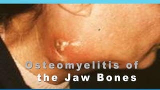
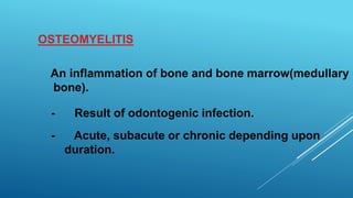
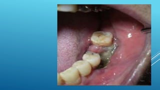
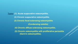
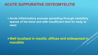
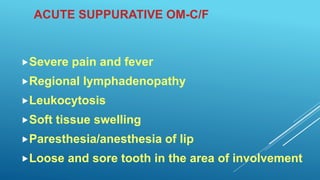
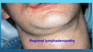
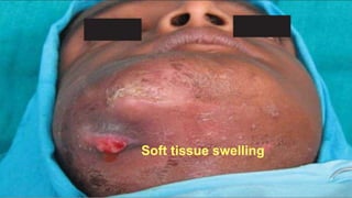
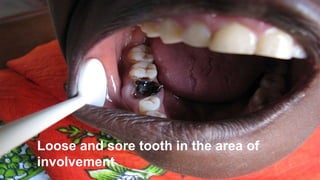
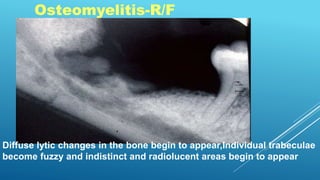
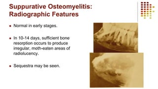
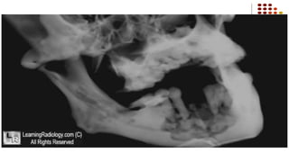
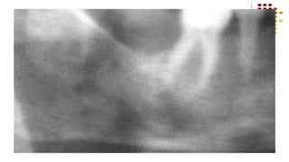
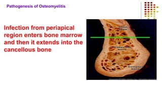
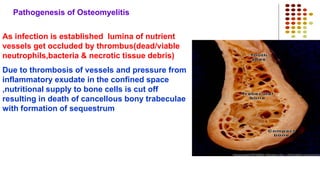
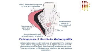
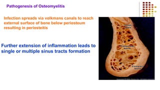
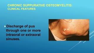
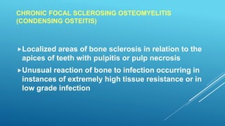
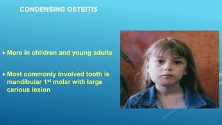
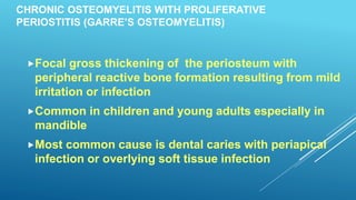
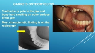
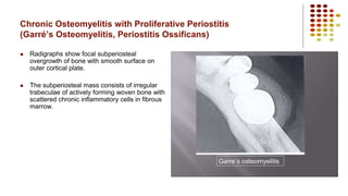
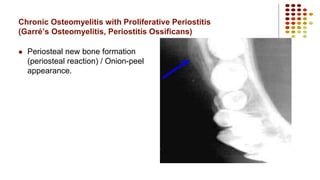
DR VINOD KUMAR R.B
PROF & HEAD
DEPT OF ORAL PATHOLOGY & MICROBIOLOGY
MALABAR DENTAL COLLEGE & RESEARCH CENTRE
OSTEOMYELITIS
An inflammation of bone and bone marrow(medullary
bone).
- Result of odontogenic infection.
- Acute, subacute or chronic depending upon
duration.
Types: (1) Acute suppurative osteomyelitis.
(2) Chronic suppurative osteomyelitis.
(3) Chronic focal sclerosing osteomyelitis
(Condensing osteitis)
(4) Chronic diffuse sclerosing osteomyelitis.
(5) Chronic osteomyelitis with proliferative periostitis
(Garre’s osteomyelitis).
ACUTE SUPPURATIVE OSTEOMYELITIS
Acute inflammatory process spreading through medullary
spaces of the bone and with insufficient time for body to
react
Well localized in maxilla ,diffuse and widespread in
mandible
ACUTE SUPPURATIVE OM-C/F
Severe pain and fever
Regional lymphadenopathy
Leukocytosis
Soft tissue swelling
Paresthesia/anesthesia of lip
Loose and sore tooth in the area of involvement
Abscessed tooth periapical radiograph
Severe pain and fever
Regional lymphadenopathy
Soft tissue swelling
Loose and sore tooth in the area of
involvement
Osteomyelitis-R/F
Diffuse lytic changes in the bone begin to appear,Individual trabeculae
become fuzzy and indistinct and radiolucent areas begin to appear
Suppurative Osteomyelitis:
Radiographic Features
 Normal in early stages.
 In 10-14 days, sufficient bone
resorption occurs to produce
irregular, moth-eaten areas of
radiolucency.
 Sequestra may be seen.
Pathogenesis of Osteomyelitis
Infection from periapical
region enters bone marrow
and then it extends into the
cancellous bone
Pathogenesis of Osteomyelitis
As infection is established lumina of nutrient
vessels get occluded by thrombus(dead/viable
neutrophils,bacteria & necrotic tissue debris)
Due to thrombosis of vessels and pressure from
inflammatory exudate in the confined space
,nutritional supply to bone cells is cut off
resulting in death of cancellous bony trabeculae
with formation of sequestrum
Pathogenesis of Osteomyelitis
Infection spreads via volkmans canals to reach
external surface of bone below periosteum
resulting in periosteitis
Further extension of inflammation leads to
single or multiple sinus tracts formation
continued……
ACUTE SUPPURATIVE OM-H/F
Bone shows loss of osteocytes
from their lacunae,peripheral
resorption and bacterial
colonization
Periphery of bone contains
necrotic debris and acute
inflammatory infiltrate
consisting of PMNL’s
Suppurative Osteomyelitis:
Histopathologic Features
 Suppurative osteomyelitis, note the
devitalized lamellar bone sequestrum with
scalloped edges and absence of stainable
osteocytes and osteoblasts. An osteoclast
in a resorption area is seen.
ACUTE SUPPURATIVE OM-PROGNOSIS
The bone that has lost its vitality separates from the
living bone.The separated fragment is called
“sequestrum”
When the sequestrum becomes surrounded by new
living bone it is called “involucrum”
Pathological #’s may occur due to weakening of the
jaw by the destructive process
ACUTE SUPPURATIVE OM-PROGNOSIS
Pathological #’s may occur due to weakening of the
jaw by the destructive process
CHRONIC SUPPURATIVE OSTEOMYELITIS:
CLINICAL FEATURES
Discharge of pus
through one or more
intraoral or extraoral
sinuses.
CHRONIC FOCAL SCLEROSING OSTEOMYELITIS
(CONDENSING OSTEITIS)
Localized areas of bone sclerosis in relation to the
apices of teeth with pulpitis or pulp necrosis
Unusual reaction of bone to infection occurring in
instances of extremely high tissue resistance or in
low grade infection
CONDENSING OSTEITIS
More in children and young adults
Most commonly involved tooth is
mandibular 1st molar with large
carious lesion
CONDENSING OSTEITIS-R/F
Localized area of increased radiodensity adjacent to the apex of the
tooth that exhibits a thickened periodontal ligament space
Differential diagnosis
Benign cementoblastoma
Radiopacity is attached to the tooth apex in benign
cementoblastoma
CHRONIC OSTEOMYELITIS WITH PROLIFERATIVE
PERIOSTITIS (GARRE’S OSTEOMYELITIS)
Focal gross thickening of the periosteum with
peripheral reactive bone formation resulting from mild
irritation or infection
Common in children and young adults especially in
mandible
Most common cause is dental caries with periapical
infection or overlying soft tissue infection
An occlusal radiograph of the mandible showed the enhancement in
peripheral subperiosteal bone on the right buccal and lingual sides.
GARRE’S OSTEOMYELITIS
Toothache or pain in the jaw and
bony hard swelling on outer surface
of the jaw
Most characteristic finding is on the
radiograph
Chronic Osteomyelitis with Proliferative Periostitis
(Garré’s Osteomyelitis, Periostitis Ossificans)
 Radigraphs show focal subperiosteal
overgrowth of bone with smooth surface on
outer cortical plate.
 The subperiosteal mass consists of irregular
trabeculae of actively forming woven bone with
scattered chronic inflammatory cells in fibrous
marrow.
Chronic Osteomyelitis with Proliferative Periostitis
(Garré’s Osteomyelitis, Periostitis Ossificans)
 Periosteal new bone formation
(periosteal reaction) / Onion-peel
appearance.
Other conditions showing periosteal new bone
formation(neoperiostosis)
 Caffey’s disease(infantile cortical hyperostosis)
 Hypervitaminosis A
 Syphilitic osteomyelitis
 Ewing’s sarcoma
 Metastatic neuroblastoma
 Fracture callus

Pulp and periapical disease

  • 1.
    DR VINOD KUMARR.B PROF & HEAD DEPT OF ORAL PATHOLOGY & MICROBIOLOGY MALABAR DENTAL COLLEGE & RESEARCH CENTRE
  • 3.
    OSTEOMYELITIS An inflammation ofbone and bone marrow(medullary bone). - Result of odontogenic infection. - Acute, subacute or chronic depending upon duration.
  • 5.
    Types: (1) Acutesuppurative osteomyelitis. (2) Chronic suppurative osteomyelitis. (3) Chronic focal sclerosing osteomyelitis (Condensing osteitis) (4) Chronic diffuse sclerosing osteomyelitis. (5) Chronic osteomyelitis with proliferative periostitis (Garre’s osteomyelitis).
  • 6.
    ACUTE SUPPURATIVE OSTEOMYELITIS Acuteinflammatory process spreading through medullary spaces of the bone and with insufficient time for body to react Well localized in maxilla ,diffuse and widespread in mandible
  • 7.
    ACUTE SUPPURATIVE OM-C/F Severepain and fever Regional lymphadenopathy Leukocytosis Soft tissue swelling Paresthesia/anesthesia of lip Loose and sore tooth in the area of involvement
  • 10.
  • 11.
  • 12.
  • 13.
  • 14.
  • 15.
  • 16.
    Loose and soretooth in the area of involvement
  • 17.
    Osteomyelitis-R/F Diffuse lytic changesin the bone begin to appear,Individual trabeculae become fuzzy and indistinct and radiolucent areas begin to appear
  • 18.
    Suppurative Osteomyelitis: Radiographic Features Normal in early stages.  In 10-14 days, sufficient bone resorption occurs to produce irregular, moth-eaten areas of radiolucency.  Sequestra may be seen.
  • 21.
    Pathogenesis of Osteomyelitis Infectionfrom periapical region enters bone marrow and then it extends into the cancellous bone
  • 22.
    Pathogenesis of Osteomyelitis Asinfection is established lumina of nutrient vessels get occluded by thrombus(dead/viable neutrophils,bacteria & necrotic tissue debris) Due to thrombosis of vessels and pressure from inflammatory exudate in the confined space ,nutritional supply to bone cells is cut off resulting in death of cancellous bony trabeculae with formation of sequestrum
  • 24.
    Pathogenesis of Osteomyelitis Infectionspreads via volkmans canals to reach external surface of bone below periosteum resulting in periosteitis Further extension of inflammation leads to single or multiple sinus tracts formation
  • 25.
  • 26.
    ACUTE SUPPURATIVE OM-H/F Boneshows loss of osteocytes from their lacunae,peripheral resorption and bacterial colonization Periphery of bone contains necrotic debris and acute inflammatory infiltrate consisting of PMNL’s
  • 27.
    Suppurative Osteomyelitis: Histopathologic Features Suppurative osteomyelitis, note the devitalized lamellar bone sequestrum with scalloped edges and absence of stainable osteocytes and osteoblasts. An osteoclast in a resorption area is seen.
  • 28.
    ACUTE SUPPURATIVE OM-PROGNOSIS Thebone that has lost its vitality separates from the living bone.The separated fragment is called “sequestrum” When the sequestrum becomes surrounded by new living bone it is called “involucrum” Pathological #’s may occur due to weakening of the jaw by the destructive process
  • 29.
    ACUTE SUPPURATIVE OM-PROGNOSIS Pathological#’s may occur due to weakening of the jaw by the destructive process
  • 30.
    CHRONIC SUPPURATIVE OSTEOMYELITIS: CLINICALFEATURES Discharge of pus through one or more intraoral or extraoral sinuses.
  • 32.
    CHRONIC FOCAL SCLEROSINGOSTEOMYELITIS (CONDENSING OSTEITIS) Localized areas of bone sclerosis in relation to the apices of teeth with pulpitis or pulp necrosis Unusual reaction of bone to infection occurring in instances of extremely high tissue resistance or in low grade infection
  • 33.
    CONDENSING OSTEITIS More inchildren and young adults Most commonly involved tooth is mandibular 1st molar with large carious lesion
  • 34.
    CONDENSING OSTEITIS-R/F Localized areaof increased radiodensity adjacent to the apex of the tooth that exhibits a thickened periodontal ligament space Differential diagnosis Benign cementoblastoma
  • 35.
    Radiopacity is attachedto the tooth apex in benign cementoblastoma
  • 36.
    CHRONIC OSTEOMYELITIS WITHPROLIFERATIVE PERIOSTITIS (GARRE’S OSTEOMYELITIS) Focal gross thickening of the periosteum with peripheral reactive bone formation resulting from mild irritation or infection Common in children and young adults especially in mandible Most common cause is dental caries with periapical infection or overlying soft tissue infection
  • 38.
    An occlusal radiographof the mandible showed the enhancement in peripheral subperiosteal bone on the right buccal and lingual sides.
  • 40.
    GARRE’S OSTEOMYELITIS Toothache orpain in the jaw and bony hard swelling on outer surface of the jaw Most characteristic finding is on the radiograph
  • 41.
    Chronic Osteomyelitis withProliferative Periostitis (Garré’s Osteomyelitis, Periostitis Ossificans)  Radigraphs show focal subperiosteal overgrowth of bone with smooth surface on outer cortical plate.  The subperiosteal mass consists of irregular trabeculae of actively forming woven bone with scattered chronic inflammatory cells in fibrous marrow.
  • 42.
    Chronic Osteomyelitis withProliferative Periostitis (Garré’s Osteomyelitis, Periostitis Ossificans)  Periosteal new bone formation (periosteal reaction) / Onion-peel appearance.
  • 43.
    Other conditions showingperiosteal new bone formation(neoperiostosis)  Caffey’s disease(infantile cortical hyperostosis)  Hypervitaminosis A  Syphilitic osteomyelitis  Ewing’s sarcoma  Metastatic neuroblastoma  Fracture callus