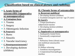
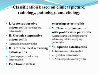
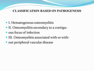
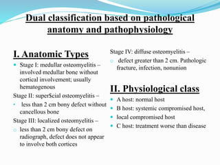
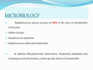
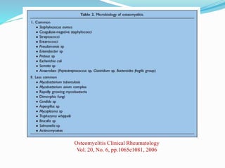
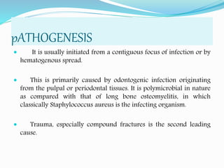
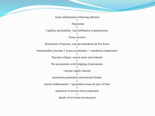
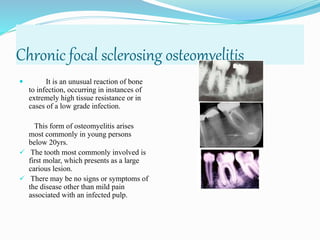
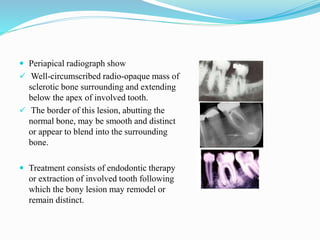
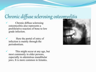
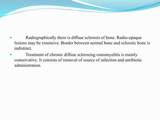
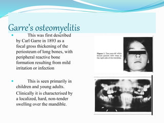
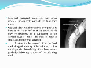
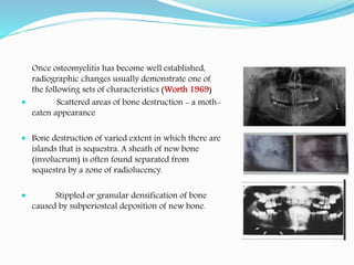
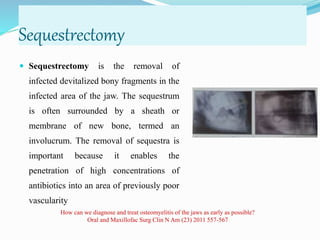
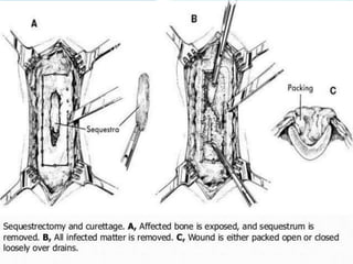
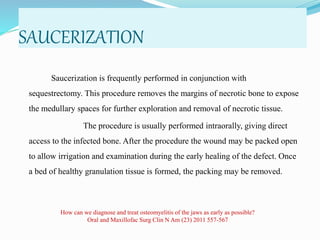
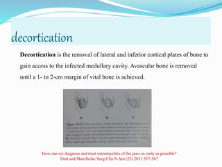
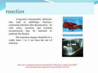
Osteomyelitis is an inflammatory condition of bone that involves the medullary cavity and has a tendency to progress along this space and involve the adjacent cortex, periosteum, and soft tissue. It is commonly caused by odontogenic infections or trauma. Staphylococcus aureus accounts for 80% of jaw osteomyelitis cases. The infection initiates from a contiguous focus or hematogenous spread and causes inflammation, tissue necrosis, pus formation, and bone destruction if not properly treated. Without treatment, it can progress to chronic stages involving bone lysis, sequestrum formation, and involucrum development.