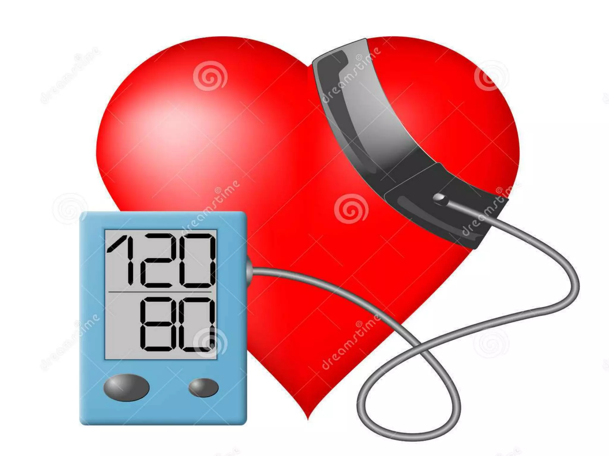
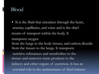
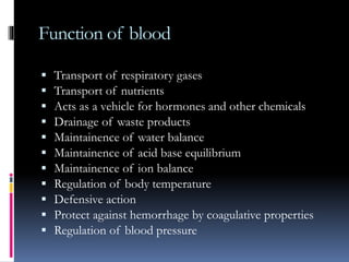
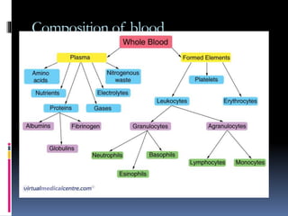
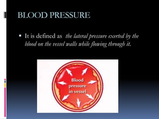
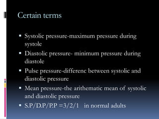
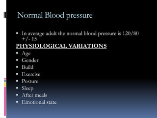
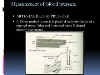
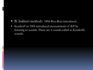
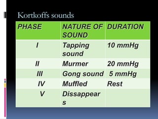
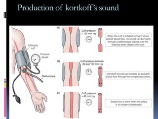
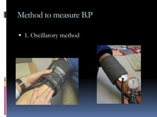
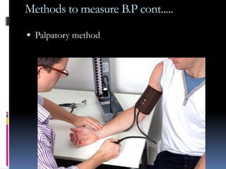
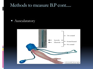
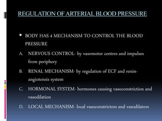
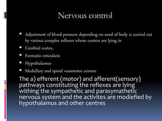
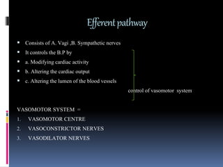
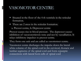
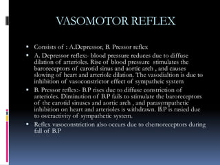
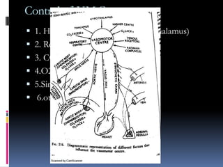
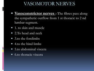
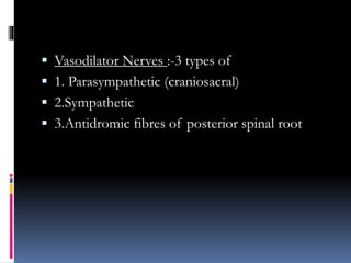
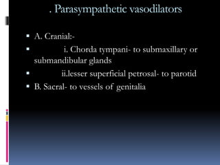
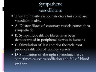
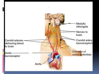
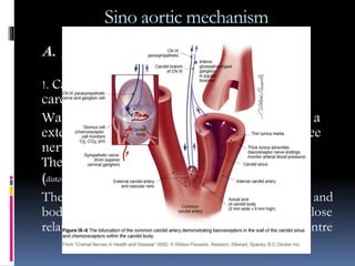
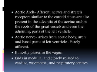
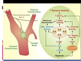
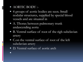
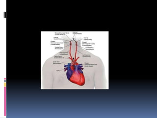
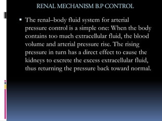
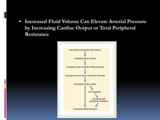
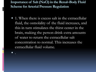
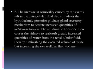
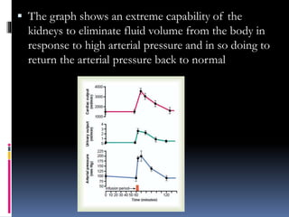
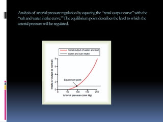
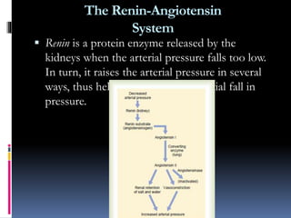
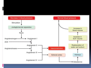
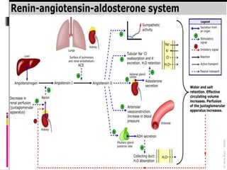
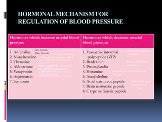
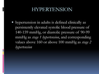
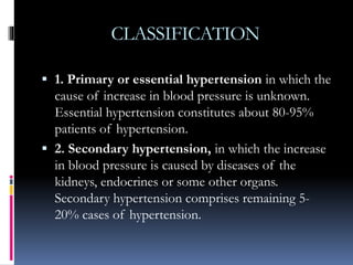
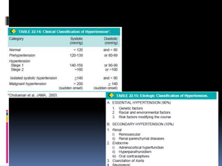
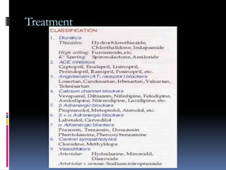
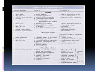
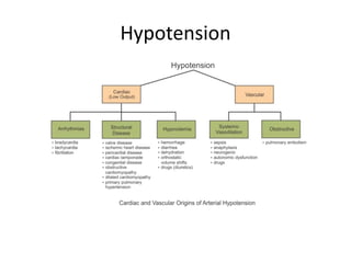
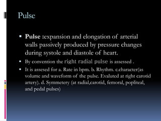
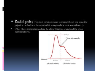
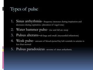
Blood pressure
PRESENTER:- DR. JAYESH
1° YR MDS
DEPTOF ORALAND MAXILLOFACIALSURGERY
1. BLOOD
2. FUNCTION OF BLOOD
3. COMPOSITION
4. BLOOD PRESSURE
5. SYSTOLE AND DIASTOLE
6. CARDIAC CYCLE
7. NORMAL BLOOD PRESSURE (B.P)
8. FUNCTIONS OF B.P
9. SIGNIFICANCE OF B.P
10. MEASUREMENT OF B.P
11. FACTORS CONTROLLING B.P
12. REGULATION OF B.P
13. HYPERTENSION
14. HYPOTENSION
15. PULSE
16. BIBLIOGRAPHY
CONTENTS
BLOOD
 It can be described as a specialized connective
tissue in which there is liquid intercellular substance
known as plasma and formed elements , the red
blood cells (RBC), the white blood cells platelets
and ions suspended in plasma
Blood
 It is the fluid that circulates through the heart,
arteries, capillaries, and veins and is the chief
means of transport within the body. It
transports oxygen
from the lungs to the body tissues, and carbon dioxide
from the tissues to the lungs. It transports
nutritive substances and metabolites to the
tissues and removes waste products to the
kidneys and other organs of excretion. It has an
essential role in the maintenance of fluid balance
Function of blood
 Transport of respiratory gases
 Transport of nutrients
 Acts as a vehicle for hormones and other chemicals
 Drainage of waste products
 Maintainence of water balance
 Maintainence of acid base equilibrium
 Maintainence of ion balance
 Regulation of body temperature
 Defensive action
 Protect against hemorrhage by coagulative properties
 Regulation of blood pressure
Composition of blood
 Formed
 Cells.- RBC , WBC, PLATELETS
 Plasma
water-91-92%
solids- 8-9%
inorganic constituents-.9% like NaCl, K,ca
organic - albumin, fibrinigen, globulins etc
proteins
urea creatininie etc
fats, carbohydrates, bile etc
BLOOD PRESSURE
 It is defined as the lateral pressure exerted by the
blood on the vessel walls while flowing through it.
CARDIAC CYCLE
Systole -
.3sec
Diastole -
.5sec
Total - .8 sec
Systole and Diastole
Significance
 Systolic pressure – it undergoes considerable
fluctuations. It indicates. 1. extent of work.2.
force with which heart is working. 3. degree of
pressure the arterial walls have to with stand
 Diastolic pressure- undergoes less fluctuations.
It is a measure of peripheral resistance. Ie the
load against which heart has to work
Cardiac cycle
Certain terms
 Systolic pressure-maximum pressure during
systole
 Diastolic pressure- minimum pressure during
diastole
 Pulse pressure-differenc between systolic and
diastolic pressure
 Mean pressure-the arithematic mean of systolic
and diastolic pressure
 S.P/D.P/P.P =3/2/1 in normal adults
Normal Blood pressure
 In average adult the normal blood pressure is 120/80
+/- 15
PHYSIOLOGICAL VARIATIONS
 Age
 Gender
 Build
 Exercise
 Posture
 Sleep
 After meals
 Emotional state
Normal function of B.P
1. To maintain sufficient presure to keep the
blood flowing.
2. To provide for the motive force of filtration at
the capillary bed- thus ensuring supplies to all
the cells and tissues, formation of lymph ,
urine and so on
Measurement of blood pressure
 ARTERIAL BLOOD PRESSURE
 A. Direct method- a canula is placed directly into lumen of a
exposed artery. Other end is inserted into a U shaped
mercury manometer.
 B. Indirect method:- 1896 Riva-Roci introduced .
 Kortkoff in 1905 introduced measurement of B.P by
listening to sounds. There are 4 sounds called as Kortkoffs
sounds.
Kortkoffs sounds
PHASE NATURE OF
SOUND
DURATION
I Tapping
sound
10 mmHg
II Murmer 20 mmHg
III Gong sound 5 mmHg
IV Muffled Rest
V Dissappear
s
Production of kortkoff’s sound
Measurement of B.P
COMMONEST METHOD – pressure is measured at
brachial artery by the use of sphygomamometer
Method to measure B.P
 1. Oscillatory method
Methods to measure B.P cont.....
 Palpatory method
Methods to measure B.P cont.....
 Ausculatatory
VENOUS BLOOD PRESSURE
Factors controllling the B.P
 Depends on
 1. Pumping action of the heart
 2. Cardiac output
 3. Peripheral resistance
 4. Elasticity of arterial wall
 5. Blood volume
 6. Viscosity of blood
REGULATION OFARTERIALBLOOD PRESSURE
 BODY HAS 4 MECHANISM TO CONTROL THE BLOOD
PRESSURE
A. NERVOUS CONTROL- by vasomotor centres and impulses
from periphery
B. RENAL MECHANISM- by regulation of ECF and renin-
angiotensis system
C. HORMONAL SYSTEM- hormones causing vasoconstriction and
vasodilation
D. LOCAL MECHANISM- local vasoconstrictors and vasodilators
Nervous control
 Adjustment of blood pressure depending on need of body is carried out
by various complex reflexes whose centres are lying in
 Cerebral cortex,
 Formatio reticularis
 Hypothalamus
 Medullary and spinal vasomotor centres
The a) efferent (motor) and afferent(sensory)
pathways constituting the reflexes are lying
withing the sympathetic and parasymathetic
nervous system and the activites are modiefied by
hypothalamus and other centres
Efferentpathway
 Consists of A. Vagi ,B. Sympathetic nerves
 It controls the B.P by
 a. Modifying cardiac activity
 b. Altering the cardiac output
 c. Altering the lumen of the blood vessels
control of vasomotor system
VASOMOTOR SYSTEM =
1. VASOMOTOR CENTRE
2. VASOCONSTRICTOR NERVES
3. VASODILATOR NERVES
VASOMOTOR CENTRE
 Situated in the floor of the 4 th ventricle in the reticular
formation.
 There are 2 areas in the reticular formation.
a. Pressor centre, b. Depressor centre
Pressor causes rise in blood pressure . The depressor causes
inhibition of vasoconstrictore tone and not by vasodilation. It
relays inhibitory impulses to pressor centre.
They form one unit and are called as vasomotoer centre.
Vasomotor centre discharges the impulse down the lateral
white column of the spinal cord in the cerviccal, thoracic and
lumbar segmetns of the spinal cord and form sypanptic
connections with lateral horn cells of spinal cord
VASOMOTOR REFLEX
 Consists of : A.Depressor, B. Pressor reflex
 A. Depressor reflex:- blood pressure reduces due to diffuse
dilation of arterioles. Rise of blood pressure stimulates the
baroreceptors of carotid sinus and aortic arch , and causes
slowing of heart and arteriole dilation. The vasodialtion is due to
inhibition of vasoconstrictor effect of sympatheic system
 B. Pressor reflex:- B.P rises due to diffuse constriction of
arterioles. Diminution of B.P fails to stimulate the baroreceptors
of the carotid sinuses and aortic arch , and parasympathetic
inhibition on heart and arterioles is withdrawn. B.P is rasied due
to overactivity of sympathetic system.
 Reflex vasoconstriction also occurs due to chemoreceptors during
fall of B.P
Control of V.M.C(vasomotor centre)
 1. Higher centres (including hypothalamus)
 2. Respiration
 3. CO2 excess
 4.O2 lack
 5.Sino- aortic nerves
 6.other afferent
VASOMOTOR NERVES
 Vasoconstrictor nerves :-The fibres pass along
the sympathetic outflow from 1 st thoracic to 2 nd
lumbar segment.
 1. to skin and muscle
 2.To head and neck
 3.to the forelimbs
 4.to the hind limbs
 5.to abdominal viscera
 6.to thoracic viscera
 Vasodilator Nerves :-3 types of
 1. Parasympathetic (craniosacral)
 2.Sympathetic
 3.Antidromic fibres of posterior spinal root
. Parasympathetic vasodilators
 A. Cranial:-
 i. Chorda tympani- to submaxillary or
submandibular glands
 ii.lesser superficial petrosal- to parotid
 B. Sacral- to vessels of genitalia
Sympathetic
vasodilators
 They are mostly vasoconstrictors but some are
vasodialtors also.
 A. Dilator fibres of coronary vessels comes thru
sympathetic
 B. Sympathetic dilator fibres have been
demonstrated in peripheral nerves in humans
 C. Stimulation of last anterior thoracic root
produces dilation of Kidney vessels
 D. Stimulation of the right splanchnic nerve
sometimes causes vasodilation and fall of blood
pressure
Antidromic vasodilators
 In the post spinal root
 When the posterior spinal root is cut, Distal to
the ganglion and peripheral end is stimulated –
although the nerve is afferent, yet the vessels in
the periphery- both skin and muscles-
dilates(axon relfex).
 In skin is due to liberation of histamin, in
muscles it liberates acetylcholine and then
vasodilation
AFFERNTPATHWAY
 They are lying in two sets of receptors that carry the
information of instantaneous circulatory system.
 These receptors are
 A. Baroreceptors
 B. Chemoreceptors
 The B.P is controlled thru different afferent pathways viz.
 ! Sino-aortic mechanism
 !! Vascular receptors other than sino aortic pathways
Sino aortic mechanism in regulation of
normal B.P
It regulates B.P by regulating the heart rate,
vasomotor centre, secretions of Adrenaline and
nor adrenaline. It also adjusts the respiratory
centre in such a way that it run parallel to heart
Sino aortic mechanism
A. BARORECEPTORS:- It includes carotid
sinus and aortic arch
!. Carotid sinus:- is a dilation at root of internal
carotid artery often involving common carotid.
Wall is thinner. Deeper part of advetitia layer is a
extensive network of nerve, the fibers end in free
nerve terminal and hava charesteristic minisci.
These pressor receptor are sensitive to stretch
(distortion effect) being stimulated by rise of B.P.
The sinus nerve(afferent) arises from carotid sinus and
body, passes along the glossopharyngeal nerve in close
relation with respiratory, cardiac and vasomotor centre
 Aortic Arch- Afferent nerves and stretch
receptors similar to the carotid sinus are also
present in the adventia of the aortuc archm
the roots of the great vessels and even the
adjoining parts of the left venticle,
 Aortic nerve- arises from aortic body, arch
and basal partu of left ventricle . Purely
afferent.
 It mostly passes in the vagus.
 Ends in medulla and closely related to
cardiac, vasomotor , and respiratory centres
 B. CHEMORECEPTORS - includes carotid
body and aortic body
 CAROTID BODY:- is a small nodule situated
close on the occipital artery, branch of external
carotid artery. Close to carotid sinus. It consist of
clumps of large polyhedral cells(Glomus cells). Is
rochly supplied with blood and nerves.
Numerous nerve fibres surround the cell clumps
and cells and terminate in special
chemoreceptors,
 AORTIC BODY :-
 4 groups of aortic bodies are seen. Small
nodular structures, supplied by special blood
vessels and are situated in
 A. Thorax between pulmonary trunk
andascending aorta
 B. Ventral surface of root of the righ subclavian
artery
 C.on the ventral surface of root of the left
subclavian artery
 D. Ventral surface of aortic arch

RENAL MECHANISMB.PCONTROL
 The renal–body fluid system for arterial
pressure control is a simple one: When the body
contains too much extracellular fluid, the blood
volume and arterial pressure rise. The rising
pressure in turn has a direct effect to cause the
kidneys to excrete the excess extracellular fluid,
thus returning the pressure back toward normal.
 Increased Fluid Volume Can Elevate Arterial Pressure
by Increasing Cardiac Output or Total Peripheral
Resistance
Importance of Salt (NaCl) in the Renal–Body Fluid
Scheme for Arterial Pressure Regulation
 1. When there is excess salt in the extracellular
fluid, the osmolality of the fluid increases, and
this in turn stimulates the thirst center in the
brain, making the person drink extra amounts
of water to return the extracellular salt
concentration to normal. This increases the
extracellular fluid volume.
 .
 2. The increase in osmolality caused by the excess
salt in the extracellular fluid also stimulates the
hypothalamic-posterior pituitary gland secretory
mechanism to secrete increased quantities of
antidiuretic hormone. The antidiuretic hormone then
causes the kidneys to reabsorb greatly increased
quantities of water from the renal tubular fluid,
thereby diminishing the excreted volume of urine
but increasing the extracellular fluid volume
 The graph shows an extreme capability of the
kidneys to eliminate fluid volume from the body in
response to high arterial pressure and in so doing to
return the arterial pressure back to normal
Analysisof arterialpressureregulationbyequatingthe“renaloutputcurve”withthe
“saltandwaterintakecurve.”Theequilibriumpoint describestheleveltowhichthe
arterialpressurewill be regulated.
The Renin-Angiotensin
System
 Renin is a protein enzyme released by the
kidneys when the arterial pressure falls too low.
In turn, it raises the arterial pressure in several
ways, thus helping to correct the initial fall in
pressure.
 Renin is synthesized and stored in an inactive
form called prorenin in the juxtaglomerular cells (JG
cells) of the kidneys.
 When the arterial pressure falls, intrinsic
reactions in the kidneys themselves cause many
of the prorenin molecules in the JG cells to split
and release renin. Most of the renin enters the
renal blood and then passes out of the kidneys
to circulate throughout the entire body
 Renin acts enzymatically on another plasma
protein, a globulin called renin substrate (or
angiotensinogen), to release a 10-amino acid
peptide, angiotensin I. Angiotensin I has mild
vasoconstrictor properties but not enough to
cause significant changes in circulatory function.
The renin persists in the blood for 30 minutes
to 1 hour and continues to cause formation of
still more angiotensin I during this entire time.
 Within a few seconds to minutes after formation of
angiotensin I, two additional amino acids are split
from the angiotensin I to form the 8-amino acid peptide
angiotensin II. This conversion occurs almost entirely
in the lungs while the blood flows through the small
vessels of the lungs, catalyzed by an enzyme called
converting enzyme that is present in the endothelium of the
lung vessels. Angiotensin II is an extremely powerful
vasoconstrictor, and it also affects circulatory function in
other ways as well. However, it persists in the blood only
for 1 or 2 minutes because it is rapidly inactivated by
multiple blood and tissue enzymes collectively called
angiotensinases
 Angiotensin II has 3 principal effects
 1. vasoconstriction in many areas of the body, occurs
rapidly
 2. decrease excretion of both salt and water by the
kidneys.
 3. is also one of the most powerful stimulators of
aldosterone secretion by the adrenal glands.
Aldosterone cause marked increase in sodium
reabsorption by the kidney tubules, thus increasing
the total body extracellular fluid sodium. This
increased sodium then causes water retention
HORMONALMECHANISM FOR
REGULATION OFBLOOD PRESSURE
Hormones which increase arterial blood
pressure
Hormones which decrease arterial
blood pressure
1. Adrenaline
2. Noradrenaline
3. Thyroxine
4. Aldosterone
5. Vasopressin
6. Angiotensin
7. Serotonin
1. Vasoactive intestinal
polypeptide (VIP)
2. Bradykinin
3. Prostaglandin
4. Histamine
5. Acetylcholine
6. Atrial natriuretic peptide
7. Brain natriuretic peptide
8. C type natriuretic peptide
Inc. systolic
Dec. diastolicActs mainly through alpha recp.
General vasoconstrictor effect
Inc. in rate & force of contraction
Inc. in C.O
SBP inc. & DBP dec.
Dec. in DBP is due to release of
metabolites due to inc. metabolic
activity & dec. TPR
Causes retention of salt & water
Inc. ECF vol. & blood vol.
Retn. Of water
Vasopressor actn.
Constriction of systemic arterioles
Secreted in stomach
Vasodilatn.
During inflammation
vasodilator
Allergic condn, inflammation or
damage
Produced by atrial
musculature of
heart
HYPERTENSION
 hypertension in adults is defined clinically as
persistently elevated systolic blood pressure of
140-159 mmHg, or diastolic pressure of 90-99
mmHg as stage 1 hypertension, and corresponding
values above 160 or above 100 mmHg as stage 2
hypertension
CLASSIFICATION
 1. Primary or essential hypertension in which the
cause of increase in blood pressure is unknown.
Essential hypertension constitutes about 80-95%
patients of hypertension.
 2. Secondary hypertension, in which the increase
in blood pressure is caused by diseases of the
kidneys, endocrines or some other organs.
Secondary hypertension comprises remaining 5-
20% cases of hypertension.
 According to the clinical course, both essential and secondary
hypertension may be benign or malignant.
 1.Benign hypertension is moderate elevation of blood
pressure and the rise is slow over the years. About 90-95%
patients of hypertension have benign hypertension.
 2.Malignant hypertension is marked and sudden increase of
blood pressure to 200/140 mmHg or more in a known case of
hypertension or in a previously normotensive individual; the
patients develop papilloedema, retinal haemorrhages and
hypertensive encephalopathy. Less than 5% of hypertensive
patients develop malignant hypertension
EFFECTS OF HYPERTENSION
 hypertension causes major effects in four main
organs
1.Heart and its blood vessels
2.Nervous system
3.Kidneys
4.Eyes
EFFECTS OF HYPERTENSION
on KIDNEYS
 The renal effects in the form of benign and
malignant nephrosclerosis
 Benign Nephrosclerosis:-
 C/f- There is variable elevation of the blood
pressure with headache, dizziness, palpitation
and nervousness. Eye ground changes may be
found but papilloedema is absent. Renal
function tests and urine examination are normal
in early stage. In long-standing cases, there may
be mild proteinuria with some hyaline or
granular casts. Rarely, renal failure and uraemia
may occur.
 Malignant Nephrosclerosis:-
 The patients of malignant nephrosclerosis have
malignant or accelerated hypertension with
blood pressure of 200/140 mmHg or higher.
Headache, dizziness and impaired vision are
commonly found. The presence of
papilloedema distinguishes malignant from
benign phase of hypertension. The urine
frequently shows haematuria and proteinuria.
Renal function tests show deterioration during
the course of the illness. Azotaemia (high BUN
and serum creatinine) and uraemia develop soon
if malignant hypertension is not treated
aggressively.
EFFECTS OF HYPERTENSION
on EYE
 HYPERTENSIVE RETINOPATHY:-
 In hypertensive retinopathy, the retinal arterioles are reduced
in their diameter leading to retinal ischaemia
 Malignant hypertension is characterised by necrotising
arteriolitis and fibrinoid necrosis of retinal arterioles.
EFFECTS OF HYPERTENSION
on HEART
 HYPERTENSIVE HEART DISEASE:-
 The stress of pressure on the ventricular wall
causes increased production of myofilaments,
myofibrils, other cell organelles and nuclear
enlargement. Since the adult myocardial fibres
do not divide, the fibres are hypertrophied
EFFECTS OF HYPERTENSION
on nervous system
 INTRACRANIAL HAEMORRHAGE:-
 a. Intracerebral Haemorrhage
 b. Subarachnoid Haemorrhage
INVESTIGATIONS OF
HYPERTENSION
Treatment
Dental aspect
Hypotension
Pulse
 Pulse :expansion and elongation of arterial
walls passively produced by pressure changes
during systole and diastole of heart.
 By convention the right radial pulse is assessed .
 It is assesed for a. Rate in bpm. b. Rhythm. c.character(as
volume and waveform of the pulse. Evaluted at right carotid
artery). d. Symmetery (at radial,carotid, femoral, popliteal,
and pedal pulses)
 Radial pulse The most common places to measure heart rate using the
palpation method is at the wrist (radial artery) and the neck (carotid artery).
 Other places sometimes used are the elbow (brachial artery) and the groin
(femoral artery).
Types of pulse
1. Sinus arrhythmia- frequency increases during inspiration and
decreases during expiration. (alteration of vagal tone)
2. Water hammer pulse- rise and fall are steep
3. Pulsus alterans–alt large and small. (myocardial infarctions)
4. Weak pulse- amount of blood ejected bty left ventricle to arteries is
less than normal
5. Pulsus paradoxicus –reverse of sinus arrhythmia.
BIBLIOGRAPHY
 Thank you
 Thank you
 Thank you
 Thank you
 Thank you
 Thank you
 Thank you

Blood pressure

  • 2.
    Blood pressure PRESENTER:- DR.JAYESH 1° YR MDS DEPTOF ORALAND MAXILLOFACIALSURGERY
  • 3.
    1. BLOOD 2. FUNCTIONOF BLOOD 3. COMPOSITION 4. BLOOD PRESSURE 5. SYSTOLE AND DIASTOLE 6. CARDIAC CYCLE 7. NORMAL BLOOD PRESSURE (B.P) 8. FUNCTIONS OF B.P 9. SIGNIFICANCE OF B.P 10. MEASUREMENT OF B.P 11. FACTORS CONTROLLING B.P 12. REGULATION OF B.P 13. HYPERTENSION 14. HYPOTENSION 15. PULSE 16. BIBLIOGRAPHY CONTENTS
  • 4.
    BLOOD  It canbe described as a specialized connective tissue in which there is liquid intercellular substance known as plasma and formed elements , the red blood cells (RBC), the white blood cells platelets and ions suspended in plasma
  • 5.
    Blood  It isthe fluid that circulates through the heart, arteries, capillaries, and veins and is the chief means of transport within the body. It transports oxygen from the lungs to the body tissues, and carbon dioxide from the tissues to the lungs. It transports nutritive substances and metabolites to the tissues and removes waste products to the kidneys and other organs of excretion. It has an essential role in the maintenance of fluid balance
  • 6.
    Function of blood Transport of respiratory gases  Transport of nutrients  Acts as a vehicle for hormones and other chemicals  Drainage of waste products  Maintainence of water balance  Maintainence of acid base equilibrium  Maintainence of ion balance  Regulation of body temperature  Defensive action  Protect against hemorrhage by coagulative properties  Regulation of blood pressure
  • 7.
    Composition of blood Formed  Cells.- RBC , WBC, PLATELETS  Plasma water-91-92% solids- 8-9% inorganic constituents-.9% like NaCl, K,ca organic - albumin, fibrinigen, globulins etc proteins urea creatininie etc fats, carbohydrates, bile etc
  • 8.
    BLOOD PRESSURE  Itis defined as the lateral pressure exerted by the blood on the vessel walls while flowing through it.
  • 9.
  • 10.
  • 11.
    Significance  Systolic pressure– it undergoes considerable fluctuations. It indicates. 1. extent of work.2. force with which heart is working. 3. degree of pressure the arterial walls have to with stand  Diastolic pressure- undergoes less fluctuations. It is a measure of peripheral resistance. Ie the load against which heart has to work
  • 12.
  • 13.
    Certain terms  Systolicpressure-maximum pressure during systole  Diastolic pressure- minimum pressure during diastole  Pulse pressure-differenc between systolic and diastolic pressure  Mean pressure-the arithematic mean of systolic and diastolic pressure  S.P/D.P/P.P =3/2/1 in normal adults
  • 14.
    Normal Blood pressure In average adult the normal blood pressure is 120/80 +/- 15 PHYSIOLOGICAL VARIATIONS  Age  Gender  Build  Exercise  Posture  Sleep  After meals  Emotional state
  • 15.
    Normal function ofB.P 1. To maintain sufficient presure to keep the blood flowing. 2. To provide for the motive force of filtration at the capillary bed- thus ensuring supplies to all the cells and tissues, formation of lymph , urine and so on
  • 16.
    Measurement of bloodpressure  ARTERIAL BLOOD PRESSURE  A. Direct method- a canula is placed directly into lumen of a exposed artery. Other end is inserted into a U shaped mercury manometer.
  • 17.
     B. Indirectmethod:- 1896 Riva-Roci introduced .  Kortkoff in 1905 introduced measurement of B.P by listening to sounds. There are 4 sounds called as Kortkoffs sounds.
  • 18.
    Kortkoffs sounds PHASE NATUREOF SOUND DURATION I Tapping sound 10 mmHg II Murmer 20 mmHg III Gong sound 5 mmHg IV Muffled Rest V Dissappear s
  • 19.
  • 20.
    Measurement of B.P COMMONESTMETHOD – pressure is measured at brachial artery by the use of sphygomamometer
  • 21.
    Method to measureB.P  1. Oscillatory method
  • 22.
    Methods to measureB.P cont.....  Palpatory method
  • 23.
    Methods to measureB.P cont.....  Ausculatatory
  • 24.
  • 25.
    Factors controllling theB.P  Depends on  1. Pumping action of the heart  2. Cardiac output  3. Peripheral resistance  4. Elasticity of arterial wall  5. Blood volume  6. Viscosity of blood
  • 26.
    REGULATION OFARTERIALBLOOD PRESSURE BODY HAS 4 MECHANISM TO CONTROL THE BLOOD PRESSURE A. NERVOUS CONTROL- by vasomotor centres and impulses from periphery B. RENAL MECHANISM- by regulation of ECF and renin- angiotensis system C. HORMONAL SYSTEM- hormones causing vasoconstriction and vasodilation D. LOCAL MECHANISM- local vasoconstrictors and vasodilators
  • 28.
    Nervous control  Adjustmentof blood pressure depending on need of body is carried out by various complex reflexes whose centres are lying in  Cerebral cortex,  Formatio reticularis  Hypothalamus  Medullary and spinal vasomotor centres The a) efferent (motor) and afferent(sensory) pathways constituting the reflexes are lying withing the sympathetic and parasymathetic nervous system and the activites are modiefied by hypothalamus and other centres
  • 29.
    Efferentpathway  Consists ofA. Vagi ,B. Sympathetic nerves  It controls the B.P by  a. Modifying cardiac activity  b. Altering the cardiac output  c. Altering the lumen of the blood vessels control of vasomotor system VASOMOTOR SYSTEM = 1. VASOMOTOR CENTRE 2. VASOCONSTRICTOR NERVES 3. VASODILATOR NERVES
  • 30.
    VASOMOTOR CENTRE  Situatedin the floor of the 4 th ventricle in the reticular formation.  There are 2 areas in the reticular formation. a. Pressor centre, b. Depressor centre Pressor causes rise in blood pressure . The depressor causes inhibition of vasoconstrictore tone and not by vasodilation. It relays inhibitory impulses to pressor centre. They form one unit and are called as vasomotoer centre. Vasomotor centre discharges the impulse down the lateral white column of the spinal cord in the cerviccal, thoracic and lumbar segmetns of the spinal cord and form sypanptic connections with lateral horn cells of spinal cord
  • 31.
    VASOMOTOR REFLEX  Consistsof : A.Depressor, B. Pressor reflex  A. Depressor reflex:- blood pressure reduces due to diffuse dilation of arterioles. Rise of blood pressure stimulates the baroreceptors of carotid sinus and aortic arch , and causes slowing of heart and arteriole dilation. The vasodialtion is due to inhibition of vasoconstrictor effect of sympatheic system  B. Pressor reflex:- B.P rises due to diffuse constriction of arterioles. Diminution of B.P fails to stimulate the baroreceptors of the carotid sinuses and aortic arch , and parasympathetic inhibition on heart and arterioles is withdrawn. B.P is rasied due to overactivity of sympathetic system.  Reflex vasoconstriction also occurs due to chemoreceptors during fall of B.P
  • 32.
    Control of V.M.C(vasomotorcentre)  1. Higher centres (including hypothalamus)  2. Respiration  3. CO2 excess  4.O2 lack  5.Sino- aortic nerves  6.other afferent
  • 33.
    VASOMOTOR NERVES  Vasoconstrictornerves :-The fibres pass along the sympathetic outflow from 1 st thoracic to 2 nd lumbar segment.  1. to skin and muscle  2.To head and neck  3.to the forelimbs  4.to the hind limbs  5.to abdominal viscera  6.to thoracic viscera
  • 34.
     Vasodilator Nerves:-3 types of  1. Parasympathetic (craniosacral)  2.Sympathetic  3.Antidromic fibres of posterior spinal root
  • 35.
    . Parasympathetic vasodilators A. Cranial:-  i. Chorda tympani- to submaxillary or submandibular glands  ii.lesser superficial petrosal- to parotid  B. Sacral- to vessels of genitalia
  • 36.
    Sympathetic vasodilators  They aremostly vasoconstrictors but some are vasodialtors also.  A. Dilator fibres of coronary vessels comes thru sympathetic  B. Sympathetic dilator fibres have been demonstrated in peripheral nerves in humans  C. Stimulation of last anterior thoracic root produces dilation of Kidney vessels  D. Stimulation of the right splanchnic nerve sometimes causes vasodilation and fall of blood pressure
  • 37.
    Antidromic vasodilators  Inthe post spinal root  When the posterior spinal root is cut, Distal to the ganglion and peripheral end is stimulated – although the nerve is afferent, yet the vessels in the periphery- both skin and muscles- dilates(axon relfex).  In skin is due to liberation of histamin, in muscles it liberates acetylcholine and then vasodilation
  • 38.
    AFFERNTPATHWAY  They arelying in two sets of receptors that carry the information of instantaneous circulatory system.  These receptors are  A. Baroreceptors  B. Chemoreceptors  The B.P is controlled thru different afferent pathways viz.  ! Sino-aortic mechanism  !! Vascular receptors other than sino aortic pathways
  • 40.
    Sino aortic mechanismin regulation of normal B.P It regulates B.P by regulating the heart rate, vasomotor centre, secretions of Adrenaline and nor adrenaline. It also adjusts the respiratory centre in such a way that it run parallel to heart
  • 41.
    Sino aortic mechanism A.BARORECEPTORS:- It includes carotid sinus and aortic arch !. Carotid sinus:- is a dilation at root of internal carotid artery often involving common carotid. Wall is thinner. Deeper part of advetitia layer is a extensive network of nerve, the fibers end in free nerve terminal and hava charesteristic minisci. These pressor receptor are sensitive to stretch (distortion effect) being stimulated by rise of B.P. The sinus nerve(afferent) arises from carotid sinus and body, passes along the glossopharyngeal nerve in close relation with respiratory, cardiac and vasomotor centre
  • 42.
     Aortic Arch-Afferent nerves and stretch receptors similar to the carotid sinus are also present in the adventia of the aortuc archm the roots of the great vessels and even the adjoining parts of the left venticle,  Aortic nerve- arises from aortic body, arch and basal partu of left ventricle . Purely afferent.  It mostly passes in the vagus.  Ends in medulla and closely related to cardiac, vasomotor , and respiratory centres
  • 43.
     B. CHEMORECEPTORS- includes carotid body and aortic body  CAROTID BODY:- is a small nodule situated close on the occipital artery, branch of external carotid artery. Close to carotid sinus. It consist of clumps of large polyhedral cells(Glomus cells). Is rochly supplied with blood and nerves. Numerous nerve fibres surround the cell clumps and cells and terminate in special chemoreceptors,
  • 44.
     AORTIC BODY:-  4 groups of aortic bodies are seen. Small nodular structures, supplied by special blood vessels and are situated in  A. Thorax between pulmonary trunk andascending aorta  B. Ventral surface of root of the righ subclavian artery  C.on the ventral surface of root of the left subclavian artery  D. Ventral surface of aortic arch 
  • 46.
    RENAL MECHANISMB.PCONTROL  Therenal–body fluid system for arterial pressure control is a simple one: When the body contains too much extracellular fluid, the blood volume and arterial pressure rise. The rising pressure in turn has a direct effect to cause the kidneys to excrete the excess extracellular fluid, thus returning the pressure back toward normal.
  • 47.
     Increased FluidVolume Can Elevate Arterial Pressure by Increasing Cardiac Output or Total Peripheral Resistance
  • 48.
    Importance of Salt(NaCl) in the Renal–Body Fluid Scheme for Arterial Pressure Regulation  1. When there is excess salt in the extracellular fluid, the osmolality of the fluid increases, and this in turn stimulates the thirst center in the brain, making the person drink extra amounts of water to return the extracellular salt concentration to normal. This increases the extracellular fluid volume.  .
  • 49.
     2. Theincrease in osmolality caused by the excess salt in the extracellular fluid also stimulates the hypothalamic-posterior pituitary gland secretory mechanism to secrete increased quantities of antidiuretic hormone. The antidiuretic hormone then causes the kidneys to reabsorb greatly increased quantities of water from the renal tubular fluid, thereby diminishing the excreted volume of urine but increasing the extracellular fluid volume
  • 50.
     The graphshows an extreme capability of the kidneys to eliminate fluid volume from the body in response to high arterial pressure and in so doing to return the arterial pressure back to normal
  • 51.
  • 52.
    The Renin-Angiotensin System  Reninis a protein enzyme released by the kidneys when the arterial pressure falls too low. In turn, it raises the arterial pressure in several ways, thus helping to correct the initial fall in pressure.
  • 54.
     Renin issynthesized and stored in an inactive form called prorenin in the juxtaglomerular cells (JG cells) of the kidneys.  When the arterial pressure falls, intrinsic reactions in the kidneys themselves cause many of the prorenin molecules in the JG cells to split and release renin. Most of the renin enters the renal blood and then passes out of the kidneys to circulate throughout the entire body
  • 55.
     Renin actsenzymatically on another plasma protein, a globulin called renin substrate (or angiotensinogen), to release a 10-amino acid peptide, angiotensin I. Angiotensin I has mild vasoconstrictor properties but not enough to cause significant changes in circulatory function. The renin persists in the blood for 30 minutes to 1 hour and continues to cause formation of still more angiotensin I during this entire time.
  • 56.
     Within afew seconds to minutes after formation of angiotensin I, two additional amino acids are split from the angiotensin I to form the 8-amino acid peptide angiotensin II. This conversion occurs almost entirely in the lungs while the blood flows through the small vessels of the lungs, catalyzed by an enzyme called converting enzyme that is present in the endothelium of the lung vessels. Angiotensin II is an extremely powerful vasoconstrictor, and it also affects circulatory function in other ways as well. However, it persists in the blood only for 1 or 2 minutes because it is rapidly inactivated by multiple blood and tissue enzymes collectively called angiotensinases
  • 57.
     Angiotensin IIhas 3 principal effects  1. vasoconstriction in many areas of the body, occurs rapidly  2. decrease excretion of both salt and water by the kidneys.  3. is also one of the most powerful stimulators of aldosterone secretion by the adrenal glands. Aldosterone cause marked increase in sodium reabsorption by the kidney tubules, thus increasing the total body extracellular fluid sodium. This increased sodium then causes water retention
  • 59.
    HORMONALMECHANISM FOR REGULATION OFBLOODPRESSURE Hormones which increase arterial blood pressure Hormones which decrease arterial blood pressure 1. Adrenaline 2. Noradrenaline 3. Thyroxine 4. Aldosterone 5. Vasopressin 6. Angiotensin 7. Serotonin 1. Vasoactive intestinal polypeptide (VIP) 2. Bradykinin 3. Prostaglandin 4. Histamine 5. Acetylcholine 6. Atrial natriuretic peptide 7. Brain natriuretic peptide 8. C type natriuretic peptide Inc. systolic Dec. diastolicActs mainly through alpha recp. General vasoconstrictor effect Inc. in rate & force of contraction Inc. in C.O SBP inc. & DBP dec. Dec. in DBP is due to release of metabolites due to inc. metabolic activity & dec. TPR Causes retention of salt & water Inc. ECF vol. & blood vol. Retn. Of water Vasopressor actn. Constriction of systemic arterioles Secreted in stomach Vasodilatn. During inflammation vasodilator Allergic condn, inflammation or damage Produced by atrial musculature of heart
  • 60.
    HYPERTENSION  hypertension inadults is defined clinically as persistently elevated systolic blood pressure of 140-159 mmHg, or diastolic pressure of 90-99 mmHg as stage 1 hypertension, and corresponding values above 160 or above 100 mmHg as stage 2 hypertension
  • 61.
    CLASSIFICATION  1. Primaryor essential hypertension in which the cause of increase in blood pressure is unknown. Essential hypertension constitutes about 80-95% patients of hypertension.  2. Secondary hypertension, in which the increase in blood pressure is caused by diseases of the kidneys, endocrines or some other organs. Secondary hypertension comprises remaining 5- 20% cases of hypertension.
  • 62.
     According tothe clinical course, both essential and secondary hypertension may be benign or malignant.  1.Benign hypertension is moderate elevation of blood pressure and the rise is slow over the years. About 90-95% patients of hypertension have benign hypertension.  2.Malignant hypertension is marked and sudden increase of blood pressure to 200/140 mmHg or more in a known case of hypertension or in a previously normotensive individual; the patients develop papilloedema, retinal haemorrhages and hypertensive encephalopathy. Less than 5% of hypertensive patients develop malignant hypertension
  • 64.
    EFFECTS OF HYPERTENSION hypertension causes major effects in four main organs 1.Heart and its blood vessels 2.Nervous system 3.Kidneys 4.Eyes
  • 65.
    EFFECTS OF HYPERTENSION onKIDNEYS  The renal effects in the form of benign and malignant nephrosclerosis  Benign Nephrosclerosis:-  C/f- There is variable elevation of the blood pressure with headache, dizziness, palpitation and nervousness. Eye ground changes may be found but papilloedema is absent. Renal function tests and urine examination are normal in early stage. In long-standing cases, there may be mild proteinuria with some hyaline or granular casts. Rarely, renal failure and uraemia may occur.
  • 66.
     Malignant Nephrosclerosis:- The patients of malignant nephrosclerosis have malignant or accelerated hypertension with blood pressure of 200/140 mmHg or higher. Headache, dizziness and impaired vision are commonly found. The presence of papilloedema distinguishes malignant from benign phase of hypertension. The urine frequently shows haematuria and proteinuria. Renal function tests show deterioration during the course of the illness. Azotaemia (high BUN and serum creatinine) and uraemia develop soon if malignant hypertension is not treated aggressively.
  • 67.
    EFFECTS OF HYPERTENSION onEYE  HYPERTENSIVE RETINOPATHY:-  In hypertensive retinopathy, the retinal arterioles are reduced in their diameter leading to retinal ischaemia  Malignant hypertension is characterised by necrotising arteriolitis and fibrinoid necrosis of retinal arterioles.
  • 68.
    EFFECTS OF HYPERTENSION onHEART  HYPERTENSIVE HEART DISEASE:-  The stress of pressure on the ventricular wall causes increased production of myofilaments, myofibrils, other cell organelles and nuclear enlargement. Since the adult myocardial fibres do not divide, the fibres are hypertrophied
  • 69.
    EFFECTS OF HYPERTENSION onnervous system  INTRACRANIAL HAEMORRHAGE:-  a. Intracerebral Haemorrhage  b. Subarachnoid Haemorrhage
  • 70.
  • 71.
  • 73.
  • 74.
  • 75.
    Pulse  Pulse :expansionand elongation of arterial walls passively produced by pressure changes during systole and diastole of heart.  By convention the right radial pulse is assessed .  It is assesed for a. Rate in bpm. b. Rhythm. c.character(as volume and waveform of the pulse. Evaluted at right carotid artery). d. Symmetery (at radial,carotid, femoral, popliteal, and pedal pulses)
  • 76.
     Radial pulseThe most common places to measure heart rate using the palpation method is at the wrist (radial artery) and the neck (carotid artery).  Other places sometimes used are the elbow (brachial artery) and the groin (femoral artery).
  • 77.
    Types of pulse 1.Sinus arrhythmia- frequency increases during inspiration and decreases during expiration. (alteration of vagal tone) 2. Water hammer pulse- rise and fall are steep 3. Pulsus alterans–alt large and small. (myocardial infarctions) 4. Weak pulse- amount of blood ejected bty left ventricle to arteries is less than normal 5. Pulsus paradoxicus –reverse of sinus arrhythmia.
  • 78.
  • 80.
     Thank you Thank you  Thank you  Thank you  Thank you  Thank you  Thank you

Editor's Notes

  • #29 Efferent, or motor, nerve fibres carry impulses away from the central nervous system; afferent, or sensory, fibres carry impulses toward the central nervous system.
  • #59 Inhibition of renin-angiotensin system :: : : be achieved by: 1 . Sympathetic blockers (p blockers, adrer. -:- 􀂛 c neurone blockers, central sympatholy!": 􀊄 o - decrease renin release. 2. Renin inhibitory pep tides and renin sp.:- :. · antibodies block renin action-interfere ·. ·: · generation of A-I from angiotensinogen 􀟱 ; · · limiting step). 3. Angiotensin converting enzyme inhibit.: 􀊅 ;.prevent generation of the active prin􀂜 : : · A-II. 4. Angiotensin receptor (AT1) antagom:::block the action of A-II on target cells. 5. Aldosterone antagonists-block mineraJ.:.: ticoid receptors