Downloaded 867 times


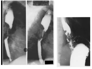
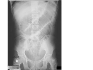
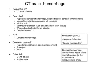
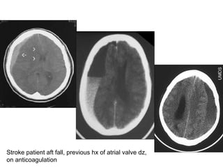

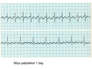
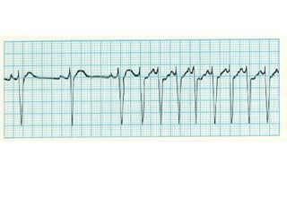

The document describes various radiological findings seen on imaging studies like CXR, CT, MRI etc. It provides descriptions of abnormalities seen in conditions like pulmonary edema, pneumonia, pneumothorax, bronchiectasis, pancreatic cancer, renal calculi, intestinal obstruction, rheumatoid arthritis, cerebral hemorrhage, subdural hemorrhage, thymoma, atrial flutter and atrial tachycardia among others. It also lists the possible causes and differentials for these conditions.


