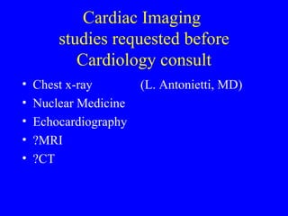
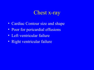
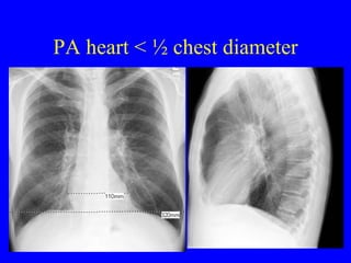
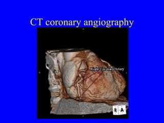
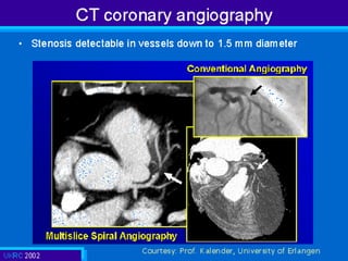
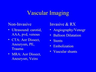
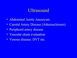
Cardiovascular Imaging M. J. McCowin, MD Clinical Professor of Radiology, UCSF G. Caputo, MD Professor of Radiology, UCD
Cardiac Imaging  studies requested before Cardiology consult Chest x-ray   (L. Antonietti, MD) Nuclear Medicine Echocardiography ?MRI ?CT
Chest x-ray Cardiac Contour size and shape Poor for pericardial effusions Left ventricular failure Right ventricular failure
PA heart < ½ chest diameter
Chamber Enlargement LA LV RV RA LV RV la append LA
Cardiac Contours: enlarged RA, LA, and RV in mitral stenosis
New enlarged LV due to aneurysm
Aortic Valve calcified due to AS
4 chamber enlargement due to MS/MI
PA and Lateral vs AP portable
Magnification of the heart with AP portable x-ray PA in Radiology AP as a portable
 
Large Cardiac Contour
Large cardiac contour AP portable magnifies Lordotic film magnif Kyphotic film minim Rotation changes Pericardial effusion looks like cardiomegaly
Heart Failure RIGHT HEART LE edema Ascites Cxr large rt heart Cxr large azygous v Often due to LHF or Pulmonary dx LEFT HEART Cardiomegaly Pulmonary Edema Pleural Fluid
Relationship of aortic arch,pa,azyg v. to trach carina
Heart Failure
Interstitial Edema Bronchovascular thickening and indistinctness Vessels get bigger and busier Vessels seen more to the periphery Kerley lines (less common)
Which vessels are bigger, busier,more peripheral?
Dry Wet
Airspace Edema May be cardiogenic or non-cardiogenic Very non-specific: aspiration, atypical pna, hemorrhage, etc. ARDS will persist and become coarse over time
Air-space edema
Cardiac imaging requested prior to Cardiology consult Nuclear Medicine Perfusion Wall motion Viability Shunts ?MRI  Pericard, chd, valv, shunts, perfusion, wm ?CT  Pericard calc, CA calc, cta for CA Echocardiography Pericardial fluid Valve function, integrity/vegetations Wall motion Shunts Congenital heart dx ?contrast agents
Nuclear Med. Perfusion Studies:  Thallium (potassium-like is extracted in K-ATPase pump), Sestamibi etc. Normal Stress Rest Perfusion stress rest stress rest
Cardiac anatomy as seen in SPECT nuclear imaging short axis, horiz. long., vert .long.
Diagram of short axis perfusion images: Would you pay full price for this donut? YES! the “ donut” is all  there. (normal septal thinning)
Thallium stress/rest: reversible ischemia inf/septum c/w RCA disease stress rest stress rest large “bite” out of donut! “ donut bite” fills in at rest
Circumferential data confirms reversible inferoseptal ischemia
Reversible ischemia ( ant/sept/apex) & stress-induced lv dilation Transient ischemic myocardial dysfunction S R R S R S
Fixed lateral perfusion deficit. ( fixed “bite out of donut”)   stress rest stress rest
Cardiac Wall Motion with quantitative ejection fraction (chemo rx)
Regional wall motion
Imaging requests by Cardiology Nuclear Medicine Myocardial Viability FDG study MR for perfusion, viability, myocardial function, CHD, evaluation of anatomy and flow, shunts, wm, pericardial dx Cardiac angiography for coronary artery assessment, CHD, valve and shunt assess  Cardiac angio for RX! plasty, stent, ASD,chd CT: CABG eval, CTA for coronary as. , contrast agents for ischemia
MRA cong double arch post-op ligation of left arch
Dilated LV and RA
Ao valv, pap muscle, rt pleural fluid, dilated lv
Coronal MRI shows aorta, av, lv (can eval for stenosis and regurg) Spin echo “black blood” anatomy Gradient echo “white blood” function & flow


CT coronary angiography
 
Vascular Imaging Non-Invasive Ultrasound: carotid, AAA, pvd, venous CTA: Aor Dissect, Aneurysm, PE, Trauma MRA: Aor Dissect, Aneurysm, Veins Invasive & RX Angiography/Venogr Balloon Dilatation Stents Embolization Vascular shunts
Ultrasound Abdominal Aortic Aneurysm Carotid Artery Disease (Atherosclerosis) Peripheral artery disease Vascular shunt evaluation Venous disease: DVT etc.
Ultrasound of Carotid Artery CCA ICA ECA
Ultrasound of Carotid Artery  note: brain, kidneys, heart must have both systolic & diastolic flow systole diastole
Atherosclerotic Plaque
Ultrasound for Venous Dx
CTA and MRA for Vascular Disease CTA Aortic Dissection Aortic Aneurysm Peripheral Vascular Disease Aortic Trauma Pulmonary Emboli MRA Aortic Dissection Aortic Aneurysm Peripheral Vascular Disease
Abdominal Aortic Aneurysm
Abdominal Aortic Aneurysm MR
Abdominal Aortic Aneurysm CTA with 3D Rendering
R/o aortic dissection CTA MRA TEE
MRA for central and peripheral Arteries and veins
 
Aortic Trauma
Aortic Trauma
Intravenous Contrast A large-bore (>22g, preferably an 18g or >) peripheral IV is required and is best placed in the right arm because the venous drainage is closer to the heart than the left arm.  PICC lines and many other central lines cannot be used for this rapid power injection.  Note also that intravenous iodinated contrast may  be contraindicated in some patients, particularly those with a history of contrast allergy and patients with renal insufficiency (creatinine > 1.5).   Additional caution regarding contrast is needed for patients in heart failure, a history of a serious allergy of any kind, multiple myeloma, diabetes particularly if on metformin (glucophage), or if a recent large contrast bolus has not yet been cleared from the body.
Pulmonary Artery Emboli Nuclear Medicine Perfusion Scan
CTAngiography for pulmonary artery emboli
Peripheral Vascular Disease
PVD after balloon Rx
Endovascular Stents
Endovascular repair of aneurysm
Acute Chest Pain:  will CTA become the one stop shop?
CTAngiography for pulmonary artery emboli
Aortic dissection
CT coronary angiography
CTA: one stop shop for chest pain? Same CTA: Rules out aortic dissection Rules out pulmonary emboli ? Rules out coronary disease? Perhaps with 64 slice and up CT and image processing Currently our Radiology Resident’s worst nightmare!
Cardiovascular Imaging NON-INVASIVE for DX Ultrasound Nuclear Medicine CTA (inc. coronary) MRA Chest x-ray INVASIVE for DX & TX Coronary arteries Fine detail of arteries Lots of Therapeutic Possibilities !

Cardio