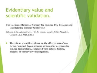
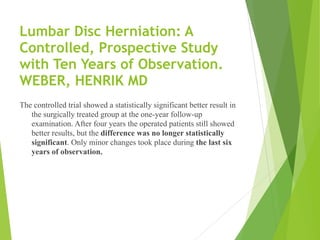
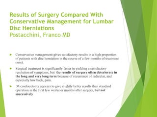
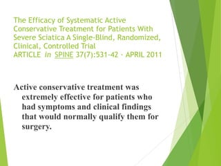
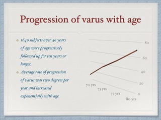
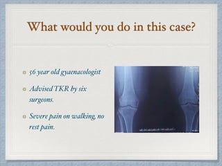
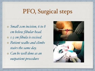
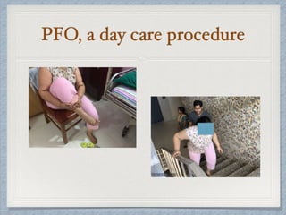
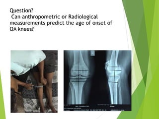
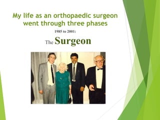
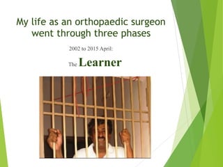
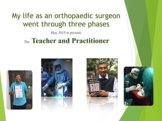
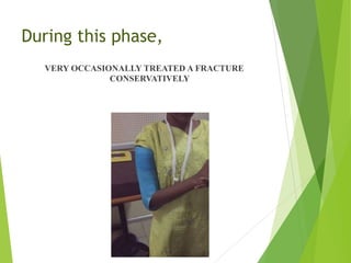
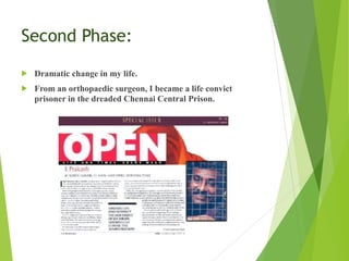
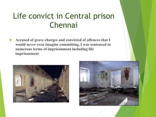
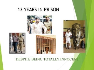
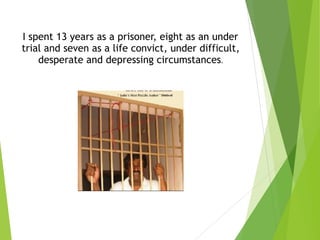
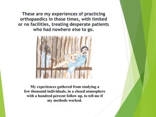
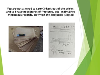
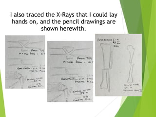
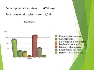
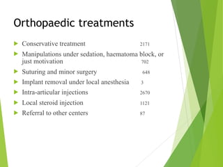
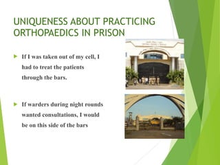
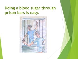
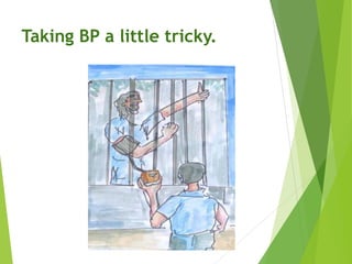
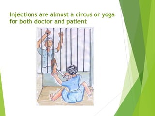
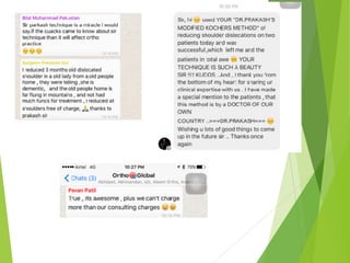
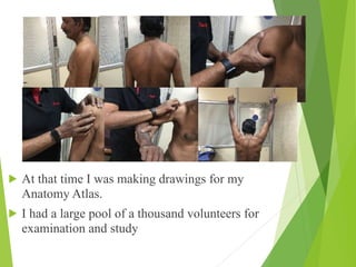
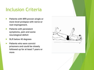
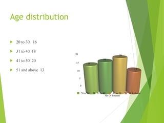
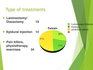
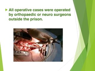
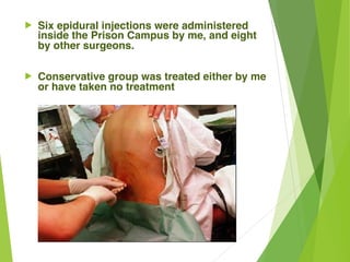
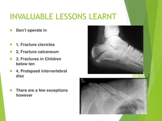
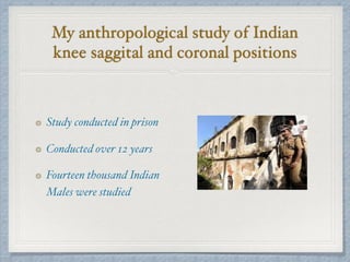
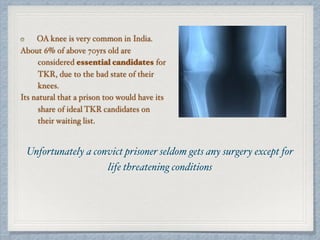
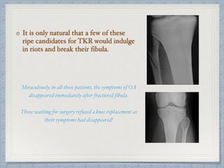
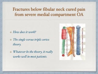
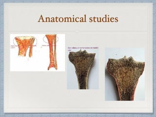
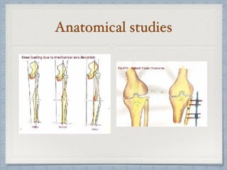
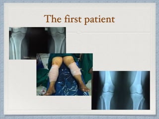
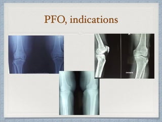
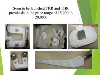
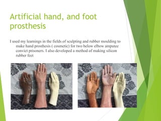
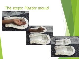
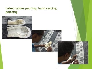
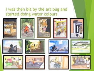
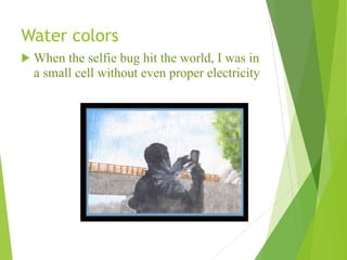
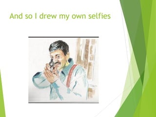
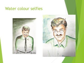
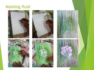
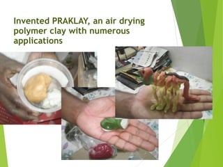
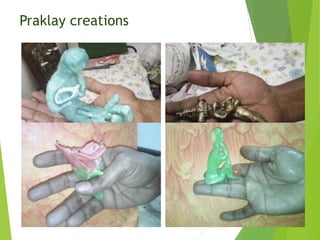
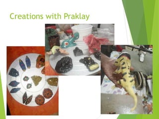
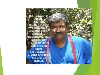
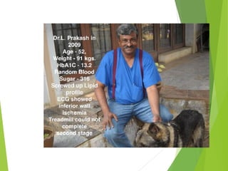
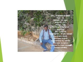
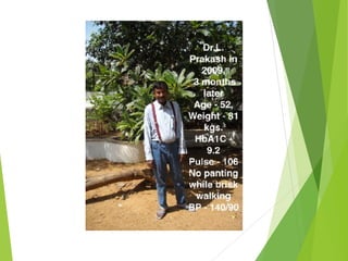
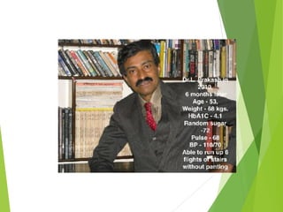
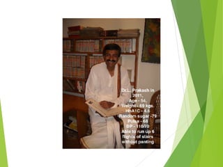
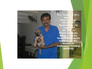
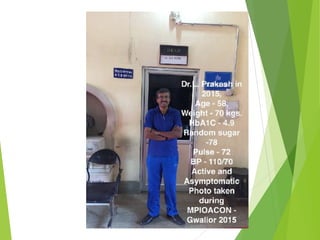
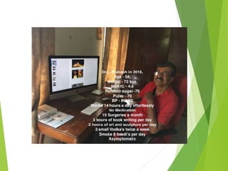
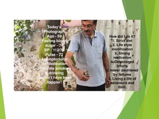
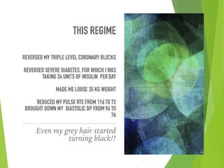
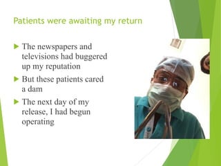
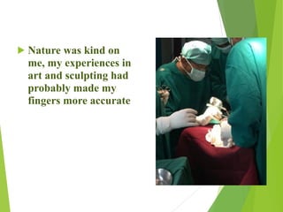
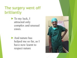
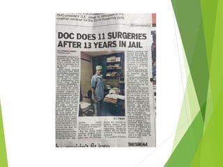
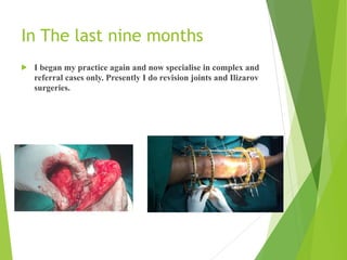
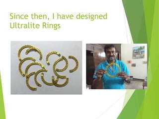
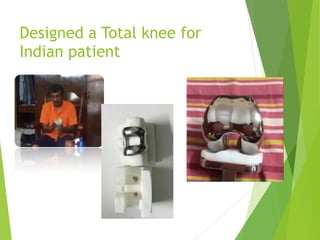
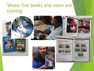
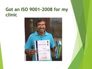
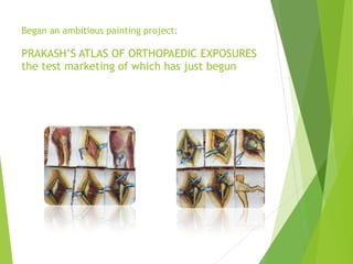
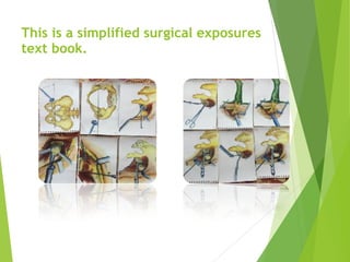
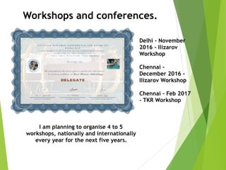
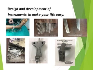
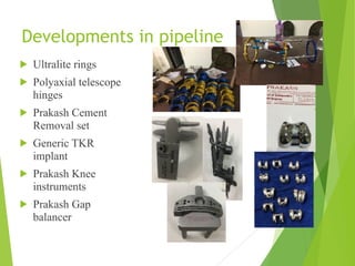
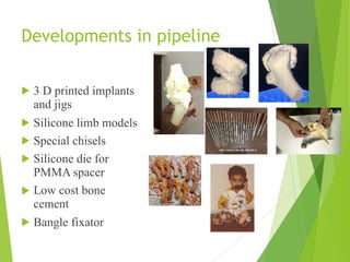
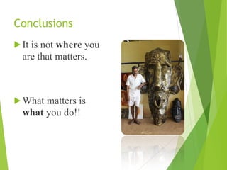
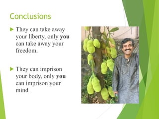
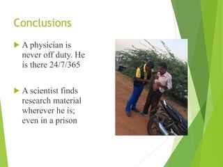
Orthopaedics, usual
and unusual
Thirty years of experience
Dr L.Prakash M.S., M.Ch (orth) Liverpool
Director. Institute for Special Orthopaedics Chennai 600106
(ISO 9001-2008 certified)


Orthopaedics, usual, and unusual

Thirty years of experience
Dr L.Prakash M.S., M.Ch (orth) Liverpool
My life as an orthopaedic surgeon
went through three phases
1985 to 2001:
The Surgeon
My life as an orthopaedic surgeon
went through three phases
2002 to 2015 April:
The Learner
My life as an orthopaedic surgeon
went through three phases
May 2015 to present:
The Teacher and Practitioner
First Phase
! Routine beginnings. M.S.,
M.Ch. etc,
! Training under greats like
Freeman, Muller, Goodfellow,
Monk, Wroblowski, Boyle,
Klenerman, Owen, Taylor etc.
! Career as a Trauma and Joint
replacement surgeon.
During this phase, I was:
A prolific surgeon, innovator, and wrote four books and
conducted 80 workshops.
During this phase,
I operated in 106 operation theatres across the country and
abroad, demonstrating surgical techniques and was the
Secretary and Vice President of Indian Orthopaedic
Association.
During this phase,
VERY OCCASIONALLY TREATED A FRACTURE
CONSERVATIVELY
Second Phase:
! Dramatic change in my life.
! From an orthopaedic surgeon, I became a life convict
prisoner in the dreaded Chennai Central Prison.
Life convict in Central prison
Chennai
! Accused of grave charges and convicted of offences that I
would never even imagine committing, I was sentenced to
numerous terms of imprisonment including life
imprisonment.
13 YEARS IN PRISON
DESPITE BEING TOTALLY INNOCENT


I spent 13 years as a prisoner, eight as an under
trial and seven as a life convict, under difficult,
desperate and depressing circumstances.
These are my experiences of practicing 

orthopaedics in those times, with limited 

or no facilities, treating desperate patients 

who had nowhere else to go.
My experiences gathered from studying a
few thousand individuals, in a closed atmosphere
with a hundred percent follow up, to tell me if
my methods worked.
You are not allowed to carry X-Rays out of the prison,
and so I have no pictures of fractures, but I maintained
meticulous records, on which this narration is based

I also traced the X-Rays that I could lay
hands on, and the pencil drawings are
shown herewith.
Period spent in the prison 4831 days


Total number of patients seen 11,248

Conservative treatment
Manipulations
Suturing and minor surgery
Implant removal under LA
Intra-articular injections
Local steroid injection
Referral to other centers
Orthopaedic treatments
! Conservative treatment 2171
! Manipulations under sedation, haematoma block, or
just motivation 702
! Suturing and minor surgery 648
! Implant removal under local anesthesia 3
! Intra-articular injections 2670
! Local steroid injection 1121
! Referral to other centers 87
Surgeries inside the prison:
! Suturing
! Abscess drainage
! Removal of thorns
and foreign bodies
! Skeletal traction
! Implant removal
UNIQUENESS ABOUT PRACTICING
ORTHOPAEDICS IN PRISON
! No X-ray machine in prison hospital
! No POP bandages or traction items
! No Lignocaine or local anaesthesia, without
special permission.
! No anaesthetic drugs
! No sedatives or tranquillisers
! Limited suture material
! No other facilities.
UNIQUENESS ABOUT PRACTICING
ORTHOPAEDICS IN PRISON
! If I was taken out of my cell, I
had to treat the patients
through the bars.
! If warders during night rounds
wanted consultations, I would
be on this side of the bars
Doing a blood sugar through
prison bars is easy.
Taking BP a little tricky.
Injections are almost a circus or yoga
for both doctor and patient

NOW JUST IMAGINE REDUCING A DISLOCATED SHOULDER
THROUGH THE PRISON BARS!! 

IT IS INDEED UNFORTUNATE THAT I SIMPLY COULD NOT HAVE
TAKEN ANY PICTURES!!

However, my experiences, led to the
discovery of a new method of reduction
of shoulders; a simple easy method
INTERESTING PROBLEMS
The torn
ear
Shoulder dislocations
! Total 89
! Anterior 83
! Posterior 6
! Associated with
fractures 9
Patients
The Prakash method of reducing
shoulder dislocations
There is no role of traction in shoulder dislocations
because shoulder dislocations are pure rotation,
lateral translation injuries.
Needs no assistant.
Needs no anaesthesia or even analgesia.
Gives consistent, reproducible results; wherever
tried.
Works in dislocations as old as three months.
Simple effective method.
The patient sits on a chair, with scapula fixed.
The shoulder is externally rotated.
Held in external rotation for two full minutes
Keeping the shoulder in maximum external
rotation for two full minutes, to relax the muscles is
the key to the success of this procedure.
The patient is reassured by engaging him in
conversation.
Adduction followed by internal
rotation; reduces the joint
smoothly.
There are no clicks or clucks.
The head glides in majestically.
The dramatic smile on the patient’s face
is an indication of success.
PROBLEMS TREATED
! Colle’s and Smiths fractures
! Fracture clavicle
! Fracture shaft of humerus
! Fracture neck of femur
! Intertrochanteric fractures
! Fracture shaft of femur
! Calcaneal fractures
! Metacarpals and Meta tarsals
Fractures of humeral shaft
! In the past I operated on all shaft
humerus fractures by AO methods.
! Surgery is not undertaken on prisoners
unless it is a life threatening condition.
! So the burden of conservatively
treating humeral fractures fell on me.
! At that time I was making drawings
for my Anatomy Atlas.
I made some unusual
observations about Humerus
fractures, treated conservatively.
1, Fractures treated with elbow in flexion
always malunited unless undisplaced, and on
occasions went into non union due to plaster
distraction.
2, Those treated with elbow in extension in
full supination, for a short while in long arm
casts, always invariably united soon.
! At that time I was making drawings for my
Anatomy Atlas.
! I had a large pool of a thousand volunteers for
examination and study
Biceps and triceps during flexion and
extension
Displacement in flexion, stability in extension
Disruptive versus protective muscles
56 cases treated in 13 years, all followed up to
three years or longer.
All fractures united, 30 with a little shortening.
Elbow movements full at six months in every case.
Full return to function within an average of two
and a half months.
Average time of clinical union was nine weeks.
Only one complication of acute radial nerve
entrapment and post fracture wrist drop recovered
fully in six months.
Are we over-treating
fractures shaft of humerus?
Conservatively treated humeral shaft
fractures never go into non union if treated
in extension cast.
Full return to function in three months.
Light weight axilla to metacarpal head long
cast in supination and full extension.
Remove cast when patient is pain free; 3 to 4
weeks, and begin elbow movements.
Fracture unites in average eight weeks.
Fractures of the femoral
shaft
I had a wonderful
opportunity for a
study.
! Total femoral fractures
treated in twelve years
70
! Closed fractures 54
! Open fractures 11
! Fractures infected
after fixation who
came to prison
subsequently 5
Closed fractures
Open fractures
Fractures infected after fixation who came to prison subsequently
Typical femoral shaft
fractures seen in prison
! Of these; 65 cases happened
inside the prison.
! Most open fractures were
compound from within out.
! Most happened after a fall from
tree, building watch tower etc
! In 2002, a patient named
Karuppuswamy climbed up a tree
and threatened to jump down.
! He slipped, fell and broke his right
femur. With a splint, he was sent to
the Government hospital, where as a
first aid, he was immobilized in a
Thomas’s splint.
! He was posted for surgery in a few
days.
! He was in the general
ward. And on both his
sides were patients with
old fractured femurs.
Discharging sinuses, foul
smell, exposed metal, and
heart wrenching stories of
months or years of misery.
! Karuppuswamy was
petrified. He did not allow
any surgeon to touch him
and was brought back to
the prison in a Thomas’s
splint
When I was
summoned to see
him, his initial X-Ray
looked something
liked this.

! I thought about it for a
day and then asked my
assistant outside to get
me an Ilizarov half ring,
couple of olive wires,
couple of wire fixation
bolts, and a length of
clothesline.
I used double olive wires on upper
tibia under local anaesthesia
! The prison authorities allowed me
to use traction on him and I gave
an upper tibial traction somewhat
like a Fisk traction using fan
hooks and locally designed
pulleys.
! The prison plumber and
electrician helped.
! Traction was provided by three
concrete bricks each about four
kilos.
Fisk Traction
The Karuppuswamy story
! Traction was provided by
three concrete bricks each
about four kilos.
! I would measure the femoral
length daily, and ensure that
there was no rotation.
! Knee was kept flexed most of
the time over pillows to
traction level.
! He was sitting up in a
week and moving in the
bed in two. He was out of
the bed by the fifth week,
walking with a six feet
bamboo cane.
! In three months he was
walking and by fourth
month back to playing
football.
! No locking plates, no
flexible nails, no rigid
locking nails, no
protruding stubs, no
scars, full function, three
degree valgus and half an
inch shortening
The fourth
month X-ray
was
somewhat
like this
Femoral fractures
! From that time on no convict would get
his femur operated.
! I managed eleven open (punctured
wounds from inside) and 54 closed
femoral fractures all with excellent
results.
! The scientific data is being analysed for
publication. It is a real pity that the
circumstances and situations did not
allow me to get or copy the radiographs.
PROBLEMS TREATED
! Low back ache
! Knee arthritis
! Knee injuries
! Frozen shoulders
! Fracture both bones forearm
! Fracture tibia/fibula
THE BACKACHE STUDY
! By luck, I had an
opportunity to monitor
and study a group of
67 patients with CT or
MRI proven Prolapsed
intervertebral discs
treated by various
means and could
study them for
periods up to 13
years, with a mean
follow-up of 10 years.
Inclusion Criteria

! Patients with MRI proven single or
more level prolapse with nerve or
root impingement.
! Patients with persistent
symptoms, pain and some
neurological deficit
! SLR below 40 degrees
! Patients who were convict
prisoners and could be closely
followed up for at least 7 years or
more
Age distribution
! 20 to 30 16
! 31 to 40 18
! 41 to 50 20
! 51 and above 13
20 to 30 31 to 40 41 to 50 51 & above
Type of treatments

! Laminectomy/
Discectomy 19
! Epidural injection 14
! Pain killers,
physiotherapy,
exercises 34
Laminectomy/diskectom
Epidural
painkillers, physio
! All operative cases were operated
by orthopaedic or neuro surgeons
outside the prison.
! Six epidural injections were administered
inside the Prison Campus by me, and eight
by other surgeons.
! Conservative group was treated either by me
or have taken no treatment
Though this is a prospective study, it is
neither double blind nor controlled. I
had no control over patients falling into
a particular group. Situations
determined the group into which the
patient fell.
Final evaluation was done by

! Visual Rating
Scale
! Visual analogue
scale
! Oswestry
disability index
Visual Rating Scale
Visual analogue scale
Oswestry disability index
Conclusions:
! No difference between Epidural injection and No
treatment
Conclusions:
! Surgery gave the worst results with
permanent complications in 19% of the
patients
Moral of the story:
! Never operate on any prolapsed
disc.
! Even those with neurological
deficit or bladder problems get well
after some time.
Evidentiary value and
scientific validation.
Conservatively treated massive prolapsed discs: a 7-year follow-up
RT Benson1, SP Tavares1, SC Robertson2, R Sharp1, and RW Marshall1
RCS Annals: Volume: 92 Issue: 2, March 2010, pp. 147-153
A massive disc herniation can pursue a favourable clinical
course. The long-term prognosis is very good and even
massive disc herniations can be treated conservatively
Evidentiary value and
scientific validation.
The Cochrane Review of Surgery for Lumbar Disc Prolapse and
Degenerative Lumbar Spondylosis
Gibson, J. N. Alastair MD, FRCS; Grant, Inga C. MSc; Waddell,
Gordon DSc, MD, FRCS
! There is no scientific evidence on the effectiveness of any
form of surgical decompression or fusion for degenerative
lumbar disc prolapse, compared with natural history,
placebo, or conservative management.
Lumbar Disc Herniation: A
Controlled, Prospective Study
with Ten Years of Observation.

WEBER, HENRIK MD
The controlled trial showed a statistically significant better result in
the surgically treated group at the one-year follow-up
examination. After four years the operated patients still showed
better results, but the difference was no longer statistically
significant. Only minor changes took place during the last six
years of observation.
Results of Surgery Compared With
Conservative Management for Lumbar
Disc Herniations

Postacchini, Franco MD

! Conservative management gives satisfactory results in a high proportion
of patients with disc herniation in the course of a few months of treatment
onset.
! Surgical treatment is significantly faster in yielding a satisfactory
resolution of symptoms, but the results of surgery often deteriorate in
the long and very long term because of recurrence of radicular, and
especially low back, pain.
! Microdiscetomy appears to give slightly better results than standard
operation in the first few weeks or months after surgery, but not
successively
The Efficacy of Systematic Active
Conservative Treatment for Patients With
Severe Sciatica A Single-Blind, Randomized,
Clinical, Controlled Trial

ARTICLE in SPINE 37(7):531-42 · APRIL 2011 
Active conservative treatment was
extremely effective for patients who
had symptoms and clinical findings
that would normally qualify them for
surgery.
Principal indications for disc
surgery:
! Holiday for the surgeon and spouse
! College admission for surgeon’s son
! A new car or holiday home
! Or possibly even a speed boat if he operates
every back!!
Dr Prakash’s SC index or B/B
Ratio
! Scrotum/cerebrum×100
! Balls over brains ratio
! While our testosterone urges us to rush into surgery,
cerebral serotonin urges caution
! We must make our choices wisely
INVALUABLE LESSONS LEARNT
! Don’t operate in
! 1, Fracture clavicles
! 2, Fracture calcaneum
! 3, Fractures in Children
below ten
! 4, Prolapsed intervertebral
disc
! There are a few exceptions
however
I had a large pool of subjects in a
closed environment, enabling me
to conduct numerous studies.
Amongst other studies, I also performed physical
and anthropometric measurements of over ten
thousand inmates and prison officials.
One of the studies enabled me to understand knee
sizes, and plan the ideal dimensions for a TKR
implant for the Indian population spread and
also device a new operation for medial
compartment arthritis, the PFO or Prakash
Fibular Osteotomy.
But before that
But why Varus scenario in Indian knees?
Are our knees different?
Is there a racial variation in coronal deviation
of the knee joint?
Apparently yes. Apart from my studies I have found two
references. One for adults and one for children.
Varus and valgus deformities in knee osteoarthritis among different ethnic
groups (Indian, Portuguese and Canadians) within an urban Canadian
rheumatology practice
Raman Joshi1, Nimu Ganguli2, Christopher Carvalho3, Faye de Leon4, Janet
Pope5
Significantly more varus deformity was noted in the Indian-born group than
the Canadian-born group (P = 0.002), and more valgus deformity was noted in
the Portuguese-born than Canadian-born group (P = 0.009).
Conclusions: Patient populations differed significantly in terms of varus and
valgus deformities at the knee.
I could locate one study, in children
Normal development of the
knee angle in healthy Indian
children: a clinical study of 215
children
Uttam Chand Saini, Kamal
Bali, Binoti Sheth, Nitesh
Gahlot, andArushi Gahlot
Normal development of the knee angle in healthy
Indian children: a clinical study of 215 children
The overall pattern of development might be slightly
different in Indian children, especially in Indian girls, with
early reversal of physiological varus (<2 years of age) and a
late peak of maximal valgus at the knee (6 years of age).
Varus after 3 years seems atypical for Indian
children. We provide an elaborate set of data for the mean
TFA of different age groups and believe that this data
could be of potential benefit to the physicians while
evaluating lower limb alignment in Indian children aged 2–
15 years.
Knee varus in Indian population
There have been few demographic or
anthropometric studies of the Indian
knees in adult population.
No study has been done on long term
measurements of knee saggital
deviation with an increase in age.
My anthropological study of Indian
knee saggital and coronal positions
Study conducted in prison
Conducted over 12 years
Fourteen thousand Indian
Males were studied
Methods
Instruments used were a Galton
calliper, Long scale, Wall, Pencil
and long sheets of paper.
Methods
Patient stood with back to the
wall.
Second toe faced straight towards
the observer
Line drawn from hip centre to
knee centre was allowed to bisect
the line from centre of ankle to
centre of knee.
Material
All subjects were male
Convict and under-trial prisoners.
Age from 18 to 91
Only 9% complained of any
problems
Except for Sex, they represented the
average Indian population
Results Saggital plane
Total Knees
studied
14,321
Varus
alignment
7642
Valgus
alignment
1387
Neutral
alignment
5292
Varus knees
Total knees
Neutral knees
Valgus knees
Relationship between age and Varus
Correlation between symptoms of medial
compartment OA and knee varus
Progression of varus with age
1640 subjects over 40 years
of age were progressively
followed up for ten years or
longer.
Average rate of progression
of varus was two degrees per
year and increased
exponentially with age.
Varus progresses with age and the progress is
more rapid in obese individuals
While less than 15% of those in twenties had varus
knees, over 60% of those above 70 had varus knees.
Varus progressed most rapidly between the ages of
60 to 80.
Once a knee gets into varus disposition, it continues
to progress till the patient’s death.
Important observation
More than 50% Indian knees had a varus disposition, though
not all varus knees had symptoms of medial compartment
OA.
Severity of symptoms had a direct correlation to the degree
of varus, and after 30 degrees, all knees were symptomatic.
Varus of the knee gradually and progressively increases with
age.
Limitations of the study
No facilities for radiographic co-relation
It was only a clinico-anthropometric study.
Large numbers give the findings credibility
Study will be published soon.
What would you do in this case?
56 year old gyaenacologist
Advised TKR by six
surgeons.
Severe pain on walking, no
rest pain.
Pre and post surgery
PFO, something new
Accidental discovery in 2004
Based on my experiences in prison
An out of the box method which produces remarkable and
startling results both functionally and radiologically.
How??
Prison riots happen infrequently.
Wardens are instructed to hit
rioting prisoners with a Lathi,
Below the knees, to avoid
grievous injuries.
Fracture proximal fibula is an
usual consequence.
Unique aspects of practicing
orthopaedics in the prison
Every single inmate has to come to you if he breaks a bone.
Most orthopaedic problems come to you.
There are absolutely no facilities available.
But for a pure scientist, this is a wonderful opportunity for
study.
And these are my studies that lead to PFO
OA knee is very common in India.
About 6% of above 70yrs old are
considered essential candidates for
TKR, due to the bad state of their
knees.
Its natural that a prison too would have its
share of ideal TKR candidates on
their waiting list.
Unfortunately a convict prisoner seldom gets any surgery except for
life threatening conditions
It is only natural that a few of these
ripe candidates for TKR would indulge
in riots and break their fibula.
Miraculously, in all these patients, the symptoms of OA
disappeared immediately after fractured fibula.
Those waiting for surgery refused a knee replacement as
their symptoms had disappeared!
Fractures below fibular neck cured pain
from severe medial compartment OA
How does it work?
The single versus triple cortex
theory.
Whatever be the theory, it really
works well in most patients.
Anatomical studies
Anatomical studies
The first patient
PFO, indications
PFO, Surgical steps
Small 2cm incision, 6 to 8
cm below fibular head.
1.5 cm fibula is excised.
Patient walks and climbs
stairs the same day.
Can be well done as an
outpatient procedure
PFO, a day care procedure
PFO, observations so far
Effective in all patients, even those with
patellofemoral OA
Patient remains pain free for three years or longer.
My first patient operated in 2004, is still happy and
refuses knee replacement.
A multi-centre trial is being conducted presently
and about 1800 surgeries have been done in the last
one year.
PFO, the first case, nine years follow up
PFO, other references
Zong-You Yang, MD; Wei Chen, MD; Cun-Xiang Li, MD; Juan Wang, MD; De-Cheng Shao, MD; Zhi-Yong hou, MD; Shi-
Jun gao, MD; Fei Wang, MD; Ji-Dong Li, MD; Jian-Dong hao, MD; Bai-Cheng Chen, MD; Ying-Ze Zhang, MD
It is a safe, simple, and effective procedure that is an alternative to total knee
arthroplasty for medial compartment OA of the knee joint. Care must be taken to
avoid potential nerve injuries.
Proximal fibular osteotomy may reduce knee pain significantly in the varus
osteoarthritic knee and improve the radiographic appearance and functional recovery
of the knee joint.
How I tackled the
knee design of an
Indian Knee??
Many questions
had intrigued me
for a long time.
Now was the time
to search for
answers.
Question?

Are Indian knees Narrower front to back,
than their Caucasian counterparts?
Question?

Does Squatting since childhood alter
the condylar shape?
Question?

What is the normal orientation of the femoral condyles
in relation to the femoral head? What is the Varus/
valgus spread in average population?
Question?

Is there a normal Tibial Varus?
Question?

Are Indian upper tibias wider from side
to side, but narrower from front to back?
Question?


What is the tilt
of the tibial
articular surface
vis a vis the
ankle joint in the
anterio-posterior
direction?
Question?

Can anthropometric or Radiological
measurements predict the age of onset of
OA knees?
Question?

Relation between the severity of OA and the state of
ligaments in and around the knee?

Methodology
employed
! Measurement of actual
bones 640
! Anthropometric
measurements 8400 knees
(4200 patients)
! Radiological measurement
of 1300 Radiographs
! Total knee joints measured
10340
Osteology
Radiography
Clinical
At one stage my bedroom had more
bones than the Anatomy department
Measurements taken
FEMORAL CONDYLE
! Mediolateral dimensions
! Anterioposterior dimension of medial condyle
! Anterioposterior dimension of lateral condyle
! Femoral valgus/varus
Measurements taken
UPPER TIBIA
! Mediolateral dimension
! Medial anterioposterior dimension
! Lateral Anterioposterior dimension
! Tibial varus/valgus
Achieving consistency in radiographic
measurements, and ensuring that they
correspond to actual knee measurements.
Each X-ray was scanned and digitized.
The patients knee girth, mid thigh girth and upper calf girth were
measured.
The AP and Side to side dimensions of the limb were thus calculated.
Using computer software, the X-rays were resized so that the outer
soft tissue shadow measured the exact dimensions of the limb in
each case.
Consistency in measurements was thus ensured.
Dr Prakash’s modified Galton
anthropometric calliper
Conclusions of the above study
LOWER FEMORAL
DIMENSIONS WITH SPREAD
! Mediolateral 64.5 to 91.2
! So Femoral component should be available in 53mm, 56mm
59mm 62mm 64mm 67mm 70mm and 75mm medio-lateral
dimensions.
Mediolateral
Numbers
LOWER FEMORAL DIMENSIONS WITH SPREAD

Anterioposterior 63.1mm to 86.4mm

! So Femoral component should be available in 50mm, 53mm,
56mm 58mm 59mm 63mm 66mm and 70mm AP dimensions.
Femur AP
inmm
0
22.5
45
67.5
90
1 51 101151201251
Anterioposterio
Numbers
Conclusions of the above study
UPPER TIBIAL DIMENSIONS WITH SPREAD
! Mediolateral 55.2 to 81.3mm, and thus the tibial trays should
be available in 55mm, 60mm, 63mm, 66mm, 71mm, 75mm
and 81mm
Tibial dimensions
M-Ldimension
0
22.5
45
67.5
90
1 20 39 58 77 96 115134153172191210229248267
UPPER TIBIAL DIMENSIONS WITH SPREAD


Anterioposterior 38.1 to 55.4

So tibial trays should come in AP dimensions of 38,
40, 43, 45, 47, 51 and 55mm


Anterioposterior
Numbers
These dimensional studies helped
me to design my knee joint
Soon to be launched TKR and THR
prosthesis in the price range of 15,000 to
20,000,
Artificial hand, and foot
prosthesis
I used my learnings in the fields of sculpting and rubber moulding to
make hand prosthesis ( cosmetic) for two below elbow amputee
convict prisoners. I also developed a method of making silicon
rubber feet
The steps: Plaster mould
Latex rubber pouring, hand casting,
painting
This helped me to make models for this
conference.
The worlds first silicone flexible
workshop models.
Other things besides
orthopaedics
! 106 books, 25 million hand written words.
! Fiction, non fiction, mythological, adventure stories, legal
thrillers, sensuous, and detective novels.
Twenty five million words look
like this
Twenty five million words
look like this as books
Twenty five million words
look like this as books
I was then bit by the art bug and
started doing water colours
More water colors
Water colors
! When the selfie bug hit the world, I was in
a small cell without even proper electricity
And so I drew my own selfies
Water colour selfies
I progressed in art to acrylic
colours
Acrylic paintings
Charcoal Sketches
Caricatures
Paintings from photographs
Charcoal and oil painting
Next stage was sculpting
Dr L.Prakash’s
Hundred sculpture
project
Clay, Plaster of 

Paris, Resins, Acrylic, Marble,
Granite, Epoxy, PMMA, Bronze
Aluminium, Dental cement,
Gypsum.
I experimented with all materials.
Bronze sculpture work
My experience in metallurgy helped
me to cast my TKR prototypes.
I also did a lot of Material
research
Masking fluid
Invented PRAKLAY, an air drying
polymer clay with numerous
applications
Praklay creations
Creations with Praklay
Latex moulding compound
Latex and silicone masks and cinema
special effects
My experiments in non
surgical reversal of heart
disease and diabetes
Knowledge under duress
And then at last I won!!

! I was acquitted in all cases that were foisted on me
When I reached home I was really surprised

Five patients were waiting for me!!

Patients were awaiting my return
! The newspapers and
televisions had buggered
up my reputation
! But these patients cared
a dam
! The next day of my
release, I had begun
operating
! Nature was kind on
me, my experiences in
art and sculpting had
probably made my
fingers more accurate
The surgery went off
brilliantly
! To my luck, I
attracted only
complex and unusual
cases.
! And nature has
helped me so far, as I
have now learnt to
respect nature
My Colleagues
! I also received a
wholehearted
welcome from my
orthopaedic
colleagues and the
Indian Orthopaedic
Association
In The last nine months
! I began my practice again and now specialise in complex and
referral cases only. Presently I do revision joints and Ilizarov
surgeries.
Since then, I have designed
Ultralite Rings
Designed a Total knee for
Indian patient
Wrote five books and more are
coming
Got an ISO 9001-2008 for my
clinic
Began an ambitious painting project:


PRAKASH’S ATLAS OF ORTHOPAEDIC EXPOSURES
the test marketing of which has just begun
This is a simplified surgical exposures
text book.
Future plans
Orthopaedic teaching and education
Workshops and conferences.
Design and development of instruments to make your life easy.
World Class made in India knees for below 20,0000 Rs
Museum of orthopaedic implants
IOA orthopaedic history museum
Streamlining orthopaedic teaching and fellowships
Sculpting the future of IOA
Orthopaedic teaching and education
Books
Surgical DVDs, and technique
videos
Workshops and conferences.
Delhi - November
2016 - Ilizarov
Workshop
Chennai -
December 2016 -
Ilizarov Workshop
Chennai - Feb 2017
- TKR Workshop
I am planning to organise 4 to 5
workshops, nationally and internationally
every year for the next five years.
Design and development of
instruments to make your life easy.
Developments in pipeline
! Ultralite rings
! Polyaxial telescope
hinges
! Prakash Cement
Removal set
! Generic TKR
implant
! Prakash Knee
instruments
! Prakash Gap
balancer
Developments in pipeline
! 3 D printed implants
and jigs
! Silicone limb models
! Special chisels
! Silicone die for
PMMA spacer
! Low cost bone
cement
! Bangle fixator
World Class made in India knees for below
20,0000 Rs
Museum of orthopaedic implants
A travelling museum to be donated to IOA
IOA orthopaedic history museum
Streamlining orthopaedic teaching and
fellowships, in India
Sculpting the future of IOA
Conclusions
! It is not where you
are that matters.
! What matters is
what you do!!
Conclusions
! They can take away
your liberty, only you
can take away your
freedom.
! They can imprison
your body, only you
can imprison your
mind
Conclusions
! A physician is
never off duty. He
is there 24/7/365
! A scientist finds
research material
wherever he is;
even in a prison
Conclusions
! Keep meticulous records, you don’t know
when they will be useful
Conclusions
! Keep smiling, for no trouble lasts for
ever. The rainbow is out there.
Thank You

Orthopaedics usual and unusual

  • 1.
    Orthopaedics, usual and unusual Thirtyyears of experience Dr L.Prakash M.S., M.Ch (orth) Liverpool Director. Institute for Special Orthopaedics Chennai 600106 (ISO 9001-2008 certified) 

  • 2.
    Orthopaedics, usual, andunusual
 Thirty years of experience Dr L.Prakash M.S., M.Ch (orth) Liverpool
  • 3.
    My life asan orthopaedic surgeon went through three phases 1985 to 2001: The Surgeon
  • 4.
    My life asan orthopaedic surgeon went through three phases 2002 to 2015 April: The Learner
  • 5.
    My life asan orthopaedic surgeon went through three phases May 2015 to present: The Teacher and Practitioner
  • 6.
    First Phase ! Routinebeginnings. M.S., M.Ch. etc, ! Training under greats like Freeman, Muller, Goodfellow, Monk, Wroblowski, Boyle, Klenerman, Owen, Taylor etc. ! Career as a Trauma and Joint replacement surgeon.
  • 7.
    During this phase,I was: A prolific surgeon, innovator, and wrote four books and conducted 80 workshops.
  • 8.
    During this phase, Ioperated in 106 operation theatres across the country and abroad, demonstrating surgical techniques and was the Secretary and Vice President of Indian Orthopaedic Association.
  • 9.
    During this phase, VERYOCCASIONALLY TREATED A FRACTURE CONSERVATIVELY
  • 10.
    Second Phase: ! Dramaticchange in my life. ! From an orthopaedic surgeon, I became a life convict prisoner in the dreaded Chennai Central Prison.
  • 11.
    Life convict inCentral prison Chennai ! Accused of grave charges and convicted of offences that I would never even imagine committing, I was sentenced to numerous terms of imprisonment including life imprisonment.
  • 12.
    13 YEARS INPRISON DESPITE BEING TOTALLY INNOCENT
  • 13.
    
 I spent 13years as a prisoner, eight as an under trial and seven as a life convict, under difficult, desperate and depressing circumstances.
  • 14.
    These are myexperiences of practicing 
 orthopaedics in those times, with limited 
 or no facilities, treating desperate patients 
 who had nowhere else to go. My experiences gathered from studying a few thousand individuals, in a closed atmosphere with a hundred percent follow up, to tell me if my methods worked.
  • 15.
    You are notallowed to carry X-Rays out of the prison, and so I have no pictures of fractures, but I maintained meticulous records, on which this narration is based

  • 16.
    I also tracedthe X-Rays that I could lay hands on, and the pencil drawings are shown herewith.
  • 17.
    Period spent inthe prison 4831 days
 
 Total number of patients seen 11,248
 Conservative treatment Manipulations Suturing and minor surgery Implant removal under LA Intra-articular injections Local steroid injection Referral to other centers
  • 18.
    Orthopaedic treatments ! Conservativetreatment 2171 ! Manipulations under sedation, haematoma block, or just motivation 702 ! Suturing and minor surgery 648 ! Implant removal under local anesthesia 3 ! Intra-articular injections 2670 ! Local steroid injection 1121 ! Referral to other centers 87
  • 19.
    Surgeries inside theprison: ! Suturing ! Abscess drainage ! Removal of thorns and foreign bodies ! Skeletal traction ! Implant removal
  • 20.
    UNIQUENESS ABOUT PRACTICING ORTHOPAEDICSIN PRISON ! No X-ray machine in prison hospital ! No POP bandages or traction items ! No Lignocaine or local anaesthesia, without special permission. ! No anaesthetic drugs ! No sedatives or tranquillisers ! Limited suture material ! No other facilities.
  • 21.
    UNIQUENESS ABOUT PRACTICING ORTHOPAEDICSIN PRISON ! If I was taken out of my cell, I had to treat the patients through the bars. ! If warders during night rounds wanted consultations, I would be on this side of the bars
  • 22.
    Doing a bloodsugar through prison bars is easy.
  • 23.
    Taking BP alittle tricky.
  • 24.
    Injections are almosta circus or yoga for both doctor and patient

  • 25.
    NOW JUST IMAGINEREDUCING A DISLOCATED SHOULDER THROUGH THE PRISON BARS!! 
 IT IS INDEED UNFORTUNATE THAT I SIMPLY COULD NOT HAVE TAKEN ANY PICTURES!!
 However, my experiences, led to the discovery of a new method of reduction of shoulders; a simple easy method
  • 26.
  • 27.
    Shoulder dislocations ! Total89 ! Anterior 83 ! Posterior 6 ! Associated with fractures 9 Patients
  • 28.
    The Prakash methodof reducing shoulder dislocations There is no role of traction in shoulder dislocations because shoulder dislocations are pure rotation, lateral translation injuries. Needs no assistant. Needs no anaesthesia or even analgesia. Gives consistent, reproducible results; wherever tried. Works in dislocations as old as three months. Simple effective method.
  • 29.
    The patient sitson a chair, with scapula fixed. The shoulder is externally rotated. Held in external rotation for two full minutes
  • 30.
    Keeping the shoulderin maximum external rotation for two full minutes, to relax the muscles is the key to the success of this procedure. The patient is reassured by engaging him in conversation.
  • 31.
    Adduction followed byinternal rotation; reduces the joint smoothly. There are no clicks or clucks. The head glides in majestically. The dramatic smile on the patient’s face is an indication of success.
  • 35.
    PROBLEMS TREATED ! Colle’sand Smiths fractures ! Fracture clavicle ! Fracture shaft of humerus ! Fracture neck of femur ! Intertrochanteric fractures ! Fracture shaft of femur ! Calcaneal fractures ! Metacarpals and Meta tarsals
  • 36.
    Fractures of humeralshaft ! In the past I operated on all shaft humerus fractures by AO methods. ! Surgery is not undertaken on prisoners unless it is a life threatening condition. ! So the burden of conservatively treating humeral fractures fell on me. ! At that time I was making drawings for my Anatomy Atlas.
  • 37.
    I made someunusual observations about Humerus fractures, treated conservatively. 1, Fractures treated with elbow in flexion always malunited unless undisplaced, and on occasions went into non union due to plaster distraction. 2, Those treated with elbow in extension in full supination, for a short while in long arm casts, always invariably united soon.
  • 38.
    ! At thattime I was making drawings for my Anatomy Atlas. ! I had a large pool of a thousand volunteers for examination and study
  • 39.
    Biceps and tricepsduring flexion and extension
  • 40.
    Displacement in flexion,stability in extension Disruptive versus protective muscles
  • 41.
    56 cases treatedin 13 years, all followed up to three years or longer. All fractures united, 30 with a little shortening. Elbow movements full at six months in every case. Full return to function within an average of two and a half months. Average time of clinical union was nine weeks. Only one complication of acute radial nerve entrapment and post fracture wrist drop recovered fully in six months.
  • 42.
    Are we over-treating fracturesshaft of humerus? Conservatively treated humeral shaft fractures never go into non union if treated in extension cast. Full return to function in three months. Light weight axilla to metacarpal head long cast in supination and full extension. Remove cast when patient is pain free; 3 to 4 weeks, and begin elbow movements. Fracture unites in average eight weeks.
  • 45.
    Fractures of thefemoral shaft I had a wonderful opportunity for a study. ! Total femoral fractures treated in twelve years 70 ! Closed fractures 54 ! Open fractures 11 ! Fractures infected after fixation who came to prison subsequently 5 Closed fractures Open fractures Fractures infected after fixation who came to prison subsequently
  • 46.
    Typical femoral shaft fracturesseen in prison ! Of these; 65 cases happened inside the prison. ! Most open fractures were compound from within out. ! Most happened after a fall from tree, building watch tower etc
  • 47.
    ! In 2002,a patient named Karuppuswamy climbed up a tree and threatened to jump down. ! He slipped, fell and broke his right femur. With a splint, he was sent to the Government hospital, where as a first aid, he was immobilized in a Thomas’s splint. ! He was posted for surgery in a few days.
  • 48.
    ! He wasin the general ward. And on both his sides were patients with old fractured femurs. Discharging sinuses, foul smell, exposed metal, and heart wrenching stories of months or years of misery. ! Karuppuswamy was petrified. He did not allow any surgeon to touch him and was brought back to the prison in a Thomas’s splint
  • 49.
    When I was summonedto see him, his initial X-Ray looked something liked this.

  • 50.
    ! I thoughtabout it for a day and then asked my assistant outside to get me an Ilizarov half ring, couple of olive wires, couple of wire fixation bolts, and a length of clothesline.
  • 51.
    I used doubleolive wires on upper tibia under local anaesthesia
  • 52.
    ! The prisonauthorities allowed me to use traction on him and I gave an upper tibial traction somewhat like a Fisk traction using fan hooks and locally designed pulleys. ! The prison plumber and electrician helped. ! Traction was provided by three concrete bricks each about four kilos.
  • 53.
  • 54.
    The Karuppuswamy story !Traction was provided by three concrete bricks each about four kilos. ! I would measure the femoral length daily, and ensure that there was no rotation. ! Knee was kept flexed most of the time over pillows to traction level.
  • 55.
    ! He wassitting up in a week and moving in the bed in two. He was out of the bed by the fifth week, walking with a six feet bamboo cane. ! In three months he was walking and by fourth month back to playing football. ! No locking plates, no flexible nails, no rigid locking nails, no protruding stubs, no scars, full function, three degree valgus and half an inch shortening
  • 56.
  • 57.
    Femoral fractures ! Fromthat time on no convict would get his femur operated. ! I managed eleven open (punctured wounds from inside) and 54 closed femoral fractures all with excellent results. ! The scientific data is being analysed for publication. It is a real pity that the circumstances and situations did not allow me to get or copy the radiographs.
  • 58.
    PROBLEMS TREATED ! Lowback ache ! Knee arthritis ! Knee injuries ! Frozen shoulders ! Fracture both bones forearm ! Fracture tibia/fibula
  • 59.
    THE BACKACHE STUDY !By luck, I had an opportunity to monitor and study a group of 67 patients with CT or MRI proven Prolapsed intervertebral discs treated by various means and could study them for periods up to 13 years, with a mean follow-up of 10 years.
  • 60.
    Inclusion Criteria
 ! Patientswith MRI proven single or more level prolapse with nerve or root impingement. ! Patients with persistent symptoms, pain and some neurological deficit ! SLR below 40 degrees ! Patients who were convict prisoners and could be closely followed up for at least 7 years or more
  • 61.
    Age distribution ! 20to 30 16 ! 31 to 40 18 ! 41 to 50 20 ! 51 and above 13 20 to 30 31 to 40 41 to 50 51 & above
  • 62.
    Type of treatments
 !Laminectomy/ Discectomy 19 ! Epidural injection 14 ! Pain killers, physiotherapy, exercises 34 Laminectomy/diskectom Epidural painkillers, physio
  • 63.
    ! All operativecases were operated by orthopaedic or neuro surgeons outside the prison.
  • 64.
    ! Six epiduralinjections were administered inside the Prison Campus by me, and eight by other surgeons. ! Conservative group was treated either by me or have taken no treatment
  • 65.
    Though this isa prospective study, it is neither double blind nor controlled. I had no control over patients falling into a particular group. Situations determined the group into which the patient fell.
  • 66.
    Final evaluation wasdone by
 ! Visual Rating Scale ! Visual analogue scale ! Oswestry disability index Visual Rating Scale Visual analogue scale Oswestry disability index
  • 67.
    Conclusions: ! No differencebetween Epidural injection and No treatment
  • 68.
    Conclusions: ! Surgery gavethe worst results with permanent complications in 19% of the patients
  • 69.
    Moral of thestory: ! Never operate on any prolapsed disc. ! Even those with neurological deficit or bladder problems get well after some time.
  • 70.
    Evidentiary value and scientificvalidation. Conservatively treated massive prolapsed discs: a 7-year follow-up RT Benson1, SP Tavares1, SC Robertson2, R Sharp1, and RW Marshall1 RCS Annals: Volume: 92 Issue: 2, March 2010, pp. 147-153 A massive disc herniation can pursue a favourable clinical course. The long-term prognosis is very good and even massive disc herniations can be treated conservatively
  • 71.
    Evidentiary value and scientificvalidation. The Cochrane Review of Surgery for Lumbar Disc Prolapse and Degenerative Lumbar Spondylosis Gibson, J. N. Alastair MD, FRCS; Grant, Inga C. MSc; Waddell, Gordon DSc, MD, FRCS ! There is no scientific evidence on the effectiveness of any form of surgical decompression or fusion for degenerative lumbar disc prolapse, compared with natural history, placebo, or conservative management.
  • 72.
    Lumbar Disc Herniation:A Controlled, Prospective Study with Ten Years of Observation.
 WEBER, HENRIK MD The controlled trial showed a statistically significant better result in the surgically treated group at the one-year follow-up examination. After four years the operated patients still showed better results, but the difference was no longer statistically significant. Only minor changes took place during the last six years of observation.
  • 73.
    Results of SurgeryCompared With Conservative Management for Lumbar Disc Herniations
 Postacchini, Franco MD
 ! Conservative management gives satisfactory results in a high proportion of patients with disc herniation in the course of a few months of treatment onset. ! Surgical treatment is significantly faster in yielding a satisfactory resolution of symptoms, but the results of surgery often deteriorate in the long and very long term because of recurrence of radicular, and especially low back, pain. ! Microdiscetomy appears to give slightly better results than standard operation in the first few weeks or months after surgery, but not successively
  • 74.
    The Efficacy ofSystematic Active Conservative Treatment for Patients With Severe Sciatica A Single-Blind, Randomized, Clinical, Controlled Trial
 ARTICLE in SPINE 37(7):531-42 · APRIL 2011  Active conservative treatment was extremely effective for patients who had symptoms and clinical findings that would normally qualify them for surgery.
  • 75.
    Principal indications fordisc surgery: ! Holiday for the surgeon and spouse ! College admission for surgeon’s son ! A new car or holiday home ! Or possibly even a speed boat if he operates every back!!
  • 76.
    Dr Prakash’s SCindex or B/B Ratio ! Scrotum/cerebrum×100 ! Balls over brains ratio ! While our testosterone urges us to rush into surgery, cerebral serotonin urges caution ! We must make our choices wisely
  • 77.
    INVALUABLE LESSONS LEARNT !Don’t operate in ! 1, Fracture clavicles ! 2, Fracture calcaneum ! 3, Fractures in Children below ten ! 4, Prolapsed intervertebral disc ! There are a few exceptions however
  • 78.
    I had alarge pool of subjects in a closed environment, enabling me to conduct numerous studies. Amongst other studies, I also performed physical and anthropometric measurements of over ten thousand inmates and prison officials. One of the studies enabled me to understand knee sizes, and plan the ideal dimensions for a TKR implant for the Indian population spread and also device a new operation for medial compartment arthritis, the PFO or Prakash Fibular Osteotomy.
  • 79.
    But before that Butwhy Varus scenario in Indian knees? Are our knees different? Is there a racial variation in coronal deviation of the knee joint?
  • 80.
    Apparently yes. Apartfrom my studies I have found two references. One for adults and one for children. Varus and valgus deformities in knee osteoarthritis among different ethnic groups (Indian, Portuguese and Canadians) within an urban Canadian rheumatology practice Raman Joshi1, Nimu Ganguli2, Christopher Carvalho3, Faye de Leon4, Janet Pope5 Significantly more varus deformity was noted in the Indian-born group than the Canadian-born group (P = 0.002), and more valgus deformity was noted in the Portuguese-born than Canadian-born group (P = 0.009). Conclusions: Patient populations differed significantly in terms of varus and valgus deformities at the knee.
  • 81.
    I could locateone study, in children Normal development of the knee angle in healthy Indian children: a clinical study of 215 children Uttam Chand Saini, Kamal Bali, Binoti Sheth, Nitesh Gahlot, andArushi Gahlot
  • 82.
    Normal development ofthe knee angle in healthy Indian children: a clinical study of 215 children The overall pattern of development might be slightly different in Indian children, especially in Indian girls, with early reversal of physiological varus (<2 years of age) and a late peak of maximal valgus at the knee (6 years of age). Varus after 3 years seems atypical for Indian children. We provide an elaborate set of data for the mean TFA of different age groups and believe that this data could be of potential benefit to the physicians while evaluating lower limb alignment in Indian children aged 2– 15 years.
  • 83.
    Knee varus inIndian population There have been few demographic or anthropometric studies of the Indian knees in adult population. No study has been done on long term measurements of knee saggital deviation with an increase in age.
  • 84.
    My anthropological studyof Indian knee saggital and coronal positions Study conducted in prison Conducted over 12 years Fourteen thousand Indian Males were studied
  • 85.
    Methods Instruments used werea Galton calliper, Long scale, Wall, Pencil and long sheets of paper.
  • 86.
    Methods Patient stood withback to the wall. Second toe faced straight towards the observer Line drawn from hip centre to knee centre was allowed to bisect the line from centre of ankle to centre of knee.
  • 87.
    Material All subjects weremale Convict and under-trial prisoners. Age from 18 to 91 Only 9% complained of any problems Except for Sex, they represented the average Indian population
  • 88.
    Results Saggital plane TotalKnees studied 14,321 Varus alignment 7642 Valgus alignment 1387 Neutral alignment 5292
  • 89.
  • 90.
  • 91.
    Correlation between symptomsof medial compartment OA and knee varus
  • 92.
    Progression of varuswith age 1640 subjects over 40 years of age were progressively followed up for ten years or longer. Average rate of progression of varus was two degrees per year and increased exponentially with age.
  • 93.
    Varus progresses withage and the progress is more rapid in obese individuals While less than 15% of those in twenties had varus knees, over 60% of those above 70 had varus knees. Varus progressed most rapidly between the ages of 60 to 80. Once a knee gets into varus disposition, it continues to progress till the patient’s death.
  • 94.
    Important observation More than50% Indian knees had a varus disposition, though not all varus knees had symptoms of medial compartment OA. Severity of symptoms had a direct correlation to the degree of varus, and after 30 degrees, all knees were symptomatic. Varus of the knee gradually and progressively increases with age.
  • 95.
    Limitations of thestudy No facilities for radiographic co-relation It was only a clinico-anthropometric study. Large numbers give the findings credibility Study will be published soon.
  • 96.
    What would youdo in this case? 56 year old gyaenacologist Advised TKR by six surgeons. Severe pain on walking, no rest pain.
  • 97.
    Pre and postsurgery
  • 98.
    PFO, something new Accidentaldiscovery in 2004 Based on my experiences in prison An out of the box method which produces remarkable and startling results both functionally and radiologically.
  • 99.
    How?? Prison riots happeninfrequently. Wardens are instructed to hit rioting prisoners with a Lathi, Below the knees, to avoid grievous injuries. Fracture proximal fibula is an usual consequence.
  • 100.
    Unique aspects ofpracticing orthopaedics in the prison Every single inmate has to come to you if he breaks a bone. Most orthopaedic problems come to you. There are absolutely no facilities available. But for a pure scientist, this is a wonderful opportunity for study. And these are my studies that lead to PFO
  • 101.
    OA knee isvery common in India. About 6% of above 70yrs old are considered essential candidates for TKR, due to the bad state of their knees. Its natural that a prison too would have its share of ideal TKR candidates on their waiting list. Unfortunately a convict prisoner seldom gets any surgery except for life threatening conditions
  • 102.
    It is onlynatural that a few of these ripe candidates for TKR would indulge in riots and break their fibula. Miraculously, in all these patients, the symptoms of OA disappeared immediately after fractured fibula. Those waiting for surgery refused a knee replacement as their symptoms had disappeared!
  • 103.
    Fractures below fibularneck cured pain from severe medial compartment OA How does it work? The single versus triple cortex theory. Whatever be the theory, it really works well in most patients.
  • 104.
  • 105.
  • 106.
  • 107.
  • 108.
    PFO, Surgical steps Small2cm incision, 6 to 8 cm below fibular head. 1.5 cm fibula is excised. Patient walks and climbs stairs the same day. Can be well done as an outpatient procedure
  • 109.
    PFO, a daycare procedure
  • 110.
    PFO, observations sofar Effective in all patients, even those with patellofemoral OA Patient remains pain free for three years or longer. My first patient operated in 2004, is still happy and refuses knee replacement. A multi-centre trial is being conducted presently and about 1800 surgeries have been done in the last one year.
  • 111.
    PFO, the firstcase, nine years follow up
  • 112.
  • 113.
    PFO, other references Zong-YouYang, MD; Wei Chen, MD; Cun-Xiang Li, MD; Juan Wang, MD; De-Cheng Shao, MD; Zhi-Yong hou, MD; Shi- Jun gao, MD; Fei Wang, MD; Ji-Dong Li, MD; Jian-Dong hao, MD; Bai-Cheng Chen, MD; Ying-Ze Zhang, MD It is a safe, simple, and effective procedure that is an alternative to total knee arthroplasty for medial compartment OA of the knee joint. Care must be taken to avoid potential nerve injuries. Proximal fibular osteotomy may reduce knee pain significantly in the varus osteoarthritic knee and improve the radiographic appearance and functional recovery of the knee joint.
  • 114.
    How I tackledthe knee design of an Indian Knee?? Many questions had intrigued me for a long time. Now was the time to search for answers.
  • 115.
    Question?
 Are Indian kneesNarrower front to back, than their Caucasian counterparts?
  • 116.
    Question?
 Does Squatting sincechildhood alter the condylar shape?
  • 117.
    Question?
 What is thenormal orientation of the femoral condyles in relation to the femoral head? What is the Varus/ valgus spread in average population?
  • 118.
    Question?
 Is there anormal Tibial Varus?
  • 119.
    Question?
 Are Indian uppertibias wider from side to side, but narrower from front to back?
  • 120.
    Question?
 
 What is thetilt of the tibial articular surface vis a vis the ankle joint in the anterio-posterior direction?
  • 121.
    Question?
 Can anthropometric orRadiological measurements predict the age of onset of OA knees?
  • 122.
    Question?
 Relation between theseverity of OA and the state of ligaments in and around the knee?

  • 123.
    Methodology employed ! Measurement ofactual bones 640 ! Anthropometric measurements 8400 knees (4200 patients) ! Radiological measurement of 1300 Radiographs ! Total knee joints measured 10340 Osteology Radiography Clinical
  • 124.
    At one stagemy bedroom had more bones than the Anatomy department
  • 125.
    Measurements taken FEMORAL CONDYLE !Mediolateral dimensions ! Anterioposterior dimension of medial condyle ! Anterioposterior dimension of lateral condyle ! Femoral valgus/varus
  • 126.
    Measurements taken UPPER TIBIA !Mediolateral dimension ! Medial anterioposterior dimension ! Lateral Anterioposterior dimension ! Tibial varus/valgus
  • 127.
    Achieving consistency inradiographic measurements, and ensuring that they correspond to actual knee measurements. Each X-ray was scanned and digitized. The patients knee girth, mid thigh girth and upper calf girth were measured. The AP and Side to side dimensions of the limb were thus calculated. Using computer software, the X-rays were resized so that the outer soft tissue shadow measured the exact dimensions of the limb in each case. Consistency in measurements was thus ensured.
  • 128.
    Dr Prakash’s modifiedGalton anthropometric calliper
  • 129.
    Conclusions of theabove study LOWER FEMORAL DIMENSIONS WITH SPREAD ! Mediolateral 64.5 to 91.2 ! So Femoral component should be available in 53mm, 56mm 59mm 62mm 64mm 67mm 70mm and 75mm medio-lateral dimensions. Mediolateral Numbers
  • 130.
    LOWER FEMORAL DIMENSIONSWITH SPREAD
 Anterioposterior 63.1mm to 86.4mm
 ! So Femoral component should be available in 50mm, 53mm, 56mm 58mm 59mm 63mm 66mm and 70mm AP dimensions. Femur AP inmm 0 22.5 45 67.5 90 1 51 101151201251 Anterioposterio Numbers
  • 131.
    Conclusions of theabove study UPPER TIBIAL DIMENSIONS WITH SPREAD ! Mediolateral 55.2 to 81.3mm, and thus the tibial trays should be available in 55mm, 60mm, 63mm, 66mm, 71mm, 75mm and 81mm Tibial dimensions M-Ldimension 0 22.5 45 67.5 90 1 20 39 58 77 96 115134153172191210229248267
  • 132.
    UPPER TIBIAL DIMENSIONSWITH SPREAD
 
 Anterioposterior 38.1 to 55.4
 So tibial trays should come in AP dimensions of 38, 40, 43, 45, 47, 51 and 55mm
 
 Anterioposterior Numbers
  • 133.
    These dimensional studieshelped me to design my knee joint
  • 134.
    Soon to belaunched TKR and THR prosthesis in the price range of 15,000 to 20,000,
  • 135.
    Artificial hand, andfoot prosthesis I used my learnings in the fields of sculpting and rubber moulding to make hand prosthesis ( cosmetic) for two below elbow amputee convict prisoners. I also developed a method of making silicon rubber feet
  • 136.
  • 137.
    Latex rubber pouring,hand casting, painting
  • 139.
    This helped meto make models for this conference. The worlds first silicone flexible workshop models.
  • 140.
    Other things besides orthopaedics !106 books, 25 million hand written words. ! Fiction, non fiction, mythological, adventure stories, legal thrillers, sensuous, and detective novels.
  • 141.
    Twenty five millionwords look like this
  • 142.
    Twenty five millionwords look like this as books
  • 143.
    Twenty five millionwords look like this as books
  • 144.
    I was thenbit by the art bug and started doing water colours
  • 145.
  • 146.
    Water colors ! Whenthe selfie bug hit the world, I was in a small cell without even proper electricity
  • 147.
    And so Idrew my own selfies
  • 148.
  • 149.
  • 150.
    I progressed inart to acrylic colours
  • 151.
  • 152.
  • 153.
  • 154.
  • 156.
  • 157.
    Next stage wassculpting
  • 158.
    Dr L.Prakash’s Hundred sculpture project Clay,Plaster of 
 Paris, Resins, Acrylic, Marble, Granite, Epoxy, PMMA, Bronze Aluminium, Dental cement, Gypsum. I experimented with all materials.
  • 159.
  • 160.
    My experience inmetallurgy helped me to cast my TKR prototypes.
  • 161.
    I also dida lot of Material research
  • 162.
  • 163.
    Invented PRAKLAY, anair drying polymer clay with numerous applications
  • 164.
  • 165.
  • 166.
  • 167.
    Latex and siliconemasks and cinema special effects
  • 168.
    My experiments innon surgical reversal of heart disease and diabetes Knowledge under duress
  • 188.
    And then atlast I won!!
 ! I was acquitted in all cases that were foisted on me
  • 189.
    When I reachedhome I was really surprised
 Five patients were waiting for me!!

  • 190.
    Patients were awaitingmy return ! The newspapers and televisions had buggered up my reputation ! But these patients cared a dam ! The next day of my release, I had begun operating
  • 191.
    ! Nature waskind on me, my experiences in art and sculpting had probably made my fingers more accurate
  • 192.
    The surgery wentoff brilliantly ! To my luck, I attracted only complex and unusual cases. ! And nature has helped me so far, as I have now learnt to respect nature
  • 194.
    My Colleagues ! Ialso received a wholehearted welcome from my orthopaedic colleagues and the Indian Orthopaedic Association
  • 195.
    In The lastnine months ! I began my practice again and now specialise in complex and referral cases only. Presently I do revision joints and Ilizarov surgeries.
  • 196.
    Since then, Ihave designed Ultralite Rings
  • 197.
    Designed a Totalknee for Indian patient
  • 198.
    Wrote five booksand more are coming
  • 199.
    Got an ISO9001-2008 for my clinic
  • 200.
    Began an ambitiouspainting project:
 
 PRAKASH’S ATLAS OF ORTHOPAEDIC EXPOSURES the test marketing of which has just begun
  • 201.
    This is asimplified surgical exposures text book.
  • 202.
    Future plans Orthopaedic teachingand education Workshops and conferences. Design and development of instruments to make your life easy. World Class made in India knees for below 20,0000 Rs Museum of orthopaedic implants IOA orthopaedic history museum Streamlining orthopaedic teaching and fellowships Sculpting the future of IOA
  • 203.
  • 204.
  • 205.
    Surgical DVDs, andtechnique videos
  • 206.
    Workshops and conferences. Delhi- November 2016 - Ilizarov Workshop Chennai - December 2016 - Ilizarov Workshop Chennai - Feb 2017 - TKR Workshop I am planning to organise 4 to 5 workshops, nationally and internationally every year for the next five years.
  • 207.
    Design and developmentof instruments to make your life easy.
  • 208.
    Developments in pipeline !Ultralite rings ! Polyaxial telescope hinges ! Prakash Cement Removal set ! Generic TKR implant ! Prakash Knee instruments ! Prakash Gap balancer
  • 209.
    Developments in pipeline !3 D printed implants and jigs ! Silicone limb models ! Special chisels ! Silicone die for PMMA spacer ! Low cost bone cement ! Bangle fixator
  • 210.
    World Class madein India knees for below 20,0000 Rs
  • 211.
    Museum of orthopaedicimplants A travelling museum to be donated to IOA
  • 212.
  • 213.
    Streamlining orthopaedic teachingand fellowships, in India
  • 214.
  • 215.
    Conclusions ! It isnot where you are that matters. ! What matters is what you do!!
  • 216.
    Conclusions ! They cantake away your liberty, only you can take away your freedom. ! They can imprison your body, only you can imprison your mind
  • 217.
    Conclusions ! A physicianis never off duty. He is there 24/7/365 ! A scientist finds research material wherever he is; even in a prison
  • 218.
    Conclusions ! Keep meticulousrecords, you don’t know when they will be useful
  • 219.
    Conclusions ! Keep smiling,for no trouble lasts for ever. The rainbow is out there.
  • 220.