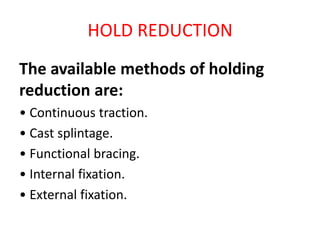
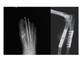
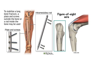
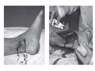
Treatment of fractures
Treat the patient ,not only the
fracture
1/ABC.
2/ MANAGE MORE SERIOUS INJURIES.
3/LIMB INJURY MANAGEMENT ACCORDING TO
PRIORITIES (BONE AND JOINT IS THE THIRD
PRIORITY AFTER VASCULAR AND SOFT TISSUES
Treatment of fracture
1/early measures(adopted for all fractures)
-ice packs
-elevation to decrease the edema
-analgesics , avoid narcotics
-temporary immobilization to
decrease pain, prevent soft tissue injury and
facilitate patient transfer.
2/definitive treatment (reduction + hold + exercise)
-it is either conservative or operative
• Reduce.(closed or open reduction)
• Hold.(till fracture union)
• Exercise.,(of the non immobilized joints,
including weight bearing
HOLD VERSUS MOVE
SPEED VERSUS SAFETY
REDUCTION
reduction is unnecessary in :
(1) when there is little or no displacement.
(2) when displacement does not matter initially
(e.g. in fractures of the clavicle) .
(3) when reduction is unlikely to succeed
(e.g. with compression fractures of the
vertebrae).
Reduction should aim for :
1/adequate apposition:
-gap may cause delayed union and non union,
the greater contact area the faster union
2/normal alignment of the bone fragments:
-some types of mal alignment is acceptable in
some bones(like clavicle) and in children due to
remodelling, but not the rotation.
CLOSED REDUCTION
Under appropriate anaesthesia and muscle relaxation:-
three-fold manoeuvre:
(1) the distal part of the limb is pulled in the line of the
Bone.(traction and counter traction)
(2) as the fragments disengage, they are repositioned
(by reversing the original direction of force if
this can be reduced)
(3) alignment is adjusted in each plane.
Closed reduction is used for:
1/ all minimally displaced fractures.
2/ for most fractures in children
3/ for fractures that are stable after reduction and can
be held in some form of splint or cast.
4/Unstable fractures can also be reduced using
closed methods prior to stabilization with internal or
external fixation. (eg. percut. Wiring)
This avoids direct manipulation of the fracture site by
open reduction, which damages the local blood supply
and may lead to slower healing times; increasingly
OPEN REDUCTION
(1) when closed reduction fails, either because of
difficulty in controlling the fragments or
because soft tissues are interposed between them.
(2) when there is a large articular fragment that
needs accurate positioning .
(3) for traction (avulsion) fractures in which the
fragments are held apart..
HOLD REDUCTION
The available methods of holding
reduction are:
• Continuous traction.
• Cast splintage.
• Functional bracing.
• Internal fixation.
• External fixation.
CONTINUOUS TRACTION
-Traction is safe enough, provided it is not
excessive
-The problem is speed: not because the fracture
unite slowly (it does not) but because lower limb
traction keeps the patient in hospital.
-as soon as the fracture is ‘sticky’ (deformable
but not displaceable), traction should be
replaced by bracing.
It is a method for reduction and
holding the fracture
Types of traction
• 1/Traction by gravity – This applies only to
upper limb injuries. with a wrist sling the weight
of the arm provides continuous traction to the
humerus.
2/Skin traction – Skin traction will sustain a pull of
no more than 4 or 5 kg. strapping or one way
stretch Elastoplast is stuck to the shaved skin and
held on with a bandage. The malleoli are protected
, and cords or tapes are used for traction
3/Skeletal traction –
-A stiff wire or pin is inserted usually behind the
tibial tubercle for hip, thigh and knee injuries, or
through the calcaneum for tibial fractures – and
cords tied to them for applying traction.
Fixed traction
-The pull is exerted against a fixed point.
-The usual method is to tie the traction cords to
the distal end of a Thomas’ splint and pull the
leg down until the proximal padded ring of the
splint abuts firmly against the pelvis.
Balanced traction
- The traction cords are guided over pulleys at
the foot of the bed and loaded with weights; --
counter-traction is provided by the weight of the
body when the foot of the bed is raised.
Complications of traction
1/Circulatory embarrassment In children especially,
traction tapes and circular bandages may constrict the
circulation;
2/Nerve injury In older people, leg traction may
predispose to peroneal nerve injury and cause a
dropfoot;
the limb should be checked repeatedly
3/Pin site infection Pin sites must be kept clean and
should be checked daily.
4/skin laceration especially in lax skin in elderly people.
CAST SPLINTAGE
-Plaster of Paris is still widely used as a splint.
-It is safe enough, so long as one is alert to the
danger of a tight cast and provided pressure
sores are prevented.
-The speed of union is neither greater nor less
than with traction, but the patient can go home
sooner.
-joints encased in plaster cannot move and are
liable to stiffen;
-While the swelling and haematoma resolve,
adhesions may form that bind muscle fibers to
each other and to the bone.
-Newer substitutes have some advantage over
plaster (they are impervious to water, and also
lighter and more radiolucent.)
Stiffness can be minimized by:
(1) delayed splintage that is, by using traction
until movement has been regained, and only
then applying plaster.
(2) starting with a conventional cast but, after a
few weeks, when the limb can be handled
without too much discomfort, replacing the cast
by a functional brace which permits joint
movement.
Complications
1/Tight cast :
-The cast may be put on too tightly, or it may
become tight if the limb swells. The patient complains
of diffuse pain; only later – sometimes much later – do
the signs of vascular compression appear. The limb
should be elevated, but if the pain persists, the only safe
course is to split the cast and ease it open:
(1) throughout its length and (2) through all the padding
down to skin.
2/Pressure sores
-Even a well-fitting cast may press upon the skin
over a bony prominence (the patella, heel,
elbow or head of the ulna).
-The patient complains of localized pain
precisely over the pressure spot.
-Such localized pain demands immediat
inspection through a window in the cast
3/Skin abrasion or laceration
-This is really a complication of removing plasters,
especially if an electric saw is used.
4/Loose cast
-Once the swelling has subsided, the cast
may no longer hold the fracture securely. If it is
loose,the cast should be replaced.
INTERNAL FIXATION
-Bone fragments may be fixed with screws, a
metal plate held by screws, a long intramedullary
rod or nail (with or without locking screws),
circumferential bands or a combination of these
methods.
-Properly applied, internal fixation holds a
fracture securely so that movement can begin at
once; with early movement the ‘fracture disease’
(stiffness and edema) is abolished.
-As far as speed is concerned, the patient can
leave hospital as soon as the wound is healed,
but he must remember that, even though the
bone moves in one piece, the fracture is not
united – it is merely held by a metal bridge and
unprotected weight bearing is, for some time,
unsafe
Safety..The greatest danger is sepsis
-if infection supervenes, all the manifest advantages
of internal fixation(precise reduction, immediate
stability and early movement) may be lost.
-The risk of infection depends upon:
(1) the patient – devitalized tissues, a dirty wound
and an unfit patient are all dangerous.
(2) the surgeon – thorough training, a high degree of
surgical dexterity and adequate assistance are all
essential
(3) the facilities – a guaranteed aseptic routine, a
full range of implants and staff familiar with their use
are all important.
Indications of internal fixation
1. Fractures that cannot be reduced except by
operation.
2. Fractures that are inherently unstable and prone
to re-displace after reduction (e.g. mid-shaft
fractures of the forearm and some displaced ankle
fractures).
- Also included are those avulsion fractures liable
to be pulled apart by muscle action (e.g. transverse
fracture of the patella or olecranon).
3. Fractures that unite poorly and slowly, principally
fractures of the femoral neck.
4. Pathological fractures in which bone disease may
prevent healing.
5. Multiple fractures where early fixation (by either
internal or external fixation) reduces the risk of
general complications and late multisystem organ
failure
6. Fractures in patients who present nursing
difficulties (paraplegics, those with multiple injuries
and the very elderly).
Complications of internal fixation
1/Infection
Iatrogenic infection is now the most common cause of chronic
osteomyelitis, the metal does not predispose to infection but
the operation and quality of the patient’s tissues do.
2/Non-union
-If the bones have been fixed rigidly with a gap between the
ends, the fracture may fail to unite.
-This is more likely in the leg or the forearm if one
bone is fractured and the other remains intact.
-Other causes of non-union are stripping of the soft tissues
and damage to the blood supply in the course of operative
fixation.
3/Implant failure
-Metal is subject to fatigue and can fail unless some union of
the fracture has occurred.
-Stress must therefore be avoided and a patient with a broken
tibia internally fixed should walk with crutches and stay away
from partial weight bearing for 6 weeks or longer, until callus
or other radiological sign of fracture healing is seen on x-ray.
4/Refracture
-It is important not to remove metal implants too soon, or the
bone may refracture.
-A year is the minimum and 18 or 24 months safer; for several
weeks after removal the bone is weak, and care or protection
is needed.
EXTERNAL FIXATION
Indications
1. Fractures associated with severe soft-tissue damage
(including open fractures) or those that are contaminated,
where internal fixation is risky
2. Fractures around joints that are potentially suitable for
internal fixation but the soft tissues are too swollen to allow
safe surgery; here, a spanning external fixator provides
stability until soft-tissue conditions improve.
3. Patients with severe multiple injuries, especially if
there are bilateral femoral fractures, pelvic fractures
with severe bleeding, and those with limb and
associated chest or head injuries.
4. non united fractures, which can be excised and
compressed; sometimes this is combined with bone
lengthening to replace the excised segment.
5. Infected fractures, for which internal fixation might
not be suitable.
The principle of external fixation is
simple
• :
• -the bone is transfixed above and below the
fracture with screws or tensioned wires and
these are then connected to each other by
rigid bars.
• -There are numerous types of external fixation
devices; they vary in the technique of
application
• -Most of them permit adjustment of length
and alignment after application on the limb.
Complications
1/Damage to soft-tissue structures Transfixing pins or
wires may injure nerves or vessels.
2/ joint stiffness pins may tether ligaments and muscles
and inhibit joint movement..
3/Overdistraction If there is no contact between the
fragments, union is unlikely.
4/Pin-track infection is the most common complication
,but can be less likely with good
operative technique.
EXERCISE
It is essential for :
1/decrease edema.
2/prevent joint stiffness
3/prevent muscle wasting.
4/promote fracture union.
5/enhance vascularity
Open fractures
Four golden rules:
•1/ Antibiotic prophylaxis.
•2/ Urgent wound and fracture debridement.
•3/ Stabilization of the fracture.
•4/ Early definitive wound cover.
1/Sterility and antibiotic cover
-co-amoxiclav or cefuroxime (or clindamycin if
penicillin allergy) is given as soon as possible.
-cover the fracture with sterile dressing.
-
2/Debridement
-irrigation with saline aids for
removing foreign bodies,
decrease bacteria count and
prevent tissue dryness.
-wound excision removing any suspected dead
tissues , skin , muscles , bones and nerves and
tendon.
-may need wound extension, especially high
energy open fractures
Fracture stabilization
1/ internal fixation can be used if we are sure of
clean wound,usually after 5-7 days after
negative wound swab for bacteria culture.
2/ otherwise and especially in Gustilo type III
it is safer to use external fixation
4/definitive wound cover
After being sure of clean wound:
1/tissue undermining and direct suturing.
2/skin graft
3/skin flap
4/myocutaneous flap
introduccccccccccccccccccccccccction 2.pptx

introduccccccccccccccccccccccccction 2.pptx

  • 1.
    Treatment of fractures Treatthe patient ,not only the fracture
  • 2.
    1/ABC. 2/ MANAGE MORESERIOUS INJURIES. 3/LIMB INJURY MANAGEMENT ACCORDING TO PRIORITIES (BONE AND JOINT IS THE THIRD PRIORITY AFTER VASCULAR AND SOFT TISSUES
  • 3.
    Treatment of fracture 1/earlymeasures(adopted for all fractures) -ice packs -elevation to decrease the edema -analgesics , avoid narcotics -temporary immobilization to decrease pain, prevent soft tissue injury and facilitate patient transfer. 2/definitive treatment (reduction + hold + exercise) -it is either conservative or operative
  • 4.
    • Reduce.(closed oropen reduction) • Hold.(till fracture union) • Exercise.,(of the non immobilized joints, including weight bearing
  • 5.
  • 6.
    REDUCTION reduction is unnecessaryin : (1) when there is little or no displacement. (2) when displacement does not matter initially (e.g. in fractures of the clavicle) . (3) when reduction is unlikely to succeed (e.g. with compression fractures of the vertebrae).
  • 7.
    Reduction should aimfor : 1/adequate apposition: -gap may cause delayed union and non union, the greater contact area the faster union 2/normal alignment of the bone fragments: -some types of mal alignment is acceptable in some bones(like clavicle) and in children due to remodelling, but not the rotation.
  • 8.
    CLOSED REDUCTION Under appropriateanaesthesia and muscle relaxation:- three-fold manoeuvre: (1) the distal part of the limb is pulled in the line of the Bone.(traction and counter traction) (2) as the fragments disengage, they are repositioned (by reversing the original direction of force if this can be reduced) (3) alignment is adjusted in each plane.
  • 10.
    Closed reduction isused for: 1/ all minimally displaced fractures. 2/ for most fractures in children 3/ for fractures that are stable after reduction and can be held in some form of splint or cast. 4/Unstable fractures can also be reduced using closed methods prior to stabilization with internal or external fixation. (eg. percut. Wiring) This avoids direct manipulation of the fracture site by open reduction, which damages the local blood supply and may lead to slower healing times; increasingly
  • 12.
    OPEN REDUCTION (1) whenclosed reduction fails, either because of difficulty in controlling the fragments or because soft tissues are interposed between them. (2) when there is a large articular fragment that needs accurate positioning . (3) for traction (avulsion) fractures in which the fragments are held apart..
  • 13.
    HOLD REDUCTION The availablemethods of holding reduction are: • Continuous traction. • Cast splintage. • Functional bracing. • Internal fixation. • External fixation.
  • 14.
  • 15.
    -Traction is safeenough, provided it is not excessive -The problem is speed: not because the fracture unite slowly (it does not) but because lower limb traction keeps the patient in hospital. -as soon as the fracture is ‘sticky’ (deformable but not displaceable), traction should be replaced by bracing.
  • 16.
    It is amethod for reduction and holding the fracture
  • 17.
    Types of traction •1/Traction by gravity – This applies only to upper limb injuries. with a wrist sling the weight of the arm provides continuous traction to the humerus.
  • 18.
    2/Skin traction –Skin traction will sustain a pull of no more than 4 or 5 kg. strapping or one way stretch Elastoplast is stuck to the shaved skin and held on with a bandage. The malleoli are protected , and cords or tapes are used for traction
  • 19.
    3/Skeletal traction – -Astiff wire or pin is inserted usually behind the tibial tubercle for hip, thigh and knee injuries, or through the calcaneum for tibial fractures – and cords tied to them for applying traction.
  • 20.
    Fixed traction -The pullis exerted against a fixed point. -The usual method is to tie the traction cords to the distal end of a Thomas’ splint and pull the leg down until the proximal padded ring of the splint abuts firmly against the pelvis.
  • 21.
    Balanced traction - Thetraction cords are guided over pulleys at the foot of the bed and loaded with weights; -- counter-traction is provided by the weight of the body when the foot of the bed is raised.
  • 23.
    Complications of traction 1/Circulatoryembarrassment In children especially, traction tapes and circular bandages may constrict the circulation; 2/Nerve injury In older people, leg traction may predispose to peroneal nerve injury and cause a dropfoot; the limb should be checked repeatedly 3/Pin site infection Pin sites must be kept clean and should be checked daily. 4/skin laceration especially in lax skin in elderly people.
  • 24.
    CAST SPLINTAGE -Plaster ofParis is still widely used as a splint. -It is safe enough, so long as one is alert to the danger of a tight cast and provided pressure sores are prevented. -The speed of union is neither greater nor less than with traction, but the patient can go home sooner. -joints encased in plaster cannot move and are liable to stiffen;
  • 25.
    -While the swellingand haematoma resolve, adhesions may form that bind muscle fibers to each other and to the bone. -Newer substitutes have some advantage over plaster (they are impervious to water, and also lighter and more radiolucent.)
  • 26.
    Stiffness can beminimized by: (1) delayed splintage that is, by using traction until movement has been regained, and only then applying plaster. (2) starting with a conventional cast but, after a few weeks, when the limb can be handled without too much discomfort, replacing the cast by a functional brace which permits joint movement.
  • 29.
    Complications 1/Tight cast : -Thecast may be put on too tightly, or it may become tight if the limb swells. The patient complains of diffuse pain; only later – sometimes much later – do the signs of vascular compression appear. The limb should be elevated, but if the pain persists, the only safe course is to split the cast and ease it open: (1) throughout its length and (2) through all the padding down to skin.
  • 30.
    2/Pressure sores -Even awell-fitting cast may press upon the skin over a bony prominence (the patella, heel, elbow or head of the ulna). -The patient complains of localized pain precisely over the pressure spot. -Such localized pain demands immediat inspection through a window in the cast
  • 31.
    3/Skin abrasion orlaceration -This is really a complication of removing plasters, especially if an electric saw is used. 4/Loose cast -Once the swelling has subsided, the cast may no longer hold the fracture securely. If it is loose,the cast should be replaced.
  • 32.
    INTERNAL FIXATION -Bone fragmentsmay be fixed with screws, a metal plate held by screws, a long intramedullary rod or nail (with or without locking screws), circumferential bands or a combination of these methods. -Properly applied, internal fixation holds a fracture securely so that movement can begin at once; with early movement the ‘fracture disease’ (stiffness and edema) is abolished.
  • 35.
    -As far asspeed is concerned, the patient can leave hospital as soon as the wound is healed, but he must remember that, even though the bone moves in one piece, the fracture is not united – it is merely held by a metal bridge and unprotected weight bearing is, for some time, unsafe
  • 36.
    Safety..The greatest dangeris sepsis -if infection supervenes, all the manifest advantages of internal fixation(precise reduction, immediate stability and early movement) may be lost. -The risk of infection depends upon: (1) the patient – devitalized tissues, a dirty wound and an unfit patient are all dangerous. (2) the surgeon – thorough training, a high degree of surgical dexterity and adequate assistance are all essential (3) the facilities – a guaranteed aseptic routine, a full range of implants and staff familiar with their use are all important.
  • 38.
    Indications of internalfixation 1. Fractures that cannot be reduced except by operation. 2. Fractures that are inherently unstable and prone to re-displace after reduction (e.g. mid-shaft fractures of the forearm and some displaced ankle fractures). - Also included are those avulsion fractures liable to be pulled apart by muscle action (e.g. transverse fracture of the patella or olecranon).
  • 39.
    3. Fractures thatunite poorly and slowly, principally fractures of the femoral neck. 4. Pathological fractures in which bone disease may prevent healing. 5. Multiple fractures where early fixation (by either internal or external fixation) reduces the risk of general complications and late multisystem organ failure 6. Fractures in patients who present nursing difficulties (paraplegics, those with multiple injuries and the very elderly).
  • 41.
    Complications of internalfixation 1/Infection Iatrogenic infection is now the most common cause of chronic osteomyelitis, the metal does not predispose to infection but the operation and quality of the patient’s tissues do. 2/Non-union -If the bones have been fixed rigidly with a gap between the ends, the fracture may fail to unite. -This is more likely in the leg or the forearm if one bone is fractured and the other remains intact. -Other causes of non-union are stripping of the soft tissues and damage to the blood supply in the course of operative fixation.
  • 42.
    3/Implant failure -Metal issubject to fatigue and can fail unless some union of the fracture has occurred. -Stress must therefore be avoided and a patient with a broken tibia internally fixed should walk with crutches and stay away from partial weight bearing for 6 weeks or longer, until callus or other radiological sign of fracture healing is seen on x-ray. 4/Refracture -It is important not to remove metal implants too soon, or the bone may refracture. -A year is the minimum and 18 or 24 months safer; for several weeks after removal the bone is weak, and care or protection is needed.
  • 43.
  • 44.
    Indications 1. Fractures associatedwith severe soft-tissue damage (including open fractures) or those that are contaminated, where internal fixation is risky 2. Fractures around joints that are potentially suitable for internal fixation but the soft tissues are too swollen to allow safe surgery; here, a spanning external fixator provides stability until soft-tissue conditions improve.
  • 45.
    3. Patients withsevere multiple injuries, especially if there are bilateral femoral fractures, pelvic fractures with severe bleeding, and those with limb and associated chest or head injuries. 4. non united fractures, which can be excised and compressed; sometimes this is combined with bone lengthening to replace the excised segment. 5. Infected fractures, for which internal fixation might not be suitable.
  • 46.
    The principle ofexternal fixation is simple • : • -the bone is transfixed above and below the fracture with screws or tensioned wires and these are then connected to each other by rigid bars. • -There are numerous types of external fixation devices; they vary in the technique of application • -Most of them permit adjustment of length and alignment after application on the limb.
  • 47.
    Complications 1/Damage to soft-tissuestructures Transfixing pins or wires may injure nerves or vessels. 2/ joint stiffness pins may tether ligaments and muscles and inhibit joint movement.. 3/Overdistraction If there is no contact between the fragments, union is unlikely. 4/Pin-track infection is the most common complication ,but can be less likely with good operative technique.
  • 48.
    EXERCISE It is essentialfor : 1/decrease edema. 2/prevent joint stiffness 3/prevent muscle wasting. 4/promote fracture union. 5/enhance vascularity
  • 49.
    Open fractures Four goldenrules: •1/ Antibiotic prophylaxis. •2/ Urgent wound and fracture debridement. •3/ Stabilization of the fracture. •4/ Early definitive wound cover.
  • 50.
    1/Sterility and antibioticcover -co-amoxiclav or cefuroxime (or clindamycin if penicillin allergy) is given as soon as possible. -cover the fracture with sterile dressing. -
  • 51.
    2/Debridement -irrigation with salineaids for removing foreign bodies, decrease bacteria count and prevent tissue dryness. -wound excision removing any suspected dead tissues , skin , muscles , bones and nerves and tendon. -may need wound extension, especially high energy open fractures
  • 53.
    Fracture stabilization 1/ internalfixation can be used if we are sure of clean wound,usually after 5-7 days after negative wound swab for bacteria culture. 2/ otherwise and especially in Gustilo type III it is safer to use external fixation
  • 54.
    4/definitive wound cover Afterbeing sure of clean wound: 1/tissue undermining and direct suturing. 2/skin graft 3/skin flap 4/myocutaneous flap