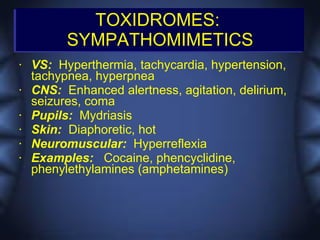
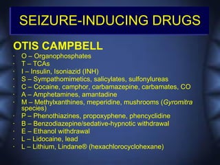
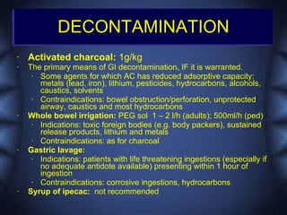
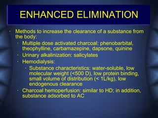
Toxicologic
   Emergencies


Emergency Medicine Clerkship Lecture Series
             Primary Authors:
 Michael Levine, MD, Susan E. Farrell, MD
     Reviewer: Michael Beeson, MD
EPIDEMIOLOGY
•   In 2004, more than 2.4 million toxic
    exposures reported to U.S. Poison
    Control Centers
    •   1183 deaths
•   Over half of poisonings occur in
    children under 5 years of age
EVALUATION OF THE
            POISONED PATIENT
•   History
•   Physical Exam
    •   Vital signs
    •   Pupil exam
    •   Skin findings
    •   Mental status
    •   Search for a toxidrome
MANAGEMENT OF THE
            POISONED PATIENT
•   A-B-C-D-E’s: ACLS measures as appropriate
•   IV, O2, cardiac monitoring, ECG
•   Determine blood glucose in all “intoxicated”
    patients. (Empiric dextrose administration is indicated for all
    patients with altered mental status if bedside glucose determination
    is not available)
•   Thiamine and naloxone empirically as indicated
•   Decontamination
•   Enhanced elimination
•   Antidotal therapy
•   Supportive care
HISTORY
•   Name and amount of agent(s)
•   Type of agent (immediate release, sustained
    release)
•   Time of ingestion/exposure
•   Route of ingestion/exposure
•   Any co-ingestants (including prescription, OTC’s,
    recreational drugs, herbals, chemicals, metals)
•   Reason for ingestion/exposure (e.g. accident,
    suicide attempt, therapeutic misuse,
    occupational)
•   Search exposure environment for pill bottles,
    drug paraphernalia, suicide note, chemical
    containers
PHYSICAL EXAM: VITAL SIGNS
•   Assess and manage the A-B-Cs:
•   Blood pressure
•   Heart rate
•   Respiratory rate
    •   Tachypnea: Salicylates
    •   Bradypnea: Opioids
•   Respiratory depth
    •   Hyperpnea: Salicylates
    •   Shallow respirations: Opioids
•   Temperature
    •   Hyperthermia: Serotonin syndrome, NMS, malignant
        hyperthermia, anti-cholinergic toxidromes, salicylates
    •   Hypothermia: Narcotic or sedative-hypnotic agents
PHYSICAL EXAM: PUPILS
•   Size
    •  Large: Anticholinergic or
       sympathomimetic toxidrome
     • Small: Cholinergic toxidrome

     • Pinpoint: Opioid toxidrome

•   Nystagmus: Check for horizontal,
    vertical, or rotatory (ethanol, phenytoin,
    ketamine, PCP)
PHYSICAL EXAM: SKIN
•   Temperature:
    •   Hyperpyrexia: Anticholinergic or
        sympathomimetic toxidromes,
        salicylates
•   Moisture:
    •   Dry: Anticholinergic toxidrome
    •   Moist: Cholinergic, sympathomimetic
•   Color: Cyanosis, pallor, erythema
PHYSICAL EXAM: OVERALL EXAM
•   Physiologic stimulation: Everything is “up”:
     • Elevated temperature, HR, BP, RR, agitated

       mental status
         • Sympathomimetics, anticholinergics, central

           hallucinogens, some drug withdrawal states
•   Physiologic depression: Everything is “down”:
     • Depressed temperature, HR, BP, RR,

       lethargy/coma
         • Sympatholytics, cholinergics, opioids,

           sedative-hypnotics
•   Mixed effects: Polysubstance overdose,
    metabolic poisons (hypoglycemic agents,
    salicylates, toxic alcohols)
TOXIDROMES
•   Anticholinergic
•   Cholinergic
•   Opioid
•   Sympathomimetic
•   Serotonin syndrome
•   Sympatholytic
•   Sedative-hypnotic
TOXIDROMES:
            ANTICHOLINERGIC
•   VS: Hyperthermia, tachycardia, elevated BP
•   CNS: Agitation, delirium, psychomotor activity,
    hallucinations, mumbling speech, unresponsive
•   Pupils: Mydriasis (minimally reactive to light)
•   Skin: Dry, warm, and flushed
•   GI/GU: Diminished BS, ileus, urinary retention
•   Examples: Atropine, antihistamines, CADs,
    cyclobenzaprine, phenothiazines, Datura spp.
•   Remember: “Dry as a bone, Red as a beet,
    Blind as a bat, Mad as a hatter, and hotter
    than hell”
TOXIDROMES:
                 CHOLINERGIC
•   VS: Bradycardia, high or low BP, tachypnea or
    bradypnea
•   CNS: Agitation, confusion, seizures, coma
•   Pupils: Miosis, eye pain, lacrimation
•   Skin: Diaphoresis
•   GI/GU: Salivation, vomiting, diarrhea, incontinence
•   Musculoskeletal: muscle fasciculations, weakness,
    paralysis
•   Examples: Organophosphate and carbamate
    insecticides, nerve agents, cholinesterase inhibitors
    (physostigmine, edrophonium), nicotine
•   Remember: “SLUDGE” Salivation, Lacrimation,
    Urinary incontinence, diarrhea, Gastrointestinal emesis
TOXIDROMES:
                  OPIOID
•   VS: Hypothermia, bradycardia, normal or low
    BP, bradypnea
•   CNS: Lethargy, coma
•   Pupils: Miosis (exceptions: meperidine, DXM)
•   Skin: Cool, pale or moist, evidence of recent or
    remote needle injection possible
•   Misc: Hyporeflexia, pulmonary edema, seizures
    (meperidine and propoxyphene), ventricular
    dysrhythmias (propoxyphene)
•   Examples: Morphine and the synthetic opioids;
    (Note: clonidine can look like an opioid)
TOXIDROMES:
          SEDATIVE-HYPNOTIC
•   VS: Hypothermia, normal or bradycardic HR,
    hypotension, bradypnea
•   CNS: Drowsiness, dysarthria, ataxia, lethargy,
    coma
•   Pupils: Midsize or miosis, nystagmus
•   Misc: Hyporeflexia; (possible breath odors)
•   Examples: Alcohols, benzodiazepines,
    barbiturates, zolpidem, chloral hydrate,
    ethchlorvynol
TOXIDROMES:
       SEROTONIN SYNDROME
•   VS: Hyperthermia, tachycardia, hypertension,
    tachypnea
•   CNS: Confusion, agitation, lethargy, coma
•   Pupils: Mydriasis
•   Skin: Diaphoretic, flushed
•   Neuromuscular: Hyperreflexia, tremor, clonus,
    rigidity
•   Examples: Combinations that increase 5-HT
    stimulation (MAOIs, SSRIs, NSRIs, meperidine,
    L-tryptophan, dextromethorphan, trazadone,
    linezolid)
TOXIDROMES:
            SYMPATHOLYTICS
•   VS: Bradycardia, hypotension, bradypnea,
    hypopnea
•   CNS: Normal, lethargy, coma, seizures
•   Pupils: Mid size to miotic
•   Examples: Alpha1-adrenergic antagonists,
    beta-adrenergic antagonists, alpha2-adrenergic
    agonists, calcium channel blockers
TOXIDROMES:
          SYMPATHOMIMETICS
•   VS: Hyperthermia, tachycardia, hypertension,
    tachypnea, hyperpnea
•   CNS: Enhanced alertness, agitation, delirium,
    seizures, coma
•   Pupils: Mydriasis
•   Skin: Diaphoretic, hot
•   Neuromuscular: Hyperreflexia
•   Examples: Cocaine, phencyclidine,
    phenylethylamines (amphetamines)
SEIZURE-INDUCING DRUGS
OTIS CAMPBELL
•   O – Organophosphates
•   T – TCAs
•   I – Insulin, Isoniazid (INH)
•   S – Sympathomimetics, salicylates, sulfonylureas
•   C – Cocaine, camphor, carbamazepine, carbamates, CO
•   A – Amphetamines, amantadine
•   M – Methylxanthines, meperidine, mushrooms (Gyromitra
    species)
•   P – Phenothiazines, propoxyphene, phencyclidine
•   B – Benzodiazepine/sedative-hypnotic withdrawal
•   E – Ethanol withdrawal
•   L – Lidocaine, lead
•   L – Lithium, Lindane® (hexachlorocyclohexane)
DECONTAMINATION
•   Activated charcoal: 1g/kg
•   The primary means of GI decontamination, IF it is warranted.
     • Some agents for which AC has reduced adsorptive capacity:
       metals (lead, iron), lithium, pesticides, hydrocarbons, alcohols,
       caustics, solvents
     • Contraindications: bowel obstruction/perforation, unprotected
       airway, caustics and most hydrocarbons
•   Whole bowel irrigation: PEG sol 1 – 2 l/h (adults); 500ml/h (ped)
     • Indications: toxic foreign bodies (e.g. body packers), sustained
       release products, lithium and metals
     • Contraindications: as for charcoal
•   Gastric lavage:
     • Indications: patients with life threatening ingestions (especially if
       no adequate antidote available) presenting within 1 hour of
       ingestion
     • Contraindications: corrosive ingestions, hydrocarbons
•   Syrup of ipecac: not recommended
ENHANCED ELIMINATION
•   Methods to increase the clearance of a substance from
    the body:
     • Multiple dose activated charcoal: phenobarbital,
       theophylline, carbamazepine, dapsone, quinine
     • Urinary alkalinization: salicylates
     • Hemodialysis:
         • Substance characteristics: water-soluble, low

           molecular weight (<500 D), low protein binding,
           small volume of distribution (< 1L/kg), low
           endogenous clearance
     • Charcoal hemoperfusion: similar to HD; in addition,
       substance adsorbed to AC
ANTIDOTES
TOXIN                      ANTIDOTE
Acetaminophen              N-Acetylcysteine

Anticholinergic agents     Physostigmine
Benzodiazepines            Flumazenil
Beta blockers or calcium   IV fluids, calcium, glucagon, insulin (HIE)
channel blockers
Carbon monoxide            O2
Cardiac glycosides         Digoxin-specific Fab fragments

Cocaine (or other          Benzodiazepines
sympathomimetics)
Cyanide                    Amyl nitrate, sodium nitrate, sodium
                           thiosulfate, hydroxycobalamin
Ethylene glycol            4-Methylpyrazole, ethanol
ANTIDOTES
TOXIN                       ANTIDOTE
Heparin                     Protamine sulfate
Hydrofluoric acid           Calcium gluconate
Iron                        Desferoxamine
Isoniazid                   Pyridoxine
Lead                        DMSA or BAL/CaNa2-EDTA
Mercury                     BAL
Methanol                    4-Methylpyrazole, ethanol

Opioids                     Naloxone
Organophosphates/           Atropine + pralidoxime
carbamates
Sulfonylureas               Glucose + octreotide
(or meglitinides)
Tricyclic antidepressants   Sodium bicarbonate, benzodiazepines
TOXICOLOGY CASE 1
•   A 23 year old female presents via EMS after
    ingesting 100 tablets of acetaminophen (APAP)
    immediate release preparation, 500mg tablets
•   The ingestion occurred 24 hours ago
•   She has had several episodes of non-bloody,
    non-biliary emesis
•   Serum acetaminophen level drawn on arrival:
    40mg/dL
TOXICOLOGY CASE 1(cont’d)
•   Vital signs: T 98.5˚F, HR: 110 bpm, RR 20,
        BP 110/68, SaO2: 97% on RA
•   Labs include:
     • PT/INR/PTT: 14.2s/1.4; PTT: 80s

     • BUN/Creat: 47mg/dL/1.8mg/dL

     • Serum glucose: 80mg/dL

     • AST: 5,423 IU/L ALT: 6,087 IU/L
APAP TOXICITY
•   Four stages to toxicity:
     • I: 0-24 hours: Asymptomatic, or mild anorexia,
       nausea, vomiting, malaise
     • II: 24-48 hours: Transaminase levels start to rise at
       12 hours; Abdominal pain, RUQ tenderness, vomiting,
       oliguria
     • III: 72-96 hours: Transaminases peak at 72 hours;
       PT rises, multi-system organ failure or recovery
     • IV: 4d-2 weeks: Resolution of hepatotoxicity
•   Toxicity results from accumulation of a toxic metabolite:
    N-acetyl-para-benzoquinoneimine (NAPQI) relative to
    endogenous glutathione
•   Toxic single ingestion is 150 mg/kg
APAP TOXICITY
•   At therapeutic doses:
     • 90% of APAP is conjugated and renally

       excreted
     • 2-4% is metabolized via P450 enzymes to

       NAPQI
     • NAPQI is quickly conjugated to glutathione to

       a non-toxic metabolite
•   In an overdose, glutathione stores are depleted,
    NAPQI accumulates leading to hepatotoxicity
RUMACK-MATTHEW
  NOMOGRAM
N-Acetylcysteine
•   PO dosing: 140 mg/kg load, followed by
    70mg/kg q4h x17 doses
•   IV dosing: 150 mg/kg load over 15 min,
    followed by 50mg/kg over 4 hours,
    followed by 100 mg/kg over 16 hours
•   Prolonging the initial loading period for IV
    NAC may reduce the incidence of
    anaphylactoid reactions
APAP TRANSPLANT
                GUIDELINES
•   King’s College guidelines
    •   pH < 7.3 after fluid resuscitation
           or
    •   PT > 100
    •   Creatinine > 3.4
    •   Grade III or IV encephalopathy
    •   Lactate > 3.5mmol/L
TOXICOLOGY CASE 2
•   A 20 year old male presents via EMS after his
    neighbor found him unresponsive. The patient is
    comatose
•   The neighbor developed a headache and
    nausea after spending 10 minutes in the
    patient’s house
•   It is winter, and the patient had been using a
    camp stove for heat
TOXICOLOGY CASE 2 (cont’d)
•   VS: T: 98.9˚F, HR: 110 bpm, RR: 6,
        BP: 150/100 mmHg, SaO2: 99%.
•   Moans to painful stimuli with no focal neurologic
    deficits
•   Pupils 4mm, sluggishly reactive
•   Skin notable for central cyanosis
•   Blood glucose: 90mg/dL
•   ECG: Sinus tachycardia, normal intervals,no
    evidence of acute ischemia
•   Labs include: COHb: 60%
CO TOXICITY
•   17,115 cases of CO exposure reported to US
    Poison Control Centers in 2004
•   CO is a colorless, odorless, non-irritating gas
•   Sources of CO exposure include:
     • Smoke

     • Car exhaust

     • Propane powered vehicles or engines

     • Hibachi grills and kerosene heaters

     • Methylene chloride
CO TOXICITY
•   CO combines with Hgb to form
    carboxyhemoglobin (COHb)
•   COHb has 240 X the affinity for O2
•   CO + Hgb  shifts the O2 dissociation curve to
    the left: oxygen delivery to tissues is reduced
•   CO can cause hypotension via CO-induced
    cGMP production and increased NO production
•   CO can inhibit electron transport which limits
    ATP production
•   CO is associated with microvascular damage
    and inflammation in the CNS
CLINICAL EFFECTS OF CO

 COHb%         Signs/Symptoms
<5%           None or mild HA
10%           Slight HA, dyspnea on vigorous exertion
20%           Throbbing headache, dyspnea with
              moderate exertion

30%           Severe HA, irritability, fatigue, dim vision
40-50%        Tachycardia, confusion, lethargy, syncope
50-70%        Coma, seizures, death
> 70%         Rapidly fatal
CO TOXICITY
•   CO poisoning is frequently misdiagnosed: symptoms are
    nonspecific
•   Need a high index of suspicion
•   Consider CO poisoning:
     • Multiple patients with similar complaints, especially
       from the same household
     • Vague, flu like symptoms without fever or
       lymphadenopathy
     • Winter, environmental history and exposures
     • Uncommon presentation of syncope
•   Normal COHb levels
     • 0-5% in non-smokers
     • up to 10% in smokers > 1ppd
PULSE OXIMETRY
•   Noninvasive measure of functional
    hemoglobin oxygen saturation
•   Does not measure hemoglobin
    species that cannot carry oxygen
    •   MetHb
    •   COHb
•   Co-oximeter measures fractional
    hemoglobin oxygen saturation
PULSE OXIMETRY GAP
Severe CO poisoning
• Significant dyshemoglobinemia results in a

  divergence between functional and
  fractional hemoglobin oxygen saturation
• In patients with markedly elevated COHb

  levels, pulse oximetry can overestimate
  O2Hb%
•   In severe CO poisoning, the pulse
    oximetry gap approaches the COHb level
CO TREATMENT
•   Oxygen!!
•   The half life of COHb decreases with
    inspired O2 concentration:
    •   t1/2 at room air: 4-6 hours
    •   t1/2 at “100%” O2 via NRB at 1 ATM: 90 min
    •   t1/2 at 100% O2 via ETT at 1 ATM: 60 min
    •   t1/2 at 100% O2 at 3 ATM (HBO): 23 minutes
HYPERBARIC OXYGEN
•   The rationale behind HBO therapy for CO:
    •   Decrease the incidence of delayed neurologic
        sequelae
    •   Should be started within 6 hours
•   HBO indications are controversial, but generally
    include:
    •   COHgb > 25-40%
    •   Altered Mental Status or history of same (syncope)
    •   Arrhythmias
    •   Symptoms of cardiac ischemia
    •   COHgb > 15% if pregnant
TOXICOLOGY CASE 3
•   A 22 year old male brought via EMS after being
    found “drunk.” He was found near an empty
    bottle of window-washer fluid
•   The patient had threatened suicide earlier in the
    day
TOXICOLOGY CASE 3 (cont’d)
•   Labs include:
     • Serum glucose: 124 mg/dL

     • Sodium: 130 mEq/L; K: 3.7 mEq/L; Cl: 88

       mEq/L; Bicarbonate: 12 mEq/L; BUN: 22
       mg/dL; Creatinine 1.5 mg/dL
     • Anion gap of 30

     • Serum ethanol: non-detectable

     • Serum APAP/ASA: non-detectable

     • Serum osmolality: 324 mOsm/kg
TOXIC ALCOHOLS
•   Most commonly: methanol, isopropanol,
    and ethylene glycol (EG)
•   Should be suspected based on:
     • history, physical exam, lab

       abnormalities
•   The degree of intoxication correlates with
    the number of carbons in the alcohol:
     • Methanol < ethanol or ethylene glycol

       < isopropanol
TOXIC ALCOHOL LABS
•   All toxic alcohols cause an osmolar gap
•   Methanol and EG cause an increased anion gap
    acidosis
•   Isopropanol causes ketosis without acidosis
•   Osmolar gaps can be present early after
    ingestion, but will be absent after the alcohol is
    metabolized
•   Anion gap acidosis can be absent early after
    ingestion, but will develops after methanol or EG
    metabolism
“GAPS”


                 Anion gap


Gap’s
                   Osmolar gap


         Time
METHANOL
•   Methanol (CH3OH):
    • window-washer fluid, anti-icing agents,

      solvents, varnish/paint removers, some
      anti-freezes
•   Methanol intoxication:
    • “Snow storm” blindness (edema of the

      optic disk/nerve)
    • Abdominal pain, nausea, vomiting

    • Lethargy, coma
METHANOL METABOLISM
                              Methanol
                                Alcohol      dehydrogenase*


                              Formaldehyde
                             Aldehyde         dehydrogenase


                              Formic acid
                                 Folate

                              CO2 + H2O
* Inhibited by 4-methylpyrazole or ethanol
ISOPROPANOL
•   Isopropanol (CH3-CHOH-CH3):
    •   The most intoxicating alcohol
    •   Osmolar gap, followed by ketosis
    •   Metabolized to acetone by alcohol
        dehydrogenase
ETHYLENE GLYCOL
•   Ethylene glycol C(OH2) – C(OH2) sources:
    •   Antifreeze, brake fluid, anti-icing solutions, solvents
•   If fluorescein has been added to an EG-
    containing antifreeze, the patient’s urine may
    fluoresce under Wood’s lamp
•   Metabolized to:
    •   Glycolic acid: anion gap acidosis
    •   Oxalic acid, combines with calcium, causing calcium
        oxylate crystal deposition and hypocalcemia
•   Calcium oxylate deposition in the renal tubules
    causes acute renal failure
ETHYLENE GLYCOL
                    METABOLISM
                               Ethylene glycol
                               Alcohol      dehydrogenase*


                               Glycoaldehyde
                               Aldehyde     dehydrogenase


                               Glycolic acid
                               Lactate       dehydrogenase


                               Glyoxylic acid
              Pyridoxine, Mg                          Thiamine

   Glycine +                                                  α-OH-β-
   Benzoic acid                Oxalic acid                    ketoadipic acid
*Inhibited by 4-methylpyrazole or ethanol

Pyridoxine, Mg, and thiamine are co-factors for their respective reactions
TREATMENT
•   Methanol or EG: 4-methyl-pyrazole (4-MP,
    fomepizole)
•   4-MP inhibits alcohol dehydrogenase activity
•   Ethanol also competes for active sites on alcohol
    dehydrogenase and inhibits methanol and EG
    metabolism
•   Potential adverse effects of ethanol infusion:
    •   Intoxication, hypotension, pancreatitis, gastritis,
        hypoglycemia, or phlebitis
•   Hemodialysis clears the toxic alcohol and
    corrects acid/base abnormalities
TREATMENT (cont’d)
•   EG: Other cofactors to enhance nontoxic
    metabolism:
    •   thiamine, pyridoxine, magnesium
•   Methanol: Other cofactors to enhance
    nontoxic metabolism:
    •   folic acid (or folinic acid)
•   Treatment of Isopropanol ingestion:
    •   Supportive care
    •   H2 blockers or proton-pump inhibitors
    •   Ensure that no other toxic alcohol is present
TOXICOLOGY CASE 4
•   A 3 year old male is brought by his parents 1
    hour after he is found with one of his
    grandmother’s sustained – release verapamil
    tablets in his mouth
•   A pill count shows 1 additional tablet might be
    missing
•
    The child is asymptomatic
TOXICOLOGY CASE 4 (cont’d)
•   Vital signs: T: 98.6˚F, HR: 80 bpm, RR: 22,
    BP:100/60, SaO2: 99%
•   Initial labs:
     •   Na: 140 mEq/L; K: 3.7 mEq/L; Cl: 113 mEq/L;
         Bicarbonate: 22 mEq/L; BUN: 12 mg/dL; Creatinine
         0.8 mg/dL. Serum glucose: 120mg/dL
     •   ECG: normal sinus rhythm, normal intervals.
•   Two hours later: the patient is less arousable
•   Vital signs: HR: 50 bpm, RR: 18, BP: 70/40
    SaO2: 99%
     •   ECG: junctional bradycardia, normal QRS and QTc
         intervals
     •   Serum glucose: 190 mg/dL
CALCIUM CHANNEL BLOCKER
              (CCB)
•   Classes of CCB approved in the US:
     • Phenylalkylamines: Verapamil
         •   Verapamil: Effects cardiac myocytes and electrical
             conduction system ( decreased contractility, AV nodal
             conduction delay and block)
    •   Benzothiazepines: Diltiazem
         •   Benzothiazepines: Effects cardiac myocytes, electrical
             conduction system, and peripheral vascular smooth muscle
             cells
    •   Dihydropyridines: Nifedipine, amlodipine, nicardipine
         •   Dihydropyridines: Effects peripheral vascular smooth muscle
             cells ( peripheral vasodilation, decreased peripheral
             vascular resistance)
•   In overdose, the selectivity of the CCB classes may be
    lost
CCB TOXICITY
•   CCBs:
    •   Block L-type calcium channels
    •   Inhibit intracellular calcium influx
•   In overdose:
    •  Verapamil or diltiazem: Bradycardia and hypotension
     • Dihydropyridines: Hypotension and tachycardia
•   Insulin release from pancreatic β-cells depends on L-
    type calcium channels; hyperglycemia can occur after
    CCB overdose
•   The degree of hyperglycemia may correlate with the
    severity of the overdose
CCBs versus BETA BLOCKERS
•   β1 antagonism:
    •   Decreased cardiac contractility
    •   Reduced AV nodal conduction
•   β2 antagonism:
    •   Increased smooth muscle tone…bronchospasm
•   Labetolol:
    •   7:1 β:α antagonist activity
•   Βeta adrenergic antagonists:
    •   Inhibit gluconeogenesis and glycogenolysis
    •   Hypoglycemia can occur in overdose
    •   Seizures can occur in overdose (propranolol)
CCB and BETA BLOCKER
              TREATMENT

•   Ensure ABCs
•   Improve heart rate and blood pressure:
    •   Atropine: Often fails to improve HR
    •   Calcium: Used in both CCB and Beta blocker toxicity;
        Improves HR and contractility
    •   Glucagon: Improves myocardial contractility
    •   Direct α agonist agents: Increase peripheral vascular
        resistance
    •   (Epinephrine has both β1 and α1 agonist effects)
CCB/BETA BLOCKER TX
•   Therapies unique to CCB or β blocker involve:
     • IV fluids – Offsets hypotension induced by peripheral
       vasodilation
     • Calcium – Calcium competitively overcomes blockade
       of the voltage-sensitive calcium channels
     • Glucagon: Acts on adenylate cyclase independently
       of the β receptor to convert ATP into cAMP
     • Epinephrine: Binds to β receptors to convert
       adenylate cyclase into cAMP
     • Insulin: Promotes increased uptake and utilization of
       carbohydrates by cardiac myocytes (primarily used
       only for CCB toxicity
Hyperinsulinemic Euglycemia (HIE)
•   Normally: Cardiac myocytes preferentially
    metabolize glucose; in shock states, metabolism
    is dependent on free fatty acids
•   Hyperinsulinemic euglycemic (HIE) therapy:
    shifts myocardial metabolism from FFA to
    carbohydrates
•   HIE:
    •   Insulin (0.5-1 unit/kg bolus, followed by 0.5-1
        unit/kg/hr)
    •   Dextrose (1 amp D50, or continuous D10 infusion)
    •   Watch for hypokalemia and hypophosphatemia
•   HIE therapy: Associated with rapid, dramatic
    improvement in cardiovascular hemodynamics
CARDIAC GLYCOSIDES
•   Digoxin: A cardiac glycoside used for the
    treatment of CHF and atrial fibrillation
•   Mechanism of action:
    •   Inhibits Na/K/ATPase, leading to:
    •   Increased intracellular sodium/calcium exchange
    •   Increased intracellular calcium
    •   Increased extracellular potassium
•   Digoxin
    •   Increases excitability and automaticity of cardiac
        myocytes
    •   Decreases conduction velocity at the AV node
CARDIAC GLYCOSIDE TOXICITY
•    Cardiac glycosides:
     •   Foxglove, oleander, lily of the valley, red squill
     •   Secretions of Bufo toads (e.g. Colorado river toad)
•   Symptoms of toxicity:
     •   Nausea and vomiting
     •   Weakness, lethargy, confusion
     •   Visual disturbances
•   Acute toxicity:
     •   Serum potassium is elevated, predictive of mortality.
•   Chronic toxicity:
     •   Precipitated by hypokalemia, hypomagnesemia, renal failure
     •   Digoxin toxicity can occur with therapeutic digoxin levels
CARDIAC GLYCOSIDE TOXICITY:
         THE ECG
•   Nearly every dysrhythmia has been associated
    with digoxin toxicity
•   PVCs are the most common ECG abnormality
•   Bidirectional ventricular tachycardia and
    accelerated junctional rhythms with nodal block
    are relatively specific for cardiac glycoside
    toxicity, but are less common
CARDIAC GLYCOSIDE TOXICITY:
        TREATMENT
•   Digoxin-specific Fab fragments indications:
     • Hyperkalemia (K > 5.0)

     • Life-threatening arrhythmias

•   Phenytoin or lidocaine:
     • May suppress ventricular dysrhythmias if

       digoxin-specific Fab is unavailable
•   Correct hypokalemia, hypomagnesemia
•   Calcium therapy for hyperkalemia should be
    avoided with concomitant digoxin toxicity
TOXICOLOGY CASE 5
•   A 42 year old woman presents via EMS after
    she was found unresponsive at home

•   Vital signs: T 99.8˚ F, HR: 121 bpm, RR: 14; BP:
    97/52; SaO2: 93% on RA

•   PE: Disheveled, minimally responsive female;
    pupils: 8 mm, minimally reactive; dry lips and
    mucous membranes; tachycardia, absent bowel
    sounds; skin warm and flushed
TOXICOLOGY CASE 5 (cont’d)
•   The patient is placed on a cardiac monitor and
    IV access is obtained

•   Shortly after an ECG is performed, the patient
    has a brief, generalized tonic-clonic seizure
TCA ingestion


Note the tachycardia, QRS prolongation, tall R wave in aVR, and the rightward
                 deflection of the terminal 40 msec of aVR.
TRICYCLIC ANTIDEPRESSANT
             TOXICITY
•   TCA toxicity:
    •   Sodium channel blockade: conduction delay
    •   Alpha1 adrenergic blockade: hypotension
    •   Cholinergic (muscarinic) blockade: mydriasis, dry
        mucous membranes, tachycardia, ileus, urinary
        retention
    •   Histamine blockade
•   Treatment:
    •   Sodium bicarbonate
    •   Direct alpha1 adrenergic agents as pressors
    •   Benzodiazepines as seizure prophylaxis/treatment
•   NaHCO3 is indicated for any QRS > 100 ms
TRICYCLIC ANTIDEPRESSANT
             TOXICITY
•    The risk of ventricular dysrhythmias and
     seizures correlates with QRS prolongation
•    ECG findings suggestive of TCA toxicity
     include:
       Tachycardia
       Prolonged PR, QRS intervals
       Tall R wave in aVR
       Rightward deflection of terminal 40 msec in aVR
•    NaHCO3 is indicated for any QRS > 100 ms
In Summary
Approach all patients in a systematic
fashion
Toxic exposures most often only require
supportive care
Be aware of toxic exposures that require
specific antidotes
Most toxic exposures are unintentional
Consider contacting a regional poison
control center for all but the most straight
forward cases

Toxicology Emergencies CDEM

  • 1.
    Toxicologic Emergencies Emergency Medicine Clerkship Lecture Series Primary Authors: Michael Levine, MD, Susan E. Farrell, MD Reviewer: Michael Beeson, MD
  • 2.
    EPIDEMIOLOGY • In 2004, more than 2.4 million toxic exposures reported to U.S. Poison Control Centers • 1183 deaths • Over half of poisonings occur in children under 5 years of age
  • 3.
    EVALUATION OF THE POISONED PATIENT • History • Physical Exam • Vital signs • Pupil exam • Skin findings • Mental status • Search for a toxidrome
  • 4.
    MANAGEMENT OF THE POISONED PATIENT • A-B-C-D-E’s: ACLS measures as appropriate • IV, O2, cardiac monitoring, ECG • Determine blood glucose in all “intoxicated” patients. (Empiric dextrose administration is indicated for all patients with altered mental status if bedside glucose determination is not available) • Thiamine and naloxone empirically as indicated • Decontamination • Enhanced elimination • Antidotal therapy • Supportive care
  • 5.
    HISTORY • Name and amount of agent(s) • Type of agent (immediate release, sustained release) • Time of ingestion/exposure • Route of ingestion/exposure • Any co-ingestants (including prescription, OTC’s, recreational drugs, herbals, chemicals, metals) • Reason for ingestion/exposure (e.g. accident, suicide attempt, therapeutic misuse, occupational) • Search exposure environment for pill bottles, drug paraphernalia, suicide note, chemical containers
  • 6.
    PHYSICAL EXAM: VITALSIGNS • Assess and manage the A-B-Cs: • Blood pressure • Heart rate • Respiratory rate • Tachypnea: Salicylates • Bradypnea: Opioids • Respiratory depth • Hyperpnea: Salicylates • Shallow respirations: Opioids • Temperature • Hyperthermia: Serotonin syndrome, NMS, malignant hyperthermia, anti-cholinergic toxidromes, salicylates • Hypothermia: Narcotic or sedative-hypnotic agents
  • 7.
    PHYSICAL EXAM: PUPILS • Size • Large: Anticholinergic or sympathomimetic toxidrome • Small: Cholinergic toxidrome • Pinpoint: Opioid toxidrome • Nystagmus: Check for horizontal, vertical, or rotatory (ethanol, phenytoin, ketamine, PCP)
  • 8.
    PHYSICAL EXAM: SKIN • Temperature: • Hyperpyrexia: Anticholinergic or sympathomimetic toxidromes, salicylates • Moisture: • Dry: Anticholinergic toxidrome • Moist: Cholinergic, sympathomimetic • Color: Cyanosis, pallor, erythema
  • 9.
    PHYSICAL EXAM: OVERALLEXAM • Physiologic stimulation: Everything is “up”: • Elevated temperature, HR, BP, RR, agitated mental status • Sympathomimetics, anticholinergics, central hallucinogens, some drug withdrawal states • Physiologic depression: Everything is “down”: • Depressed temperature, HR, BP, RR, lethargy/coma • Sympatholytics, cholinergics, opioids, sedative-hypnotics • Mixed effects: Polysubstance overdose, metabolic poisons (hypoglycemic agents, salicylates, toxic alcohols)
  • 10.
    TOXIDROMES • Anticholinergic • Cholinergic • Opioid • Sympathomimetic • Serotonin syndrome • Sympatholytic • Sedative-hypnotic
  • 11.
    TOXIDROMES: ANTICHOLINERGIC • VS: Hyperthermia, tachycardia, elevated BP • CNS: Agitation, delirium, psychomotor activity, hallucinations, mumbling speech, unresponsive • Pupils: Mydriasis (minimally reactive to light) • Skin: Dry, warm, and flushed • GI/GU: Diminished BS, ileus, urinary retention • Examples: Atropine, antihistamines, CADs, cyclobenzaprine, phenothiazines, Datura spp. • Remember: “Dry as a bone, Red as a beet, Blind as a bat, Mad as a hatter, and hotter than hell”
  • 12.
    TOXIDROMES: CHOLINERGIC • VS: Bradycardia, high or low BP, tachypnea or bradypnea • CNS: Agitation, confusion, seizures, coma • Pupils: Miosis, eye pain, lacrimation • Skin: Diaphoresis • GI/GU: Salivation, vomiting, diarrhea, incontinence • Musculoskeletal: muscle fasciculations, weakness, paralysis • Examples: Organophosphate and carbamate insecticides, nerve agents, cholinesterase inhibitors (physostigmine, edrophonium), nicotine • Remember: “SLUDGE” Salivation, Lacrimation, Urinary incontinence, diarrhea, Gastrointestinal emesis
  • 13.
    TOXIDROMES: OPIOID • VS: Hypothermia, bradycardia, normal or low BP, bradypnea • CNS: Lethargy, coma • Pupils: Miosis (exceptions: meperidine, DXM) • Skin: Cool, pale or moist, evidence of recent or remote needle injection possible • Misc: Hyporeflexia, pulmonary edema, seizures (meperidine and propoxyphene), ventricular dysrhythmias (propoxyphene) • Examples: Morphine and the synthetic opioids; (Note: clonidine can look like an opioid)
  • 14.
    TOXIDROMES: SEDATIVE-HYPNOTIC • VS: Hypothermia, normal or bradycardic HR, hypotension, bradypnea • CNS: Drowsiness, dysarthria, ataxia, lethargy, coma • Pupils: Midsize or miosis, nystagmus • Misc: Hyporeflexia; (possible breath odors) • Examples: Alcohols, benzodiazepines, barbiturates, zolpidem, chloral hydrate, ethchlorvynol
  • 15.
    TOXIDROMES: SEROTONIN SYNDROME • VS: Hyperthermia, tachycardia, hypertension, tachypnea • CNS: Confusion, agitation, lethargy, coma • Pupils: Mydriasis • Skin: Diaphoretic, flushed • Neuromuscular: Hyperreflexia, tremor, clonus, rigidity • Examples: Combinations that increase 5-HT stimulation (MAOIs, SSRIs, NSRIs, meperidine, L-tryptophan, dextromethorphan, trazadone, linezolid)
  • 16.
    TOXIDROMES: SYMPATHOLYTICS • VS: Bradycardia, hypotension, bradypnea, hypopnea • CNS: Normal, lethargy, coma, seizures • Pupils: Mid size to miotic • Examples: Alpha1-adrenergic antagonists, beta-adrenergic antagonists, alpha2-adrenergic agonists, calcium channel blockers
  • 17.
    TOXIDROMES: SYMPATHOMIMETICS • VS: Hyperthermia, tachycardia, hypertension, tachypnea, hyperpnea • CNS: Enhanced alertness, agitation, delirium, seizures, coma • Pupils: Mydriasis • Skin: Diaphoretic, hot • Neuromuscular: Hyperreflexia • Examples: Cocaine, phencyclidine, phenylethylamines (amphetamines)
  • 18.
    SEIZURE-INDUCING DRUGS OTIS CAMPBELL • O – Organophosphates • T – TCAs • I – Insulin, Isoniazid (INH) • S – Sympathomimetics, salicylates, sulfonylureas • C – Cocaine, camphor, carbamazepine, carbamates, CO • A – Amphetamines, amantadine • M – Methylxanthines, meperidine, mushrooms (Gyromitra species) • P – Phenothiazines, propoxyphene, phencyclidine • B – Benzodiazepine/sedative-hypnotic withdrawal • E – Ethanol withdrawal • L – Lidocaine, lead • L – Lithium, Lindane® (hexachlorocyclohexane)
  • 19.
    DECONTAMINATION • Activated charcoal: 1g/kg • The primary means of GI decontamination, IF it is warranted. • Some agents for which AC has reduced adsorptive capacity: metals (lead, iron), lithium, pesticides, hydrocarbons, alcohols, caustics, solvents • Contraindications: bowel obstruction/perforation, unprotected airway, caustics and most hydrocarbons • Whole bowel irrigation: PEG sol 1 – 2 l/h (adults); 500ml/h (ped) • Indications: toxic foreign bodies (e.g. body packers), sustained release products, lithium and metals • Contraindications: as for charcoal • Gastric lavage: • Indications: patients with life threatening ingestions (especially if no adequate antidote available) presenting within 1 hour of ingestion • Contraindications: corrosive ingestions, hydrocarbons • Syrup of ipecac: not recommended
  • 20.
    ENHANCED ELIMINATION • Methods to increase the clearance of a substance from the body: • Multiple dose activated charcoal: phenobarbital, theophylline, carbamazepine, dapsone, quinine • Urinary alkalinization: salicylates • Hemodialysis: • Substance characteristics: water-soluble, low molecular weight (<500 D), low protein binding, small volume of distribution (< 1L/kg), low endogenous clearance • Charcoal hemoperfusion: similar to HD; in addition, substance adsorbed to AC
  • 21.
    ANTIDOTES TOXIN ANTIDOTE Acetaminophen N-Acetylcysteine Anticholinergic agents Physostigmine Benzodiazepines Flumazenil Beta blockers or calcium IV fluids, calcium, glucagon, insulin (HIE) channel blockers Carbon monoxide O2 Cardiac glycosides Digoxin-specific Fab fragments Cocaine (or other Benzodiazepines sympathomimetics) Cyanide Amyl nitrate, sodium nitrate, sodium thiosulfate, hydroxycobalamin Ethylene glycol 4-Methylpyrazole, ethanol
  • 22.
    ANTIDOTES TOXIN ANTIDOTE Heparin Protamine sulfate Hydrofluoric acid Calcium gluconate Iron Desferoxamine Isoniazid Pyridoxine Lead DMSA or BAL/CaNa2-EDTA Mercury BAL Methanol 4-Methylpyrazole, ethanol Opioids Naloxone Organophosphates/ Atropine + pralidoxime carbamates Sulfonylureas Glucose + octreotide (or meglitinides) Tricyclic antidepressants Sodium bicarbonate, benzodiazepines
  • 23.
    TOXICOLOGY CASE 1 • A 23 year old female presents via EMS after ingesting 100 tablets of acetaminophen (APAP) immediate release preparation, 500mg tablets • The ingestion occurred 24 hours ago • She has had several episodes of non-bloody, non-biliary emesis • Serum acetaminophen level drawn on arrival: 40mg/dL
  • 24.
    TOXICOLOGY CASE 1(cont’d) • Vital signs: T 98.5˚F, HR: 110 bpm, RR 20, BP 110/68, SaO2: 97% on RA • Labs include: • PT/INR/PTT: 14.2s/1.4; PTT: 80s • BUN/Creat: 47mg/dL/1.8mg/dL • Serum glucose: 80mg/dL • AST: 5,423 IU/L ALT: 6,087 IU/L
  • 25.
    APAP TOXICITY • Four stages to toxicity: • I: 0-24 hours: Asymptomatic, or mild anorexia, nausea, vomiting, malaise • II: 24-48 hours: Transaminase levels start to rise at 12 hours; Abdominal pain, RUQ tenderness, vomiting, oliguria • III: 72-96 hours: Transaminases peak at 72 hours; PT rises, multi-system organ failure or recovery • IV: 4d-2 weeks: Resolution of hepatotoxicity • Toxicity results from accumulation of a toxic metabolite: N-acetyl-para-benzoquinoneimine (NAPQI) relative to endogenous glutathione • Toxic single ingestion is 150 mg/kg
  • 26.
    APAP TOXICITY • At therapeutic doses: • 90% of APAP is conjugated and renally excreted • 2-4% is metabolized via P450 enzymes to NAPQI • NAPQI is quickly conjugated to glutathione to a non-toxic metabolite • In an overdose, glutathione stores are depleted, NAPQI accumulates leading to hepatotoxicity
  • 27.
  • 28.
    N-Acetylcysteine • PO dosing: 140 mg/kg load, followed by 70mg/kg q4h x17 doses • IV dosing: 150 mg/kg load over 15 min, followed by 50mg/kg over 4 hours, followed by 100 mg/kg over 16 hours • Prolonging the initial loading period for IV NAC may reduce the incidence of anaphylactoid reactions
  • 29.
    APAP TRANSPLANT GUIDELINES • King’s College guidelines • pH < 7.3 after fluid resuscitation or • PT > 100 • Creatinine > 3.4 • Grade III or IV encephalopathy • Lactate > 3.5mmol/L
  • 30.
    TOXICOLOGY CASE 2 • A 20 year old male presents via EMS after his neighbor found him unresponsive. The patient is comatose • The neighbor developed a headache and nausea after spending 10 minutes in the patient’s house • It is winter, and the patient had been using a camp stove for heat
  • 31.
    TOXICOLOGY CASE 2(cont’d) • VS: T: 98.9˚F, HR: 110 bpm, RR: 6, BP: 150/100 mmHg, SaO2: 99%. • Moans to painful stimuli with no focal neurologic deficits • Pupils 4mm, sluggishly reactive • Skin notable for central cyanosis • Blood glucose: 90mg/dL • ECG: Sinus tachycardia, normal intervals,no evidence of acute ischemia • Labs include: COHb: 60%
  • 32.
    CO TOXICITY • 17,115 cases of CO exposure reported to US Poison Control Centers in 2004 • CO is a colorless, odorless, non-irritating gas • Sources of CO exposure include: • Smoke • Car exhaust • Propane powered vehicles or engines • Hibachi grills and kerosene heaters • Methylene chloride
  • 33.
    CO TOXICITY • CO combines with Hgb to form carboxyhemoglobin (COHb) • COHb has 240 X the affinity for O2 • CO + Hgb  shifts the O2 dissociation curve to the left: oxygen delivery to tissues is reduced • CO can cause hypotension via CO-induced cGMP production and increased NO production • CO can inhibit electron transport which limits ATP production • CO is associated with microvascular damage and inflammation in the CNS
  • 34.
    CLINICAL EFFECTS OFCO COHb% Signs/Symptoms <5% None or mild HA 10% Slight HA, dyspnea on vigorous exertion 20% Throbbing headache, dyspnea with moderate exertion 30% Severe HA, irritability, fatigue, dim vision 40-50% Tachycardia, confusion, lethargy, syncope 50-70% Coma, seizures, death > 70% Rapidly fatal
  • 35.
    CO TOXICITY • CO poisoning is frequently misdiagnosed: symptoms are nonspecific • Need a high index of suspicion • Consider CO poisoning: • Multiple patients with similar complaints, especially from the same household • Vague, flu like symptoms without fever or lymphadenopathy • Winter, environmental history and exposures • Uncommon presentation of syncope • Normal COHb levels • 0-5% in non-smokers • up to 10% in smokers > 1ppd
  • 36.
    PULSE OXIMETRY • Noninvasive measure of functional hemoglobin oxygen saturation • Does not measure hemoglobin species that cannot carry oxygen • MetHb • COHb • Co-oximeter measures fractional hemoglobin oxygen saturation
  • 37.
    PULSE OXIMETRY GAP SevereCO poisoning • Significant dyshemoglobinemia results in a divergence between functional and fractional hemoglobin oxygen saturation • In patients with markedly elevated COHb levels, pulse oximetry can overestimate O2Hb% • In severe CO poisoning, the pulse oximetry gap approaches the COHb level
  • 38.
    CO TREATMENT • Oxygen!! • The half life of COHb decreases with inspired O2 concentration: • t1/2 at room air: 4-6 hours • t1/2 at “100%” O2 via NRB at 1 ATM: 90 min • t1/2 at 100% O2 via ETT at 1 ATM: 60 min • t1/2 at 100% O2 at 3 ATM (HBO): 23 minutes
  • 39.
    HYPERBARIC OXYGEN • The rationale behind HBO therapy for CO: • Decrease the incidence of delayed neurologic sequelae • Should be started within 6 hours • HBO indications are controversial, but generally include: • COHgb > 25-40% • Altered Mental Status or history of same (syncope) • Arrhythmias • Symptoms of cardiac ischemia • COHgb > 15% if pregnant
  • 40.
    TOXICOLOGY CASE 3 • A 22 year old male brought via EMS after being found “drunk.” He was found near an empty bottle of window-washer fluid • The patient had threatened suicide earlier in the day
  • 41.
    TOXICOLOGY CASE 3(cont’d) • Labs include: • Serum glucose: 124 mg/dL • Sodium: 130 mEq/L; K: 3.7 mEq/L; Cl: 88 mEq/L; Bicarbonate: 12 mEq/L; BUN: 22 mg/dL; Creatinine 1.5 mg/dL • Anion gap of 30 • Serum ethanol: non-detectable • Serum APAP/ASA: non-detectable • Serum osmolality: 324 mOsm/kg
  • 42.
    TOXIC ALCOHOLS • Most commonly: methanol, isopropanol, and ethylene glycol (EG) • Should be suspected based on: • history, physical exam, lab abnormalities • The degree of intoxication correlates with the number of carbons in the alcohol: • Methanol < ethanol or ethylene glycol < isopropanol
  • 43.
    TOXIC ALCOHOL LABS • All toxic alcohols cause an osmolar gap • Methanol and EG cause an increased anion gap acidosis • Isopropanol causes ketosis without acidosis • Osmolar gaps can be present early after ingestion, but will be absent after the alcohol is metabolized • Anion gap acidosis can be absent early after ingestion, but will develops after methanol or EG metabolism
  • 44.
    “GAPS” Anion gap Gap’s Osmolar gap Time
  • 45.
    METHANOL • Methanol (CH3OH): • window-washer fluid, anti-icing agents, solvents, varnish/paint removers, some anti-freezes • Methanol intoxication: • “Snow storm” blindness (edema of the optic disk/nerve) • Abdominal pain, nausea, vomiting • Lethargy, coma
  • 46.
    METHANOL METABOLISM Methanol Alcohol dehydrogenase* Formaldehyde Aldehyde dehydrogenase Formic acid Folate CO2 + H2O * Inhibited by 4-methylpyrazole or ethanol
  • 47.
    ISOPROPANOL • Isopropanol (CH3-CHOH-CH3): • The most intoxicating alcohol • Osmolar gap, followed by ketosis • Metabolized to acetone by alcohol dehydrogenase
  • 48.
    ETHYLENE GLYCOL • Ethylene glycol C(OH2) – C(OH2) sources: • Antifreeze, brake fluid, anti-icing solutions, solvents • If fluorescein has been added to an EG- containing antifreeze, the patient’s urine may fluoresce under Wood’s lamp • Metabolized to: • Glycolic acid: anion gap acidosis • Oxalic acid, combines with calcium, causing calcium oxylate crystal deposition and hypocalcemia • Calcium oxylate deposition in the renal tubules causes acute renal failure
  • 49.
    ETHYLENE GLYCOL METABOLISM Ethylene glycol Alcohol dehydrogenase* Glycoaldehyde Aldehyde dehydrogenase Glycolic acid Lactate dehydrogenase Glyoxylic acid Pyridoxine, Mg Thiamine Glycine + α-OH-β- Benzoic acid Oxalic acid ketoadipic acid *Inhibited by 4-methylpyrazole or ethanol Pyridoxine, Mg, and thiamine are co-factors for their respective reactions
  • 50.
    TREATMENT • Methanol or EG: 4-methyl-pyrazole (4-MP, fomepizole) • 4-MP inhibits alcohol dehydrogenase activity • Ethanol also competes for active sites on alcohol dehydrogenase and inhibits methanol and EG metabolism • Potential adverse effects of ethanol infusion: • Intoxication, hypotension, pancreatitis, gastritis, hypoglycemia, or phlebitis • Hemodialysis clears the toxic alcohol and corrects acid/base abnormalities
  • 51.
    TREATMENT (cont’d) • EG: Other cofactors to enhance nontoxic metabolism: • thiamine, pyridoxine, magnesium • Methanol: Other cofactors to enhance nontoxic metabolism: • folic acid (or folinic acid) • Treatment of Isopropanol ingestion: • Supportive care • H2 blockers or proton-pump inhibitors • Ensure that no other toxic alcohol is present
  • 52.
    TOXICOLOGY CASE 4 • A 3 year old male is brought by his parents 1 hour after he is found with one of his grandmother’s sustained – release verapamil tablets in his mouth • A pill count shows 1 additional tablet might be missing • The child is asymptomatic
  • 53.
    TOXICOLOGY CASE 4(cont’d) • Vital signs: T: 98.6˚F, HR: 80 bpm, RR: 22, BP:100/60, SaO2: 99% • Initial labs: • Na: 140 mEq/L; K: 3.7 mEq/L; Cl: 113 mEq/L; Bicarbonate: 22 mEq/L; BUN: 12 mg/dL; Creatinine 0.8 mg/dL. Serum glucose: 120mg/dL • ECG: normal sinus rhythm, normal intervals. • Two hours later: the patient is less arousable • Vital signs: HR: 50 bpm, RR: 18, BP: 70/40 SaO2: 99% • ECG: junctional bradycardia, normal QRS and QTc intervals • Serum glucose: 190 mg/dL
  • 54.
    CALCIUM CHANNEL BLOCKER (CCB) • Classes of CCB approved in the US: • Phenylalkylamines: Verapamil • Verapamil: Effects cardiac myocytes and electrical conduction system ( decreased contractility, AV nodal conduction delay and block) • Benzothiazepines: Diltiazem • Benzothiazepines: Effects cardiac myocytes, electrical conduction system, and peripheral vascular smooth muscle cells • Dihydropyridines: Nifedipine, amlodipine, nicardipine • Dihydropyridines: Effects peripheral vascular smooth muscle cells ( peripheral vasodilation, decreased peripheral vascular resistance) • In overdose, the selectivity of the CCB classes may be lost
  • 55.
    CCB TOXICITY • CCBs: • Block L-type calcium channels • Inhibit intracellular calcium influx • In overdose: • Verapamil or diltiazem: Bradycardia and hypotension • Dihydropyridines: Hypotension and tachycardia • Insulin release from pancreatic β-cells depends on L- type calcium channels; hyperglycemia can occur after CCB overdose • The degree of hyperglycemia may correlate with the severity of the overdose
  • 56.
    CCBs versus BETABLOCKERS • β1 antagonism: • Decreased cardiac contractility • Reduced AV nodal conduction • β2 antagonism: • Increased smooth muscle tone…bronchospasm • Labetolol: • 7:1 β:α antagonist activity • Βeta adrenergic antagonists: • Inhibit gluconeogenesis and glycogenolysis • Hypoglycemia can occur in overdose • Seizures can occur in overdose (propranolol)
  • 57.
    CCB and BETABLOCKER TREATMENT • Ensure ABCs • Improve heart rate and blood pressure: • Atropine: Often fails to improve HR • Calcium: Used in both CCB and Beta blocker toxicity; Improves HR and contractility • Glucagon: Improves myocardial contractility • Direct α agonist agents: Increase peripheral vascular resistance • (Epinephrine has both β1 and α1 agonist effects)
  • 58.
    CCB/BETA BLOCKER TX • Therapies unique to CCB or β blocker involve: • IV fluids – Offsets hypotension induced by peripheral vasodilation • Calcium – Calcium competitively overcomes blockade of the voltage-sensitive calcium channels • Glucagon: Acts on adenylate cyclase independently of the β receptor to convert ATP into cAMP • Epinephrine: Binds to β receptors to convert adenylate cyclase into cAMP • Insulin: Promotes increased uptake and utilization of carbohydrates by cardiac myocytes (primarily used only for CCB toxicity
  • 59.
    Hyperinsulinemic Euglycemia (HIE) • Normally: Cardiac myocytes preferentially metabolize glucose; in shock states, metabolism is dependent on free fatty acids • Hyperinsulinemic euglycemic (HIE) therapy: shifts myocardial metabolism from FFA to carbohydrates • HIE: • Insulin (0.5-1 unit/kg bolus, followed by 0.5-1 unit/kg/hr) • Dextrose (1 amp D50, or continuous D10 infusion) • Watch for hypokalemia and hypophosphatemia • HIE therapy: Associated with rapid, dramatic improvement in cardiovascular hemodynamics
  • 60.
    CARDIAC GLYCOSIDES • Digoxin: A cardiac glycoside used for the treatment of CHF and atrial fibrillation • Mechanism of action: • Inhibits Na/K/ATPase, leading to: • Increased intracellular sodium/calcium exchange • Increased intracellular calcium • Increased extracellular potassium • Digoxin • Increases excitability and automaticity of cardiac myocytes • Decreases conduction velocity at the AV node
  • 61.
    CARDIAC GLYCOSIDE TOXICITY • Cardiac glycosides: • Foxglove, oleander, lily of the valley, red squill • Secretions of Bufo toads (e.g. Colorado river toad) • Symptoms of toxicity: • Nausea and vomiting • Weakness, lethargy, confusion • Visual disturbances • Acute toxicity: • Serum potassium is elevated, predictive of mortality. • Chronic toxicity: • Precipitated by hypokalemia, hypomagnesemia, renal failure • Digoxin toxicity can occur with therapeutic digoxin levels
  • 62.
    CARDIAC GLYCOSIDE TOXICITY: THE ECG • Nearly every dysrhythmia has been associated with digoxin toxicity • PVCs are the most common ECG abnormality • Bidirectional ventricular tachycardia and accelerated junctional rhythms with nodal block are relatively specific for cardiac glycoside toxicity, but are less common
  • 63.
    CARDIAC GLYCOSIDE TOXICITY: TREATMENT • Digoxin-specific Fab fragments indications: • Hyperkalemia (K > 5.0) • Life-threatening arrhythmias • Phenytoin or lidocaine: • May suppress ventricular dysrhythmias if digoxin-specific Fab is unavailable • Correct hypokalemia, hypomagnesemia • Calcium therapy for hyperkalemia should be avoided with concomitant digoxin toxicity
  • 64.
    TOXICOLOGY CASE 5 • A 42 year old woman presents via EMS after she was found unresponsive at home • Vital signs: T 99.8˚ F, HR: 121 bpm, RR: 14; BP: 97/52; SaO2: 93% on RA • PE: Disheveled, minimally responsive female; pupils: 8 mm, minimally reactive; dry lips and mucous membranes; tachycardia, absent bowel sounds; skin warm and flushed
  • 65.
    TOXICOLOGY CASE 5(cont’d) • The patient is placed on a cardiac monitor and IV access is obtained • Shortly after an ECG is performed, the patient has a brief, generalized tonic-clonic seizure
  • 66.
    TCA ingestion Note thetachycardia, QRS prolongation, tall R wave in aVR, and the rightward deflection of the terminal 40 msec of aVR.
  • 67.
    TRICYCLIC ANTIDEPRESSANT TOXICITY • TCA toxicity: • Sodium channel blockade: conduction delay • Alpha1 adrenergic blockade: hypotension • Cholinergic (muscarinic) blockade: mydriasis, dry mucous membranes, tachycardia, ileus, urinary retention • Histamine blockade • Treatment: • Sodium bicarbonate • Direct alpha1 adrenergic agents as pressors • Benzodiazepines as seizure prophylaxis/treatment • NaHCO3 is indicated for any QRS > 100 ms
  • 68.
    TRICYCLIC ANTIDEPRESSANT TOXICITY • The risk of ventricular dysrhythmias and seizures correlates with QRS prolongation • ECG findings suggestive of TCA toxicity include: Tachycardia Prolonged PR, QRS intervals Tall R wave in aVR Rightward deflection of terminal 40 msec in aVR • NaHCO3 is indicated for any QRS > 100 ms
  • 69.
    In Summary Approach allpatients in a systematic fashion Toxic exposures most often only require supportive care Be aware of toxic exposures that require specific antidotes Most toxic exposures are unintentional Consider contacting a regional poison control center for all but the most straight forward cases