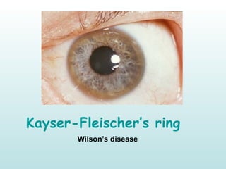
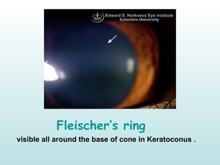
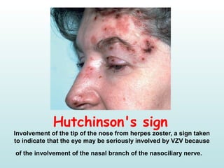
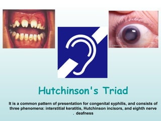
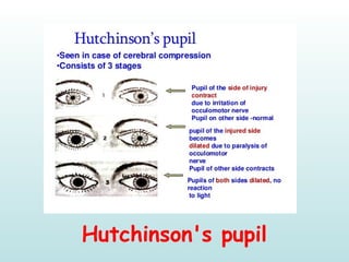
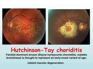
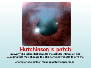
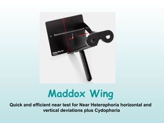
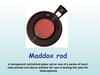
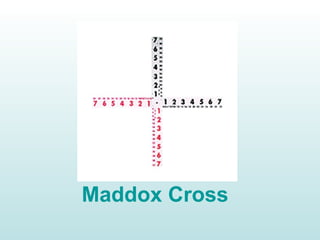
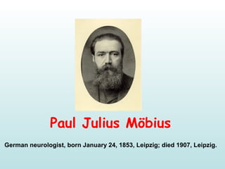
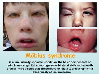
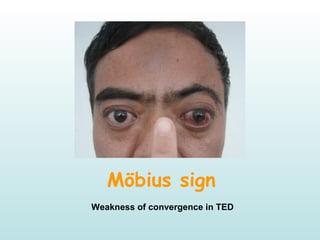
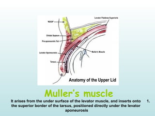
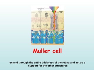
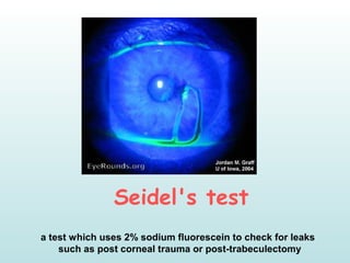
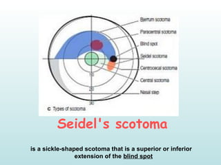
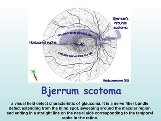
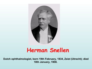
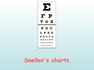
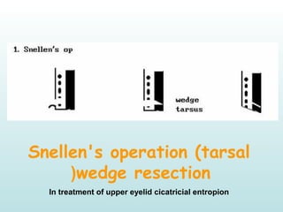
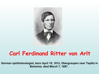
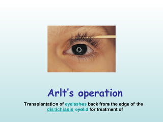
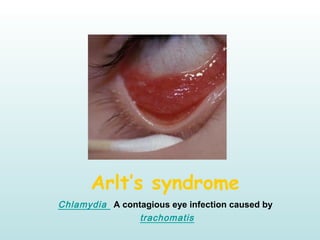
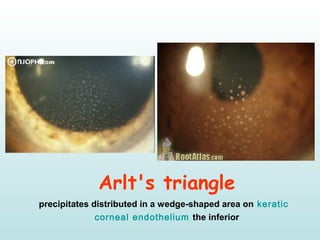
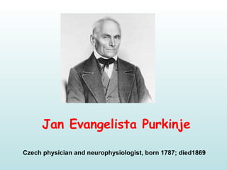
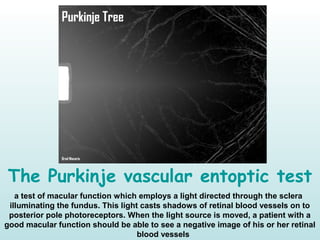
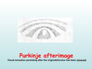
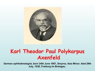
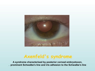
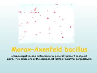
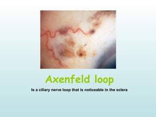
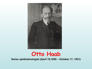
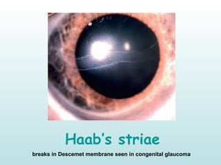
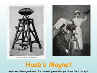
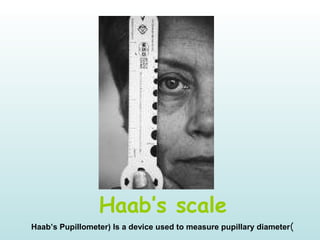
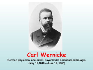
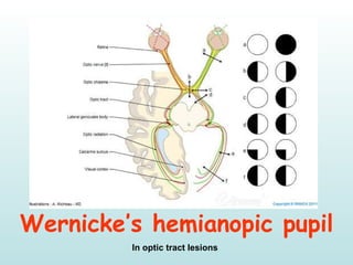
OphthalmologyOphthalmology
Eponyms II !Eponyms II !
By
Dr.Ahmed Alsherbiny
MSc. Ophth
For The Great
Ophthalmologists Whom
Paved The Way For Us , I
Dedicate This
Presentation :)
Fleischer
Bruno Fleischer 
German ophthalmologist, born May 2, 1874, Stuttgart; died 1965 
Kayser-Fleischer’s ring
Wilson’s disease
Fleischer’s ring
visible all around the base of cone in Keratoconus .
Hutchinson
Sir Jonathan Hutchinson
English ophthalmologist, born 1828 and died 1913. 
Hutchinson's sign
Involvement of the tip of the nose from herpes zoster, a sign taken 
to indicate that the eye may be seriously involved by VZV because 
of the involvement of the nasal branch of the nasociliary nerve.
Hutchinson's Triad
It is a common pattern of presentation for congenital syphilis, and consists of 
three phenomena: interstitial keratitis, Hutchinson incisors, and eighth nerve 
deafness.
Hutchinson ferckle
Lentigo maligna (melanoma in situ, intraepidermal melanoma, 
Hutchinson freckle) is an uncommon condition that develops in 
sun-damaged skin in elderly individuals. Malignant change may 
occur, with infiltration of the dermis.
Hutchinson's pupil
Hutchinson-Tay choriditis
Familial dominant drusen (Doyne honeycomb choroiditis, malattia 
leventinese) is thought to represent an early-onset variant of age-
related macular degeneration.
Hutchinson's patch
in syphylitic interstitial keratitis the cellular infiltration and 
clouding that may obscure the still-perfused vessels to give the 
characteristic pinkish ‘salmon patch’ appearance
Maddox
Ernest Edmund Maddox
British ophthalmologist  (1863-1933) . 
Maddox Wing
Quick and efficient near test for Near Heterophoria horizontal and 
vertical deviations plus Cydophoria
Maddox rod
a transparent cylindrical glass rod or one of a series of such
rods placed one above another for use in testing the eyes for
heterophoria
Maddox Cross
Möbius
Paul Julius Möbius
German neurologist, born January 24, 1853, Leipzig; died 1907, Leipzig.
Möbius syndrome
is a rare, usually sporadic, condition, the basic components of
which are congenital non-progressive bilateral sixth and seventh
cranial nerve palsies that are believed to relate to a developmental
abnormality of the brainstem 
Möbius sign
Weakness of convergence in TED
Muller
Heinrich Müller
German anatomist (1820–1864)
Muller’s muscle
1.It arises from the under surface of the levator muscle, and inserts onto
the superior border of the tarsus, positioned directly under the levator
aponeurosis
Muller cell
extend through the entire thickness of the retina and act as a
support for the other structures
Seidel
Erich Seidel 
German Ophthalmologist (1882–1948).
Seidel's test
a test which uses 2% sodium fluorescein to check for leaks
such as post corneal trauma or post-trabeculectomy
Seidel's scotoma
 is a sickle-shaped scotoma that is a superior or inferior
extension of the blind spot
Bjerrum
Jannik Peterson Bjerrum
Danish ophthalmologist. Born 1851, died 1920.
Bjerrum screen
Bjerrum scotoma
a visual field defect characteristic of glaucoma. It is a nerve fiber bundle
defect extending from the blind spot, sweeping around the macular region
and ending in a straight line on the nasal side corresponding to the temporal
raphe in the retina.
Snellen
Herman Snellen
Dutch ophthalmologist, born 19th February, 1834, Zeist (Utrecht); died
18th January, 1908.
Snellen's charts
Snellen's operation (tarsal
wedge resection)
In treatment of upper eyelid cicatricial entropion
Arlt 
Carl Ferdinand Ritter von Arlt
German ophthalmologist, born April 18, 1812, Obergraupen near Teplitz in
Bohemia; died March 7, 1887.
Arlt's Line 
Trachoma conjunctival scar in sulcus
subtarsalis. 
Arlt’s operation
Transplantation of eyelashes back from the edge of the
eyelid for treatment of distichiasis
Arlt’s syndrome
A contagious eye infection caused by Chlamydia
trachomatis
Arlt's triangle
 keratic precipitates distributed in a wedge-shaped area on
the inferior corneal endothelium
Purkinje  
Jan Evangelista Purkinje
Czech physician and neurophysiologist, born 1787; died1869
The Purkinje vascular entoptic test
a test of macular function which employs a light directed through the sclera
illuminating the fundus. This light casts shadows of retinal blood vessels on to
posterior pole photoreceptors. When the light source is moved, a patient with a
good macular function should be able to see a negative image of his or her retinal
blood vessels
Purkinje-Sanson images
At least four Purkinje images are usually visible. The first Purkinje image (P1)
is the reflection from the outer surface of the cornea. The second Purkinje
image (P2) is the reflection from the inner surface of the cornea. The third
Purkinje image (P3) is the reflection from the outer (anterior) surface of
the lens. The fourth Purkinje image (P4) is the reflection from the inner
(posterior) surface of the lens. Unlike the others, P4 is an inverted image.
Purkinje effect
is the tendency for the peak luminance sensitivity of the human eye to shift
toward the blue end of the color spectrum at low illumination levels
Purkinje afterimage
Visual sensation persisting after the originalstimulus has been removed
Axenfeld
Karl Theodor Paul Polykarpus
Axenfeld 
German ophthalmologist, born 24th June 1867, Smyrna, Asia Minor; died 29th 
July, 1930, Freiburg im Breisgau. 
Axenfeld's syndrome
A syndrome characterised by posterior corneal embryotoxon, 
prominent Schwalbe's line and iris adhesion to the Schwalbe’s line.
Morax–Axenfeld bacillus
is Gram negative, non motile bacteria, generally present as diploid 
pairs. They cause one of the commonest forms of catarrhal conjunctivitis
Axenfeld loop
Is a ciliary nerve loop that is noticeable in the sclera
Haab
Otto Haab
Swiss ophthalmologist (April 19,1850 – October 17, 1931)
Haab’s striae
breaks in Descemet membrane seen in congenital glaucoma
Haab’s reflex
Is another name for pupillary light reflex
Haab’s Magnet
A powerful magnet used for removing metallic particles from the eye
Haab’s scale
)Haab’s Pupillometer) Is a device used to measure pupillary diameter
Wernicke
Carl Wernicke
German physician, anatomist, psychiatrist and neuropathologis
 (May 15,1848 – June 15, 1905)
Wernicke’s hemianopic pupil
In optic tract lesions
Wernicke’s encephalopathy
Due to Thiamin (vitamin B1) defeciency
Thank You

Ophthalmology Eponyms II !