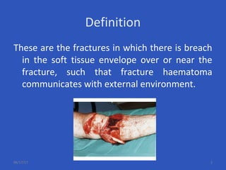
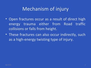
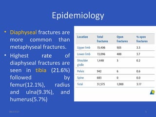
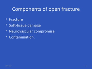
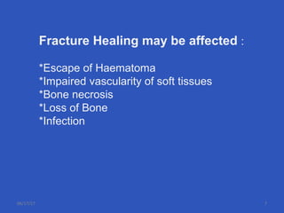
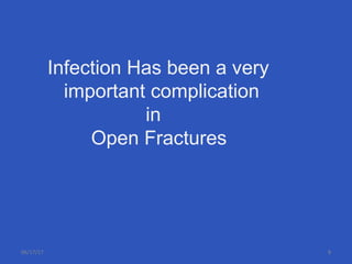
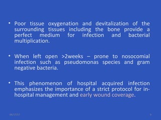
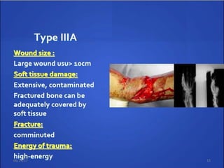
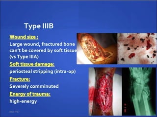
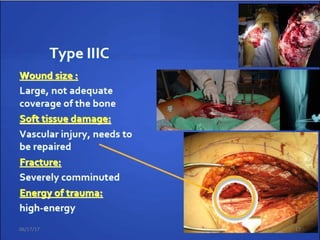
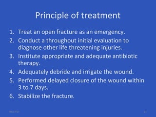
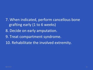
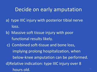
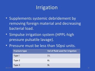
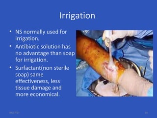
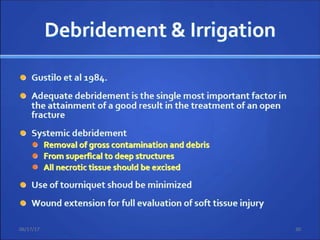
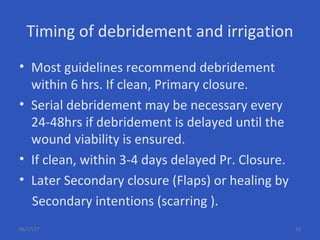
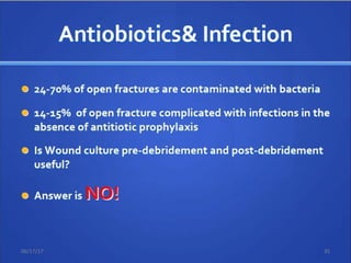
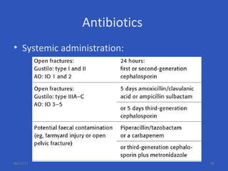
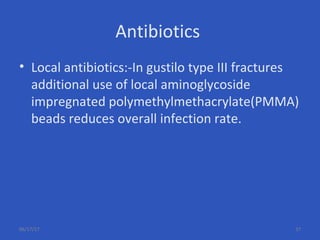
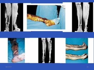
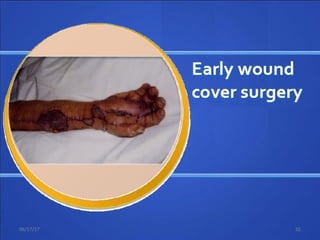
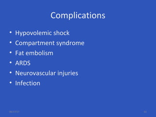
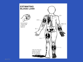
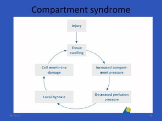
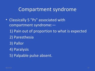
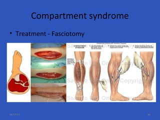
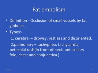
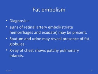
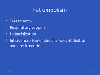
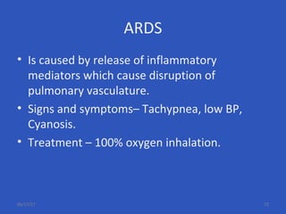
Management
of
OPEN FRACTURES
DR. SUMAN PAUL
Department of
Orthopaedics & Traumatology,
Rajshahi Medical College & Hospital
06/17/17
Definition
These are the fractures in which there is breach
in the soft tissue envelope over or near the
fracture, such that fracture haematoma
communicates with external environment.
06/17/17 2
WHO facts 2020
• WHO predicts that by the year 2020, the
causes of death and loss of health from
disability in a row:
• 1. Ischemic heart disease
• 2. Stroke
• 3. Road traffic injuries.
06/17/17 3
Mechanism of injury
• Open fractures occur as a result of direct high
energy trauma either from Road traffic
collisions or falls from height.
• These fractures can also occur indirectly, such
as a high-energy twisting type of injury.
06/17/17 4
Epidemiology
• Diaphyseal fractures are
more common than
metaphyseal fractures.
• Highest rate of
diaphyseal fractures are
seen in tibia (21.6%)
followed by
femur(12.1%), radius
and ulna(9.3%), and
humerus(5.7%)
06/17/17 5
Components of open fracture
• Fracture
• Soft-tissue damage
• Neurovascular compromise
• Contamination.
06/17/17 6
Fracture Healing may be affected :
*Escape of Haematoma
*Impaired vascularity of soft tissues
*Bone necrosis
*Loss of Bone
*Infection
06/17/17 7
Infection Has been a very
important complication
in
Open Fractures
06/17/17 8
• Poor tissue oxygenation and devitalization of the
surrounding tissues including the bone provide a
perfect medium for infection and bacterial
multiplication.
• When left open >2weeks – prone to nosocomial
infection such as pseudomonas species and gram
negative bacteria.
• This phenomenon of hospital acquired infection
emphasizes the importance of a strict protocol for in-
hospital management and early wound coverage.
06/17/17 9
OPEN FRACTURES – GRADING AND
CLASSIFICATION
TO ACCURATELY DESCRIBE SIMILAR INJURIES IN ORDER TO PROVIDE A
BASIS FOR TREATMENT, TO ESTIMATE PROGNOSIS.
06/17/17 10
Gustilo and Anderson
06/17/17 11
Gustilo and Anderson
06/17/17 12
06/17/17 13
06/17/17 14
06/17/17 15
06/17/17 16
Nerve
 Sensate No major nerve
injury
1
 Dorsal Deep peroneal
nerve
2
 Plantar partial Tibial nerve injury 3
 Plantar complete Sciatic nerve 4
Ischemia
 None Good to fair pulses,
no ischemia
0
 Mild Decreased pulses
perfusion
1
 Moderate Prolonged capillary
refill, Doppler
pulses fill
2
 Severe Pulseless, cool,
ischemic, no
doppler
3
Soft Tissue
 Grade I Minimal
contamination
0
 Grade II Moderate soft
tissue injury, low
velocity
1
 Grade IIIA Moderate crush
injury, high velocity
with Considerable
contamination
2
 Grade IIIB Massive crush
injury severe
contamination
3
Skeletal
 Spiral or oblique
fracture
0
 Transverse
fracture-minimal
contamination
1
 Moderate
displacement and
communition with
high velocity
2
 Segmental
fracture, severe
communition, bony
loss
3
Shock
 Normotensive 0
 Transient
hypotensive
1
 Persistent
hypotensive
2
06/17/17 17
Goal Of Open fractureTreatment
• Prevent sepsis.
• Heal the fracture.
• Restore function of extremity.
06/17/17 19
OPEN FRACTURES - MANAGEMENT
THE TREATMENT OF HIGH ENERGY INJURIES AIM TO PRESERVE LIFE,
LIMB AND FUNCTION.
06/17/17 20
Principle of treatment
1. Treat an open fracture as an emergency.
2. Conduct a throughout initial evaluation to
diagnose other life threatening injuries.
3. Institute appropriate and adequate antibiotic
therapy.
4. Adequately debride and irrigate the wound.
5. Performed delayed closure of the wound within
3 to 7 days.
6. Stabilize the fracture.
06/17/17 21
7. When indicated, perform cancellous bone
grafting early (1 to 6 weeks)
8. Decide on early amputation.
9. Treat compartment syndrome.
10. Rehabilitate the involved extremity.
06/17/17 22
Decide on early amputation
a) type IIIC injury with posterior tibial nerve
loss.
b) Massive soft tissue injury with poor
functional results likely.
c) Combined soft-tissue and bone loss,
implying prolong hospitalization, when
below-knee amputation can be performed.
d)Relative indication: type IIIC injury over 8
hours old. 23
06/17/17 24
Stages of care
06/17/17 25
Initial assessment
• Important components in assessing traumatized
extremity are
1. History and mechanism of injury
2. Neurovascular status
3. Size of skin wound
4. Muscle crush or loss
5. Periosteal stripping or bone loss
6. Fracture pattern, fragmentation
7. Contamination
8. Compartment syndrome.
06/17/17 26
06/17/17 27
Irrigation
• Supplements systemic debridement by
removing foreign material and decreasing
bacterial load.
• Simpulse irrigation system (HPPL-high
pressure pulsatile lavage).
• Pressure must be less than 50psi units.
Fracture type Vol of fluid used for irrigation
Type I 3 L
Type 2 6L
Type 3 9L
06/17/17 28
Irrigation
• NS normally used for
irrigation.
• Antibiotic solution has
no advantage than soap
for irrigation.
• Surfactant(non sterile
soap) same
effectiveness, less
tissue damage and
more economical.
06/17/17 29
06/17/17 30
*SKIN margins excised sparingly
06/17/17 31
“The greatest of all antiseptics is living tissue”
- Sir Alexander Fleming.
Damaged tissue should be excised cleanly with
curved scissors.
Excision must be ruthlessly pursued until only
viable, bleeding muscle remains.
06/17/17 32
Timing of debridement and irrigation
• Most guidelines recommend debridement
within 6 hrs. If clean, Primary closure.
• Serial debridement may be necessary every
24-48hrs if debridement is delayed until the
wound viability is ensured.
• If clean, within 3-4 days delayed Pr. Closure.
• Later Secondary closure (Flaps) or healing by
Secondary intentions (scarring ).
06/17/17 33
06/17/17 34
06/17/17 35
Antibiotics
• Systemic administration:
06/17/17 36
Antibiotics
• Local antibiotics:-In gustilo type III fractures
additional use of local aminoglycoside
impregnated polymethylmethacrylate(PMMA)
beads reduces overall infection rate.
06/17/17 37
Antibiotic bead pouch technique
06/17/17 38
Tetanus prophylaxis
• Tetanus Toxoid(TT), dose is 0.5ml i.m.
regardless of age.
• Immunoglobulin:-
75IU <5yrs of age
125IU 5-10yrs
250IU >10yrs.
06/17/17 39
06/17/17 40
06/17/17 41
Primary surgery
Timing :
•Surgical emergency
•Operating within 6-8hrs of injury –
contaminated wounds not treated within this
time will have sustained bacterial multiplication
to result in early infection.
06/17/17 42
Primary surgery
Fracture stabilization:
•As soon as primary wound care is completed,
treatment should proceed to fracture reduction
and fixation.
•Surgeon should rescrub and regown.
•Different set of instruments than those used for
debridement is necessary.
06/17/17 43
Fixation Options ??
External Fixation Vs Internal Fixation
06/17/17 45
Relative Indications for External
Fixation in Open Fractures
1) Severe contamination any site,
2) Periarticular fractures
– Definitive
• Distal radius
• Elbow dislocation
– Relative
• Knee
• Ankle
• Elbow
• Wrist
• Pelvis
06/17/17 46
External fixation
• Advantages of Ext Fixation:-
• Can be applied relatively easily and quickly,
• It provides relatively stable fracture fixation,
• There is no further damage done if applied
correctly,
• It avoids implantation of hardware in open
wound.
06/17/17 47
Ext Fixator
• Disadvantages:
• Major problems with external fixation are
related to pin tract infection, malalignent ,
delayed union, poor patients compliance.
• Tubular fixactors may not be the choice of
fixation but Ring fixators may be an option in
open diaphyseal fractures.
06/17/17 48
Relative Indications for Internal
Fixation in Open Fractures
1)Periarticular fractures
– Distal/proximal tibia
– Distal/proximal femur
– Distal/proximal humerus
– Proximal ulnar radius
– Selected distal radius/ulna
– Acetabulum/pelvis
2) Diaphyseal fractures
– Femur
– Tibia
– Humerus
– Radius/ulna
06/17/17 49
Plates
• Open diaphyseal fractures of the radius and ulna
as well as the humerus are best managed with
plate fixation.
• The plate fixation of lower extremity diaphyseal
fractures is generally not recommended due to
higher rate of infections.
06/17/17 50
06/17/17 51
Intramedullary nailing
• Locked intramedullary nailing has been established as
the treatment of choice for most diaphyseal fractures
in lower extremity.
• The technique has particular value for open fractures
as Intramedullary nails can be inserted with no further
disruption of the already injured soft-tissue envelope
and preserves the remaining extra osseous blood
supply to cortical bone.
06/17/17 52
IM nailing
• Data showing that solid intramedullary nails inserted
without reaming have a lower risk of infection.
• On the other hand reamed intramedullary nails can
reliably maintain fracture reduction with regards to
angulation, rotation, displacement, and length.
• Prospective randomised trails that compared reamed
with unreamed interlocked IM nails did not show any
significant difference concerning outcome and risk of
complication.
06/17/17 53
06/17/17 54
Open wound coverage after primary
surgery
06/17/17 55
Primary Closure
If it is to be done, the following criteria must be met:-
1.The original wound must have been fairly clean, and
not have occurred in a highly contaminated environment.
2.All necrotic tissue and foreign material have been
debrided.
3.Circulation to the limb is essentially normal.
4.Nerve supply to the limb is intact.
5.The patient's general condition is satisfactory and
allows careful postoperative assessment.
6.The wound can be closed without tension.
7.Closure will not create a dead space.
8.The patient does not have multisystem injuries.
06/17/17 56
Delayed primary closure
• Closure before the 5th day is termed delayed
primary closure.
• As long as closure is achieved before the fifth
day, wound strengths at 14 days are comparable
with those in wounds closed on the first day.
• Leaving the wound open minimizes the risk of
anaerobic infection, and the delay allows the
host to mount local wound defensive
mechanisms that permit safer closure than is
possible on the first day.
06/17/17 57
• Current standard of care for all open fracture
wounds is to be left open initially.
• Delayed closure is accomplished within 2-
7days
• VAC assisted wound closure is presently
recommended for temporary management of
open fracture wounds.
06/17/17 58
VAC
• The wound bed is exposed to mechanically
induced negative pressure in a closed system .
• The system removes fluid from extravascular
space, reduces edema, improves micro
circulation and enhances the proliferation of
preparative granulation tissue.
• Polyurethane foam dressing is placed in
wound and ensures an even distribution of
negative pressure.
06/17/17 59
VAC
06/17/17 60
OPEN FRACTURES:-
COMPLICATIONS
WHY OPEN FRACTURES CAN BE DANGEROUS
06/17/17 61
Complications
• Hypovolemic shock
• Compartment syndrome
• Fat embolism
• ARDS
• Neurovascular injuries
• Infection
06/17/17 62
Hypovolemic shock - management
• Two large-bore IV lines should be started.
• Once IV access is obtained, initial fluid
resuscitation is performed with an isotonic
crystalloid, such as Ringer lactate solution or
normal saline.
• An initial bolus of 1-2 L is given in an adult (20
mL/kg in a pediatric patient), and the patient's
response is assessed.
06/17/17 63
Hypovolemic shock
• Type of fluid:-
• Colloid – albumin, dextran, plasma.
• Crystalloid – NS, D5, RL.
• Blood – uncrossed ‘O’ –ve.
• Basic Rule:-
• 3:1 rule when using crystalloids.Eg. If blood loss is
100cc the patient should receive 300cc of normal
saline or Ringer lactate.
• 1:1 rule for colloids.
06/17/17 64
06/17/17
Compartment syndrome
06/17/17 66
Compartment syndrome
• Classically 5 "Ps" associated with
compartment syndrome:—
1) Pain out of proportion to what is expected
2) Paresthesia
3) Pallor
4) Paralysis
5) Palpable pulse absent.
06/17/17 67
Compartment syndrome
• Treatment - Fasciotomy
06/17/17 68
Fat embolism
• Definition - Occlusion of small vessels by fat
globules.
• Types:-
1. cerebral – drowsy, restless and disoriented.
2.pulmonary – tachypnea, tachycardia,
petechial rash(in front of neck, ant axillary
fold, chest and conjunctiva )
06/17/17 69
Fat embolism
• Diagnosis:–
• signs of retinal artery emboli(striate
hemorrhages and exudate) may be present.
• Sputum and urine may reveal presence of fat
globules.
• X-ray of chest shows patchy pulmonary
infarcts.
06/17/17 70
Fat embolism
• Treatment:-
• Respiratory support
• Heparinization
• Intravenous low-molecular weight dextran
and corticosteriods.
06/17/17 71
ARDS
• Is caused by release of inflammatory
mediators which cause disruption of
pulmonary vasculature.
• Signs and symptoms– Tachypnea, low BP,
Cyanosis.
• Treatment – 100% oxygen inhalation.
06/17/17 72
Vascular injury
• Absent peripheral pulses in an injured limb
should be considered to be due to vascular
damage unless proved otherwise.
• Classical signs of arterial injury:-
(a) absent pulses
(b) active hemorrhage
(c) expanding hematoma, and
(d) bruit or thrill.
06/17/17 73
Vascular injury
• Investigations:-
Colour doppler study
Arteriography.
• Treatment:-
Arterial reconstruction
Bypass grafts.
• Timing:- loss of total blood
supply to the limb for > 8
hours nearly always results
in amputation.
06/17/17 74
Nerve injury
• Nerve repair should be done within 3 weeks of
injury for better results
06/17/17 75
Take Home Messages
Success of open fracture management depends
on strict adherence to principles of good surgical
debridement, antibiotic therapy, fracture
stabilization, early soft tissue reconstruction and
timely bone grafting.
06/17/17 76
06/17/17 77
Thank you for your
concentration and
patience hearing..

Openfracture

  • 1.
    Management of OPEN FRACTURES DR. SUMANPAUL Department of Orthopaedics & Traumatology, Rajshahi Medical College & Hospital 06/17/17
  • 2.
    Definition These are thefractures in which there is breach in the soft tissue envelope over or near the fracture, such that fracture haematoma communicates with external environment. 06/17/17 2
  • 3.
    WHO facts 2020 •WHO predicts that by the year 2020, the causes of death and loss of health from disability in a row: • 1. Ischemic heart disease • 2. Stroke • 3. Road traffic injuries. 06/17/17 3
  • 4.
    Mechanism of injury •Open fractures occur as a result of direct high energy trauma either from Road traffic collisions or falls from height. • These fractures can also occur indirectly, such as a high-energy twisting type of injury. 06/17/17 4
  • 5.
    Epidemiology • Diaphyseal fracturesare more common than metaphyseal fractures. • Highest rate of diaphyseal fractures are seen in tibia (21.6%) followed by femur(12.1%), radius and ulna(9.3%), and humerus(5.7%) 06/17/17 5
  • 6.
    Components of openfracture • Fracture • Soft-tissue damage • Neurovascular compromise • Contamination. 06/17/17 6
  • 7.
    Fracture Healing maybe affected : *Escape of Haematoma *Impaired vascularity of soft tissues *Bone necrosis *Loss of Bone *Infection 06/17/17 7
  • 8.
    Infection Has beena very important complication in Open Fractures 06/17/17 8
  • 9.
    • Poor tissueoxygenation and devitalization of the surrounding tissues including the bone provide a perfect medium for infection and bacterial multiplication. • When left open >2weeks – prone to nosocomial infection such as pseudomonas species and gram negative bacteria. • This phenomenon of hospital acquired infection emphasizes the importance of a strict protocol for in- hospital management and early wound coverage. 06/17/17 9
  • 10.
    OPEN FRACTURES –GRADING AND CLASSIFICATION TO ACCURATELY DESCRIBE SIMILAR INJURIES IN ORDER TO PROVIDE A BASIS FOR TREATMENT, TO ESTIMATE PROGNOSIS. 06/17/17 10
  • 11.
  • 12.
  • 13.
  • 14.
  • 15.
  • 16.
  • 17.
    Nerve  Sensate No majornerve injury 1  Dorsal Deep peroneal nerve 2  Plantar partial Tibial nerve injury 3  Plantar complete Sciatic nerve 4 Ischemia  None Good to fair pulses, no ischemia 0  Mild Decreased pulses perfusion 1  Moderate Prolonged capillary refill, Doppler pulses fill 2  Severe Pulseless, cool, ischemic, no doppler 3 Soft Tissue  Grade I Minimal contamination 0  Grade II Moderate soft tissue injury, low velocity 1  Grade IIIA Moderate crush injury, high velocity with Considerable contamination 2  Grade IIIB Massive crush injury severe contamination 3 Skeletal  Spiral or oblique fracture 0  Transverse fracture-minimal contamination 1  Moderate displacement and communition with high velocity 2  Segmental fracture, severe communition, bony loss 3 Shock  Normotensive 0  Transient hypotensive 1  Persistent hypotensive 2 06/17/17 17
  • 18.
    Goal Of OpenfractureTreatment • Prevent sepsis. • Heal the fracture. • Restore function of extremity. 06/17/17 19
  • 19.
    OPEN FRACTURES -MANAGEMENT THE TREATMENT OF HIGH ENERGY INJURIES AIM TO PRESERVE LIFE, LIMB AND FUNCTION. 06/17/17 20
  • 20.
    Principle of treatment 1.Treat an open fracture as an emergency. 2. Conduct a throughout initial evaluation to diagnose other life threatening injuries. 3. Institute appropriate and adequate antibiotic therapy. 4. Adequately debride and irrigate the wound. 5. Performed delayed closure of the wound within 3 to 7 days. 6. Stabilize the fracture. 06/17/17 21
  • 21.
    7. When indicated,perform cancellous bone grafting early (1 to 6 weeks) 8. Decide on early amputation. 9. Treat compartment syndrome. 10. Rehabilitate the involved extremity. 06/17/17 22
  • 22.
    Decide on earlyamputation a) type IIIC injury with posterior tibial nerve loss. b) Massive soft tissue injury with poor functional results likely. c) Combined soft-tissue and bone loss, implying prolong hospitalization, when below-knee amputation can be performed. d)Relative indication: type IIIC injury over 8 hours old. 23
  • 23.
  • 24.
  • 25.
    Initial assessment • Importantcomponents in assessing traumatized extremity are 1. History and mechanism of injury 2. Neurovascular status 3. Size of skin wound 4. Muscle crush or loss 5. Periosteal stripping or bone loss 6. Fracture pattern, fragmentation 7. Contamination 8. Compartment syndrome. 06/17/17 26
  • 26.
  • 27.
    Irrigation • Supplements systemicdebridement by removing foreign material and decreasing bacterial load. • Simpulse irrigation system (HPPL-high pressure pulsatile lavage). • Pressure must be less than 50psi units. Fracture type Vol of fluid used for irrigation Type I 3 L Type 2 6L Type 3 9L 06/17/17 28
  • 28.
    Irrigation • NS normallyused for irrigation. • Antibiotic solution has no advantage than soap for irrigation. • Surfactant(non sterile soap) same effectiveness, less tissue damage and more economical. 06/17/17 29
  • 29.
  • 30.
    *SKIN margins excisedsparingly 06/17/17 31
  • 31.
    “The greatest ofall antiseptics is living tissue” - Sir Alexander Fleming. Damaged tissue should be excised cleanly with curved scissors. Excision must be ruthlessly pursued until only viable, bleeding muscle remains. 06/17/17 32
  • 32.
    Timing of debridementand irrigation • Most guidelines recommend debridement within 6 hrs. If clean, Primary closure. • Serial debridement may be necessary every 24-48hrs if debridement is delayed until the wound viability is ensured. • If clean, within 3-4 days delayed Pr. Closure. • Later Secondary closure (Flaps) or healing by Secondary intentions (scarring ). 06/17/17 33
  • 33.
  • 34.
  • 35.
  • 36.
    Antibiotics • Local antibiotics:-Ingustilo type III fractures additional use of local aminoglycoside impregnated polymethylmethacrylate(PMMA) beads reduces overall infection rate. 06/17/17 37
  • 37.
    Antibiotic bead pouchtechnique 06/17/17 38
  • 38.
    Tetanus prophylaxis • TetanusToxoid(TT), dose is 0.5ml i.m. regardless of age. • Immunoglobulin:- 75IU <5yrs of age 125IU 5-10yrs 250IU >10yrs. 06/17/17 39
  • 39.
  • 40.
  • 41.
    Primary surgery Timing : •Surgicalemergency •Operating within 6-8hrs of injury – contaminated wounds not treated within this time will have sustained bacterial multiplication to result in early infection. 06/17/17 42
  • 42.
    Primary surgery Fracture stabilization: •Assoon as primary wound care is completed, treatment should proceed to fracture reduction and fixation. •Surgeon should rescrub and regown. •Different set of instruments than those used for debridement is necessary. 06/17/17 43
  • 43.
    Fixation Options ?? ExternalFixation Vs Internal Fixation 06/17/17 45
  • 44.
    Relative Indications forExternal Fixation in Open Fractures 1) Severe contamination any site, 2) Periarticular fractures – Definitive • Distal radius • Elbow dislocation – Relative • Knee • Ankle • Elbow • Wrist • Pelvis 06/17/17 46
  • 45.
    External fixation • Advantagesof Ext Fixation:- • Can be applied relatively easily and quickly, • It provides relatively stable fracture fixation, • There is no further damage done if applied correctly, • It avoids implantation of hardware in open wound. 06/17/17 47
  • 46.
    Ext Fixator • Disadvantages: •Major problems with external fixation are related to pin tract infection, malalignent , delayed union, poor patients compliance. • Tubular fixactors may not be the choice of fixation but Ring fixators may be an option in open diaphyseal fractures. 06/17/17 48
  • 47.
    Relative Indications forInternal Fixation in Open Fractures 1)Periarticular fractures – Distal/proximal tibia – Distal/proximal femur – Distal/proximal humerus – Proximal ulnar radius – Selected distal radius/ulna – Acetabulum/pelvis 2) Diaphyseal fractures – Femur – Tibia – Humerus – Radius/ulna 06/17/17 49
  • 48.
    Plates • Open diaphysealfractures of the radius and ulna as well as the humerus are best managed with plate fixation. • The plate fixation of lower extremity diaphyseal fractures is generally not recommended due to higher rate of infections. 06/17/17 50
  • 49.
  • 50.
    Intramedullary nailing • Lockedintramedullary nailing has been established as the treatment of choice for most diaphyseal fractures in lower extremity. • The technique has particular value for open fractures as Intramedullary nails can be inserted with no further disruption of the already injured soft-tissue envelope and preserves the remaining extra osseous blood supply to cortical bone. 06/17/17 52
  • 51.
    IM nailing • Datashowing that solid intramedullary nails inserted without reaming have a lower risk of infection. • On the other hand reamed intramedullary nails can reliably maintain fracture reduction with regards to angulation, rotation, displacement, and length. • Prospective randomised trails that compared reamed with unreamed interlocked IM nails did not show any significant difference concerning outcome and risk of complication. 06/17/17 53
  • 52.
  • 53.
    Open wound coverageafter primary surgery 06/17/17 55
  • 54.
    Primary Closure If itis to be done, the following criteria must be met:- 1.The original wound must have been fairly clean, and not have occurred in a highly contaminated environment. 2.All necrotic tissue and foreign material have been debrided. 3.Circulation to the limb is essentially normal. 4.Nerve supply to the limb is intact. 5.The patient's general condition is satisfactory and allows careful postoperative assessment. 6.The wound can be closed without tension. 7.Closure will not create a dead space. 8.The patient does not have multisystem injuries. 06/17/17 56
  • 55.
    Delayed primary closure •Closure before the 5th day is termed delayed primary closure. • As long as closure is achieved before the fifth day, wound strengths at 14 days are comparable with those in wounds closed on the first day. • Leaving the wound open minimizes the risk of anaerobic infection, and the delay allows the host to mount local wound defensive mechanisms that permit safer closure than is possible on the first day. 06/17/17 57
  • 56.
    • Current standardof care for all open fracture wounds is to be left open initially. • Delayed closure is accomplished within 2- 7days • VAC assisted wound closure is presently recommended for temporary management of open fracture wounds. 06/17/17 58
  • 57.
    VAC • The woundbed is exposed to mechanically induced negative pressure in a closed system . • The system removes fluid from extravascular space, reduces edema, improves micro circulation and enhances the proliferation of preparative granulation tissue. • Polyurethane foam dressing is placed in wound and ensures an even distribution of negative pressure. 06/17/17 59
  • 58.
  • 59.
    OPEN FRACTURES:- COMPLICATIONS WHY OPENFRACTURES CAN BE DANGEROUS 06/17/17 61
  • 60.
    Complications • Hypovolemic shock •Compartment syndrome • Fat embolism • ARDS • Neurovascular injuries • Infection 06/17/17 62
  • 61.
    Hypovolemic shock -management • Two large-bore IV lines should be started. • Once IV access is obtained, initial fluid resuscitation is performed with an isotonic crystalloid, such as Ringer lactate solution or normal saline. • An initial bolus of 1-2 L is given in an adult (20 mL/kg in a pediatric patient), and the patient's response is assessed. 06/17/17 63
  • 62.
    Hypovolemic shock • Typeof fluid:- • Colloid – albumin, dextran, plasma. • Crystalloid – NS, D5, RL. • Blood – uncrossed ‘O’ –ve. • Basic Rule:- • 3:1 rule when using crystalloids.Eg. If blood loss is 100cc the patient should receive 300cc of normal saline or Ringer lactate. • 1:1 rule for colloids. 06/17/17 64
  • 63.
  • 64.
  • 65.
    Compartment syndrome • Classically5 "Ps" associated with compartment syndrome:— 1) Pain out of proportion to what is expected 2) Paresthesia 3) Pallor 4) Paralysis 5) Palpable pulse absent. 06/17/17 67
  • 66.
    Compartment syndrome • Treatment- Fasciotomy 06/17/17 68
  • 67.
    Fat embolism • Definition- Occlusion of small vessels by fat globules. • Types:- 1. cerebral – drowsy, restless and disoriented. 2.pulmonary – tachypnea, tachycardia, petechial rash(in front of neck, ant axillary fold, chest and conjunctiva ) 06/17/17 69
  • 68.
    Fat embolism • Diagnosis:– •signs of retinal artery emboli(striate hemorrhages and exudate) may be present. • Sputum and urine may reveal presence of fat globules. • X-ray of chest shows patchy pulmonary infarcts. 06/17/17 70
  • 69.
    Fat embolism • Treatment:- •Respiratory support • Heparinization • Intravenous low-molecular weight dextran and corticosteriods. 06/17/17 71
  • 70.
    ARDS • Is causedby release of inflammatory mediators which cause disruption of pulmonary vasculature. • Signs and symptoms– Tachypnea, low BP, Cyanosis. • Treatment – 100% oxygen inhalation. 06/17/17 72
  • 71.
    Vascular injury • Absentperipheral pulses in an injured limb should be considered to be due to vascular damage unless proved otherwise. • Classical signs of arterial injury:- (a) absent pulses (b) active hemorrhage (c) expanding hematoma, and (d) bruit or thrill. 06/17/17 73
  • 72.
    Vascular injury • Investigations:- Colourdoppler study Arteriography. • Treatment:- Arterial reconstruction Bypass grafts. • Timing:- loss of total blood supply to the limb for > 8 hours nearly always results in amputation. 06/17/17 74
  • 73.
    Nerve injury • Nerverepair should be done within 3 weeks of injury for better results 06/17/17 75
  • 74.
    Take Home Messages Successof open fracture management depends on strict adherence to principles of good surgical debridement, antibiotic therapy, fracture stabilization, early soft tissue reconstruction and timely bone grafting. 06/17/17 76
  • 75.
    06/17/17 77 Thank youfor your concentration and patience hearing..