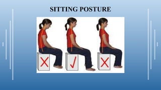
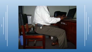
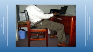
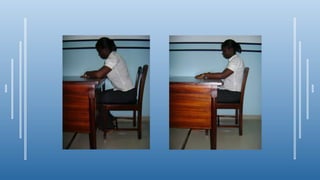
LOW BACK PAIN
Dr. SUMAN PAUL
Department of
Orthopaedics & Traumatology,
Rajshahi Medical College Hospital
CONTENT
¶Brief anatomy of the back
¶Intro to Lower Back Pain
¶Epidemiology
¶Causes of LBP
¶Presentation
¶Ways of preventing LBP
¶Treatment
THE BACK
¶ The back can be viewed as that region of the body
from the occiput to the gluteal folds (Olaogun, 1999)
¶ The back is composed of very sturdy bones and
powerful muscles.
¶ The spine or the back bone is made up of 33 bones
called vertebrae.
¶ 7 cervical, 12 thoracic and 5 lumbar vertebrae
separated by 23 intervertebral discs.
LOW BACK PAIN
Also known as lumbago (at times referred to as waist pain) is pain
(as name implies) in the lower back (lumbar) region.
Typically, the commonest area of back pain is the low back
(lumbar region) and sometimes it spreads to the buttocks or
thighs.
WHY THE LOWER BACK?
¶Lumbar region because:
¶It bears most of the body’s weight
¶Most movements of the spine occur there.
LOW BACK PAIN
¶Most bouts of back pain ease quickly, usually
within a week (acute back pain).
¶When symptoms persist for several months
(over 3 months) chronic back pain results.
¶Back pain could be mechanical, pathologic or
physiologic
FACTS:- Low Back Pain
¶Second most common cause of missed
work days
¶Leading cause of disability between ages of
19-45
¶Number one impairment in occupational
injuries
Referred LBP Is Remote From
Source of Pain
¶LBP may radiate into
• groin
• buttocks
• upper thigh (posteriorly)
areas that share an interconnecting nerve supply
¶Source of somatic referred pain is a skeletal or myofascial
structure of the lumbar spine
¶Source of visceral referred pain is within a body organ
• ovarian cysts may refer pain to low back
• cancer of head of pancreas can present as low back pain becoming
excruciating at night
Muscular Pain
¶Most back pains are caused by strain or
sprain of the back muscles & ligaments
¶Pain will be in discrete area & tender to
touch
¶It is of aching quality & may involve
muscle spasm
EPIDEMIOLOGY
¶Back pain, especially low back pain (LBP), most often affects people
between the ages of 25 and 60 years and those aged between 50 and
60 years are likely to become disabled (Corbin et al, 2002).
¶Up to 70%-85% of the population in the United Kingdom (UK)
experience back pain at some point in their lives.
¶In Ghana, over 60% of the adult population complains of back pain at
one time or the other (Osei, 2000).
¶Out of the 424 patients who presented with various conditions at the
Physiotherapy Department of KBTH, 54% were LBP cases (From June
to August 2004).
Sources of LBP
¶Damage to several structures in
the low back can result in severe
pain
• vertebrae
• thoracolumbar fascia
• ligaments
• joints
• specifically sacroiliac joint
• discs
• muscle
Deyo RA, Weinstein JN. N Engl J Med. 2001;344:363-370.
Non-Spinal Related Causes
Bladder Infection Kidney Disease
Ovarian Cancer Ovarian Cyst
Testicular Torsion Fibromyalgia
Pelvic Infections Appendicitis
Pancreatitis Prostate Disease
Gall Bladder Disease
Abdominal Aortic Aneurysm
Spine Related Causes
Arthritis
Fibromyalgia
Kyphosis
Lordosis
Rheumatoid Arthritis
Ankylosing Spondylitis
Arachnoiditis
Bone Cancer
Chiari Malformation
Compression Fractures
Discitis
Epidural Abscess
Facet Joint Syndrome
Fixed Sagittal Imbalance
Osteomyelitis
Osteophytes
Pinched Nerve
Ruptured Disc
Spina Bifida
Spinal Cord Injury
Spinal Tumor
Spondylolisthesis
Spinal Stenosis
Spinal Cord Injury
Spinal Tumor
Sprain or Strain
Synovial Cysts
Wedge Fractures
LBP Psychological Factors
¶ Psychological factors that may contribute
to or be caused by chronic LBP include
• depression
• anxiety
• post-traumatic stress disorder
• pre-existing disorders
Physiotherapy department, TQMH
Social Issues May Contribute to Chronic LBP
¶Job dissatisfaction/loss of ability to work
¶Pursuit of disability compensation
¶Substance abuse
¶Family dynamics
¶Financial issues
¶Loss of social identity or context
¶Loss of ability to participate in recreational activities
Diagnosis of LBP
History
Location
Specific Point vs. Across Back
Superficial vs. Deep
Involve Any other region (lower extremity)
History
Quality
Dull Ache (tooth ache)
Sharp/Stabbing
Burning
Tearing/Pop
History
Quality/Severity
Intermittent
Constant
Pain Scale 1-10
History
Setting
Time of day when worst/better
After strenuous activity
History
Aggravating/Relieving Factors
What Makes Better What Makes Worse
BEWARE OF THE PATIENT THAT SAYS NOTHING MAKES PAIN BETTER!
History
Associated Manifestations
Numbness
Tingling(pins/needles)
Burning
WeaknessIncontinence
Falls
OBJECTIVE ASSESSMENT
Musculoskeletal Examination
¶ Look
• pain behaviors–groaning, position changes, grimacing, etc
• atrophy, swelling, asymmetry, color changes
¶ Feel
• palpate area of pain for temperature, spasm, and pain provocation
• point palpation for trigger points/tender points
¶ Move
• active and passive
• flexion, extension, rotational, lateral bending
• leg raising
Examination
¶Posture – change in lumbar lordosis, scoliosis
¶Range of motion – flexion,extension, lateral flexion
¶SLR – seated & supine, sciatic nerve stretch test
¶Presence of paraspinal muscle spasm, trigger points
¶Tender areas –facets, sacro-iliac joints
¶Neurological deficit –
• Dermatomal hypo/hyperaesthesia
• Ability to rise from squatting position (L4),walk on heels(L5), walk on tip-
toes (S1)
• Tendon reflexes – knee jerk(L4 root), ankle jerk(S1 root)
Neurologic Exam Determines
Presence/Absence and Level of Radiculopathy and Myelopathy
¶ Motor elements
• muscle bulk/tone
• atrophy/flaccidity
• muscle strength
• coordination
• gait
¶ Sensory elements
• sensory deficits, eg, touch, position sense,
temperature, vibration
• allodynia: light touch
• hyperalgesia: single or multiple pinpricks
The exam should include
Diagnostic Studies
¶Plain X-Ray
¶MRI
¶CAT Scan
¶Myelogram
¶Discogram
¶Bone Scan
• Facet Block
• SI Joint Block
• EMG
• SSEP
• DEXAscan
• Bone Scan
Diagnostic Studies
X-Ray
¶ taken to assess the structure of the spine and to determine the
alignment of the vertebra
Diagnostic Studies
MRI
¶Extremely Sensitive for assessment of Soft
tissue structures (nerves, disc)
¶One of the most commonly ordered test to
assess low back pain
Diagnostic Studies
CAT Scan
¶ Most often used to assess bone
structures of spine.
¶ Faster and cheaper than MRI
¶ Can be very effective tool when using
reconstruction images or combined
with other modalities
Diagnostic Studies
Myelogram & Post CT
¶myelogram consists of a series of
plain xrays with a contrast agent
injected into the thecal sac.
¶The C.A.T. scan that usually
follows the myelogram depicts
this same anatomy from a C.A.T.
scan perspective
Diagnostic Studies
Myelogram & Post CT
The injection of iodine based contrast into the thecal sac
containing the nerves and/or spinal cord, promotes better
definition of those structures than the images obtained on the
regular C.A.T. scan. Cross-sections and reconstructions of the
images in different planes (including 3-D) allows different
perspectives on the anatomy. This test is often used to visualize the
spinal cord and nerves in relation to the surrounding spine
structures (bone, joint, disc, etc)
Diagnostic Studies
Discogram
¶Involves the injection of
contrast material into the disc
space
¶Concordant vs. Discordant
Pain…..??
¶Helpful in assessing discogenic
pain
¶VERY “uncomfortable” test
PREVENTION
LIFTING
¶Squat directly (by bending your knees) in front of any object to be
lifted; rise, letting your legs and thighs do the work.
¶Keep the object you’re lifting close to your body, and don’t twist.
¶Never try to lift anything you can’t easily manage – get help!
¶ Never bend over without bending your knees.
¶Avoid twisting the trunk whilst lifting
¶Lift and move the trunk in a vertical plane first
¶Turn to the desired direction with the legs afterwards
CORRECT WAY OF LIFTING
PREVENTION
STANDING
¶Standing for long periods of time can put a lot of stress on
your back.
¶If you must stand, occasionally shift your weight from
one side to the other.
¶Or, try propping one of your feet on a footstool six-to-
eight inches high.
¶Occasionally, tighten your abdominal muscles. This can
enable you to keep your back straight
STANDING
WRONG RIGHTWRONG
CARRYING BACK-PACKS
PREVENTION
SITTING
¶Sit in firm seats with straight backs, keeping your back
flat/straight with the knees bent to about 90º.
¶Rest your feet flat on the floor or on a footstool.
¶ If you cannot get the chair you prefer, learn to sit properly on whatever chair
you get.
¶ Throw head well back, then bend it forward to pull in the chin. This will
straighten the back.
¶ Now tighten abdominal muscles to raise the chest. Check position frequently.
SITTING POSTURE
SITTING POSTURE
PREVENTION
SLEEPING
¶Sleep on a firm, flat mattress.
¶It’s best to sleep on your side with your knees and hips
bent and a pillow under your head, or on your back with
pillows beneath your head and knees.
POOR SLEEPING POSITION
GOOD SLEEPING POSITION
EVEN BETTER
POOR SLEEPING POSITION
GOOD SLEEPING POSITION
EVEN BETTER
SOME GOOD AND BAD POSTURES
TREATMENT
¶Medications/ Pharmacotherapy
¶Physiotherapy
¶Surgery
Pharmacotherapy Options*
¶Antidepressants
¶Anticonvulsants
¶Muscle relaxants
¶Opioid analgesics
¶Corticosteroids
¶NSAIDs
¶Topical analgesics
* Except for certain opioids, none of these agents are indicated for chronic LBP.
PHYSIOTHERAPY
¶Exercise (stretching and strengthening of back extensors mainly)
¶Thermotherapy; Heat application in chronic lower back pain using
Infra-red, shortwave diathermy, hotpacks, etc
¶Electrotherapy; Transcutaneous Electric Nerve Stimulation, Trabert,
etc
Back Exercises
¶Ankle pump
¶Heel slides
¶Abdominal contraction
¶Wall squats
¶Heel raises
¶Straight leg raises
¶Knee to chest stretch
¶Hamstring stretch
¶Exercises with swiss ball
Surgery
1. DECOMPRESSION of spinal nerves (BURST FRACTURE, Spinal stenosis, PID)
2. Fusion & Stabilization (Instrumentation)
3. Correction of deformity
DECOMPRESSION Surgery
FUSION Surgery
References
¶ MOB Olaogun (1999):Pathomenchanics and force analysis at the low back during physical tasks (JNMRT), vol 4 (7). Pp 7-11
¶ Clark MA, Russell AM. Low back pain: a functional perspective. Thousand Oaks,
CA: National Academy of Sports Medicine; 2002.
¶ Hodges PW. Core stability exercise in chronic low back pain. Orthopedic Clinics of
North America. 2003;34:245-254.
¶ Kendall FB, McCreary EK. Muscle Testing and Function. 4th ed. Baltimore, MD:
Williams & Watkins; 1993: 215-226, 284-293.
¶Thank you……

Low BACK PAIN

  • 1.
    LOW BACK PAIN Dr.SUMAN PAUL Department of Orthopaedics & Traumatology, Rajshahi Medical College Hospital
  • 2.
    CONTENT ¶Brief anatomy ofthe back ¶Intro to Lower Back Pain ¶Epidemiology ¶Causes of LBP ¶Presentation ¶Ways of preventing LBP ¶Treatment
  • 3.
    THE BACK ¶ Theback can be viewed as that region of the body from the occiput to the gluteal folds (Olaogun, 1999) ¶ The back is composed of very sturdy bones and powerful muscles. ¶ The spine or the back bone is made up of 33 bones called vertebrae. ¶ 7 cervical, 12 thoracic and 5 lumbar vertebrae separated by 23 intervertebral discs.
  • 4.
    LOW BACK PAIN Alsoknown as lumbago (at times referred to as waist pain) is pain (as name implies) in the lower back (lumbar) region. Typically, the commonest area of back pain is the low back (lumbar region) and sometimes it spreads to the buttocks or thighs. WHY THE LOWER BACK? ¶Lumbar region because: ¶It bears most of the body’s weight ¶Most movements of the spine occur there.
  • 5.
    LOW BACK PAIN ¶Mostbouts of back pain ease quickly, usually within a week (acute back pain). ¶When symptoms persist for several months (over 3 months) chronic back pain results. ¶Back pain could be mechanical, pathologic or physiologic
  • 6.
    FACTS:- Low BackPain ¶Second most common cause of missed work days ¶Leading cause of disability between ages of 19-45 ¶Number one impairment in occupational injuries
  • 7.
    Referred LBP IsRemote From Source of Pain ¶LBP may radiate into • groin • buttocks • upper thigh (posteriorly) areas that share an interconnecting nerve supply ¶Source of somatic referred pain is a skeletal or myofascial structure of the lumbar spine ¶Source of visceral referred pain is within a body organ • ovarian cysts may refer pain to low back • cancer of head of pancreas can present as low back pain becoming excruciating at night
  • 8.
    Muscular Pain ¶Most backpains are caused by strain or sprain of the back muscles & ligaments ¶Pain will be in discrete area & tender to touch ¶It is of aching quality & may involve muscle spasm
  • 9.
    EPIDEMIOLOGY ¶Back pain, especiallylow back pain (LBP), most often affects people between the ages of 25 and 60 years and those aged between 50 and 60 years are likely to become disabled (Corbin et al, 2002). ¶Up to 70%-85% of the population in the United Kingdom (UK) experience back pain at some point in their lives. ¶In Ghana, over 60% of the adult population complains of back pain at one time or the other (Osei, 2000). ¶Out of the 424 patients who presented with various conditions at the Physiotherapy Department of KBTH, 54% were LBP cases (From June to August 2004).
  • 10.
    Sources of LBP ¶Damageto several structures in the low back can result in severe pain • vertebrae • thoracolumbar fascia • ligaments • joints • specifically sacroiliac joint • discs • muscle Deyo RA, Weinstein JN. N Engl J Med. 2001;344:363-370.
  • 11.
    Non-Spinal Related Causes BladderInfection Kidney Disease Ovarian Cancer Ovarian Cyst Testicular Torsion Fibromyalgia Pelvic Infections Appendicitis Pancreatitis Prostate Disease Gall Bladder Disease Abdominal Aortic Aneurysm
  • 12.
    Spine Related Causes Arthritis Fibromyalgia Kyphosis Lordosis RheumatoidArthritis Ankylosing Spondylitis Arachnoiditis Bone Cancer Chiari Malformation Compression Fractures Discitis Epidural Abscess Facet Joint Syndrome Fixed Sagittal Imbalance Osteomyelitis Osteophytes Pinched Nerve Ruptured Disc Spina Bifida Spinal Cord Injury Spinal Tumor Spondylolisthesis Spinal Stenosis Spinal Cord Injury Spinal Tumor Sprain or Strain Synovial Cysts Wedge Fractures
  • 13.
    LBP Psychological Factors ¶Psychological factors that may contribute to or be caused by chronic LBP include • depression • anxiety • post-traumatic stress disorder • pre-existing disorders Physiotherapy department, TQMH
  • 14.
    Social Issues MayContribute to Chronic LBP ¶Job dissatisfaction/loss of ability to work ¶Pursuit of disability compensation ¶Substance abuse ¶Family dynamics ¶Financial issues ¶Loss of social identity or context ¶Loss of ability to participate in recreational activities
  • 15.
    Diagnosis of LBP History Location SpecificPoint vs. Across Back Superficial vs. Deep Involve Any other region (lower extremity)
  • 16.
    History Quality Dull Ache (toothache) Sharp/Stabbing Burning Tearing/Pop
  • 17.
  • 18.
    History Setting Time of daywhen worst/better After strenuous activity
  • 19.
    History Aggravating/Relieving Factors What MakesBetter What Makes Worse BEWARE OF THE PATIENT THAT SAYS NOTHING MAKES PAIN BETTER!
  • 20.
  • 21.
    OBJECTIVE ASSESSMENT Musculoskeletal Examination ¶Look • pain behaviors–groaning, position changes, grimacing, etc • atrophy, swelling, asymmetry, color changes ¶ Feel • palpate area of pain for temperature, spasm, and pain provocation • point palpation for trigger points/tender points ¶ Move • active and passive • flexion, extension, rotational, lateral bending • leg raising
  • 22.
    Examination ¶Posture – changein lumbar lordosis, scoliosis ¶Range of motion – flexion,extension, lateral flexion ¶SLR – seated & supine, sciatic nerve stretch test ¶Presence of paraspinal muscle spasm, trigger points ¶Tender areas –facets, sacro-iliac joints ¶Neurological deficit – • Dermatomal hypo/hyperaesthesia • Ability to rise from squatting position (L4),walk on heels(L5), walk on tip- toes (S1) • Tendon reflexes – knee jerk(L4 root), ankle jerk(S1 root)
  • 23.
    Neurologic Exam Determines Presence/Absenceand Level of Radiculopathy and Myelopathy ¶ Motor elements • muscle bulk/tone • atrophy/flaccidity • muscle strength • coordination • gait ¶ Sensory elements • sensory deficits, eg, touch, position sense, temperature, vibration • allodynia: light touch • hyperalgesia: single or multiple pinpricks The exam should include
  • 24.
    Diagnostic Studies ¶Plain X-Ray ¶MRI ¶CATScan ¶Myelogram ¶Discogram ¶Bone Scan • Facet Block • SI Joint Block • EMG • SSEP • DEXAscan • Bone Scan
  • 25.
    Diagnostic Studies X-Ray ¶ takento assess the structure of the spine and to determine the alignment of the vertebra
  • 26.
    Diagnostic Studies MRI ¶Extremely Sensitivefor assessment of Soft tissue structures (nerves, disc) ¶One of the most commonly ordered test to assess low back pain
  • 27.
    Diagnostic Studies CAT Scan ¶Most often used to assess bone structures of spine. ¶ Faster and cheaper than MRI ¶ Can be very effective tool when using reconstruction images or combined with other modalities
  • 28.
    Diagnostic Studies Myelogram &Post CT ¶myelogram consists of a series of plain xrays with a contrast agent injected into the thecal sac. ¶The C.A.T. scan that usually follows the myelogram depicts this same anatomy from a C.A.T. scan perspective
  • 29.
    Diagnostic Studies Myelogram &Post CT The injection of iodine based contrast into the thecal sac containing the nerves and/or spinal cord, promotes better definition of those structures than the images obtained on the regular C.A.T. scan. Cross-sections and reconstructions of the images in different planes (including 3-D) allows different perspectives on the anatomy. This test is often used to visualize the spinal cord and nerves in relation to the surrounding spine structures (bone, joint, disc, etc)
  • 30.
    Diagnostic Studies Discogram ¶Involves theinjection of contrast material into the disc space ¶Concordant vs. Discordant Pain…..?? ¶Helpful in assessing discogenic pain ¶VERY “uncomfortable” test
  • 31.
    PREVENTION LIFTING ¶Squat directly (bybending your knees) in front of any object to be lifted; rise, letting your legs and thighs do the work. ¶Keep the object you’re lifting close to your body, and don’t twist. ¶Never try to lift anything you can’t easily manage – get help! ¶ Never bend over without bending your knees. ¶Avoid twisting the trunk whilst lifting ¶Lift and move the trunk in a vertical plane first ¶Turn to the desired direction with the legs afterwards
  • 32.
  • 34.
    PREVENTION STANDING ¶Standing for longperiods of time can put a lot of stress on your back. ¶If you must stand, occasionally shift your weight from one side to the other. ¶Or, try propping one of your feet on a footstool six-to- eight inches high. ¶Occasionally, tighten your abdominal muscles. This can enable you to keep your back straight
  • 35.
  • 36.
  • 37.
  • 38.
    PREVENTION SITTING ¶Sit in firmseats with straight backs, keeping your back flat/straight with the knees bent to about 90º. ¶Rest your feet flat on the floor or on a footstool. ¶ If you cannot get the chair you prefer, learn to sit properly on whatever chair you get. ¶ Throw head well back, then bend it forward to pull in the chin. This will straighten the back. ¶ Now tighten abdominal muscles to raise the chest. Check position frequently.
  • 39.
  • 43.
  • 44.
    PREVENTION SLEEPING ¶Sleep on afirm, flat mattress. ¶It’s best to sleep on your side with your knees and hips bent and a pillow under your head, or on your back with pillows beneath your head and knees.
  • 46.
  • 47.
  • 48.
  • 49.
  • 50.
  • 51.
  • 52.
    SOME GOOD ANDBAD POSTURES
  • 53.
  • 54.
    Pharmacotherapy Options* ¶Antidepressants ¶Anticonvulsants ¶Muscle relaxants ¶Opioidanalgesics ¶Corticosteroids ¶NSAIDs ¶Topical analgesics * Except for certain opioids, none of these agents are indicated for chronic LBP.
  • 55.
    PHYSIOTHERAPY ¶Exercise (stretching andstrengthening of back extensors mainly) ¶Thermotherapy; Heat application in chronic lower back pain using Infra-red, shortwave diathermy, hotpacks, etc ¶Electrotherapy; Transcutaneous Electric Nerve Stimulation, Trabert, etc
  • 56.
    Back Exercises ¶Ankle pump ¶Heelslides ¶Abdominal contraction ¶Wall squats ¶Heel raises ¶Straight leg raises ¶Knee to chest stretch ¶Hamstring stretch ¶Exercises with swiss ball
  • 57.
    Surgery 1. DECOMPRESSION ofspinal nerves (BURST FRACTURE, Spinal stenosis, PID) 2. Fusion & Stabilization (Instrumentation) 3. Correction of deformity
  • 58.
  • 59.
  • 60.
    References ¶ MOB Olaogun(1999):Pathomenchanics and force analysis at the low back during physical tasks (JNMRT), vol 4 (7). Pp 7-11 ¶ Clark MA, Russell AM. Low back pain: a functional perspective. Thousand Oaks, CA: National Academy of Sports Medicine; 2002. ¶ Hodges PW. Core stability exercise in chronic low back pain. Orthopedic Clinics of North America. 2003;34:245-254. ¶ Kendall FB, McCreary EK. Muscle Testing and Function. 4th ed. Baltimore, MD: Williams & Watkins; 1993: 215-226, 284-293.
  • 61.