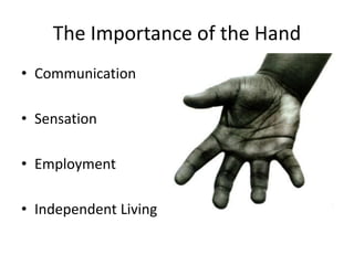
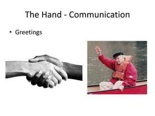
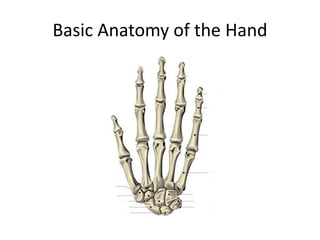
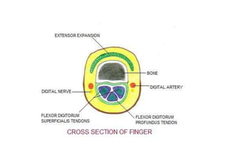
Hand Trauma
CT3 –MSK Day
Vijay Kama
Consultant EM
Peterborough City Hospital
Let us look ………
• Why is it important?
• Basic anatomy
• Clinical examination
• Common injuries
Hand Injuries
The Importance of the Hand
• Communication
• Sensation
• Employment
• Independent Living
The Hand - Communication
• Greetings
Communication…
• Gestures
Communication…
• Sign Language
Sensation
• Large area brain structure devoted to touch.
Highly sensitive.
Sensation…
• Relationships
Employment
• Use of hands fundamental to most vocations.
Independent Living
• Without the use of our hands, most people
would find independent living impossible.
• This equals 6-8 weeks off work!! No income
for 2 months. How would your finances cope?
Basic Anatomy of the Hand
Anatomy - Tendons
Anatomy - Nerves
Anatomy of the Hand
• Small area – lots to injure.
• Even small lacerations may cause functional
issues.
Assessing the Injured Hand - Look
Assess the Finger Posture
Feel
• Is it cold?
• Is sensation intact?
Frequently Presenting Hand Injuries
• Fractures
• Lacerations/Penetrating Injuries
• Amputations
• De-gloving Injuries
• Human (punch) Bites
• Animal Bites
• Hand infections
Lacerations
• Very common cause of trauma.
• Typical culprits –
Common Results
Lacerations
• Regardless of size, always have a high
suspicion for more serious injury.
• Remember, glass only ever stops cutting when
it hits bone.
• Lacerated tendon when repaired takes 6-8
weeks of healing and hand therapy to recover.
• Nerve repairs often take 3-6 months to get
some benefit from the repair
Extensor tendon Injury:
– Divided into Zones according to anatomical
location of injury
– In the hand and wrist there are 7 extensor
tendon zones
27
Tendon injuries
Ref. http://emedicine.medscape.com Orthopedic Surgery for Flexor Tendon
Lacerations Author: Michael Neumeister, MD, FRCSC, FRCSC, FACS; Chief
Editor: Harris Gellman, MD
http://www.orthobullets.com- Flexor Tendon Injuries- Derek Moore MD
Zone Presentation Management
I Mallet Deformity
•Closed: splinting 6-8 weeks
•Open: suture repair for fixation.
Soft tissue reconstruction
III
Boutonniere’s
Deformity
•Closed: splinting MCP and PIP in
hyperextension for 6 weeks
•Open: suture repair (figure of 8
suture)
V Fixed flexion of MCP
•Closed: splinting ,45 extension at
wrist and 20 flexion at MCP
•Open: suture repair.
VII Fixed flexion of MCP
•Suture repair followed by post-op
splinting
29
Deformities
can be due to tendon, bone , nerve injury and joint dislocations
– Specific types –
Tendon injuries
• Mallet finger
• Boutonniere deformity
• Z deformity of the thumb
Flexor tendon injuries –
5 zones in the hand and the wrist
Zone 1 One tendon only (FDP)
from middle of middle phalanx
distally
Zone 2 Two tendons (FDS &
FDP) from MCP joints to middle
of middle phalanx
Zone 3 Central palm
Zone 4 Tendons in the carpal
tunnel
Zone 5 Tendons proximal to the
carpal tunnel
FDS Insertion
Flexor Sheath
Presentation Flexor injury
Zone Presentation Management
I
Loss of active flexion at
DIP joint
Hyperextension of DIP
joint
(Jersey finger )
•Primary or Secondary tendon
repair
•Careful suturing prevent post-op
adhesions.
II
Loss of active flexion
at MCP joint
•Skin closure then secondary
repair by tendon grafting
•Primary repair performed by
skilled hand surgeon to minimize
post-op adhesions.
III, IV
Thumb
Same
•Primary or secondary tendon
repair
•Examine carefully for thenar
muscle injury and recurrent
branches of median nerve. 34
Amputations
• Can occur at any level.
• Ability to re-plant / re-vascularise depends on
both the level of amputation and the
mechanism.
• Once past the distal third of the distal phalanx
the vessels are too small to be anastamosed.
Finger Tip Amputation
Injured components may include skin, bone,
nail, nail bed, tendon, and the pulp, the
padded area of the fingertip .
• If just skin is removed and the defect is less than a
centimeter in diameter, it is often possible to treat these
injuries with simple dressing changes.
• If there is a little bit of bone exposed at the tip, it can
sometimes be trimmed back slightly and treated with V-Y
plasty
Amputation...
Decision is based on:
 Importance of the part,
 level of injury,
 mechanism of injury
 expected return of function.
 Recommended ischemia times for replantation:
◦ Major replant: 6 hours of warm and 12 hours of
cold ischemia.
◦ Digit: 12 hours for warm ischemia and 24 hours for
cold ischemia.
 Preoperative preparation: radiography of both
amputated and stump parts to determine the level
of injury and suitability for replantation
40
Outcome
 Overall success rates for replantation approach 80%.
 Better outcome with Guillotine (sharp) amputation
(77%) compared to severely crushed and mangled
body parts(49%). In general, the prognosis for ring
avulsion injuries is poor.
 Studies have demonstrated that patients can expect to
achieve 50% function and 50% sensation of the
replanted part.
Ref. Plastic Surgery, Goldwyn and Cohen, 3rd edition.
Plastic Surgery, Grabb and Smith, 3rd edition.
41
De-gloving Injuries
Followup
• Can get large areas of skin loss.
• Typically treated as a skin graft with original
skin, or debrided and skin grafted from the
thigh.
• Can get contraction of the scar.
Punch Bite Injuries
• Very common.
• Injury occurs after punching someone in the
mouth. Usually small laceration to the 2nd or
3rd MCPJ. Often extends into the joint with
damage to the extensor tendon.
• Always requires IVABs and a washout.
• Common consequences – septic arthritis,
extensor tendon loss.
Punch Bite
Followup
• Usually require at least 1 washout. Sometime
multiple.
• Tendon cannot be repaired if already infected.
• Tendon, although intially intact can be
completely destroyed by infection.
• Always, always refer.
Animal Bites
• Cat bites – frequently become infected. Cat
teeth puncture like a needle and deposit
bacteria at the base to then form an abscess.
• Dog bites – easier to treat than cat bites as
dog teeth typically tear leaving the wound
open and able to be irrigated. Cosmetically
more difficult to treat.
Fractures and dislocations
Diagnosis…..
• Tenderness in anatomical
Snuff box
• Xray- fracture line
Treatment
• Scaphoid cast (3-4 months)
• Dorsiflexion
& radial deviation
(glass holding
position)
•Internal fixation
• Herbert’s screw
Complications
• Avascular necrosis
• Delayed / non union
• Wrist osteoarthritis
Lunate dislocations
• Lunate dislocation
perilunate dislocation
• Open reduction
• Avascular necrosis
Bennett’s fracture dislocation
• Base of 1st
metacarapal
• Intra articular
• Longitudinal force
to thumb
Rolando Fracture
• Comminuted First Metacarpal Base #
• Presents as ‘Y’ or ‘T’ Pattern
• Differs from Bennette that usually no diaphyseal displacement
CMC dislocation 4th 5th MC
#dislocation of 5th mc , reduced and fixed with k wires
Fracture phalanges
• Fall of heavy object or crush injury
• Undisplaced
Displaced
• Strapping
• Open reduction
PIPJ dislocations/
volar plate disruption
PIPJ dislocation
• Mechanism of Injury – hyperextension of
the PIP joint
• with or without dislocation
• often initial injury seems trivial
PIPJ dislocation
• If dislocation without #
OR
If # fragment less than 30% joint
surface
→ reduce then manage
conservatively in dorsal blocking
splint (DBS)
Dorsal blocking splint
• PIPJ in 30 degrees
flexion
• volar structures
off
stretch
• slowly increase
out
to neutral
#’s
• 5th Metacarpal
– Assess ROM and digit
Rotation
– Usually managed
conservatively
#’s
• 4th Metacarpal Spiral
#
– Assess ROM and digit
Rotation
– Tendency to rotate
– Impacted #’s result in
extension lag = poor
function.
Paronychia
• infection of the finger that involves the tissue
at the edges of the fingernail
• superficial and localized to the soft tissue and
skin
• most common bacterial infection seen in the
hand ( staph; strep).
Paronychia treatment
• wound care alone.
• collection of pus - drain.
– a simple incision over the collection of pus to allow
drainage.
– scalpel may be inserted along the edge of the nail to
allow drainage.
– If the infection is large, a part of the nail may be
removed.
• oral antibiotic.
• wound care at home.
Felon
• infection of the fingertip.
• This infection is located in the fingertip pad
and soft tissue associated with it.
Felon treatment
• incision and drainage
» incision will be
made on one or
both sides of the
fingertip.
» break up the
compartments
» gauze will be
placed into the
wound to aid
the initial
drainage.
» flush out with a
sterile solution
• antibiotics.
Infectious flexor tenosynovitis &
Deep space infection
• infection involves the tendon sheaths and
deep spaces
• penetrating trauma that introduces bacteria
• surgical emergency and will require rapid
treatment with IV antibiotics.
Kanavel’s cardinal signs
• intense pain
– along the course of tendon with extension
– this is the earliest and most important sign
• flexion posture
• uniform swelling
• percussion tenderness along the course of the
tendon sheath
hand_injuries_-msk_day_2015.ppt

hand_injuries_-msk_day_2015.ppt

  • 1.
    Hand Trauma CT3 –MSKDay Vijay Kama Consultant EM Peterborough City Hospital
  • 2.
    Let us look……… • Why is it important? • Basic anatomy • Clinical examination • Common injuries
  • 4.
  • 5.
    The Importance ofthe Hand • Communication • Sensation • Employment • Independent Living
  • 6.
    The Hand -Communication • Greetings
  • 7.
  • 8.
  • 9.
    Sensation • Large areabrain structure devoted to touch. Highly sensitive.
  • 10.
  • 11.
    Employment • Use ofhands fundamental to most vocations.
  • 12.
    Independent Living • Withoutthe use of our hands, most people would find independent living impossible.
  • 13.
    • This equals6-8 weeks off work!! No income for 2 months. How would your finances cope?
  • 14.
  • 15.
  • 17.
  • 19.
    Anatomy of theHand • Small area – lots to injure. • Even small lacerations may cause functional issues.
  • 20.
  • 21.
  • 22.
    Feel • Is itcold? • Is sensation intact?
  • 23.
    Frequently Presenting HandInjuries • Fractures • Lacerations/Penetrating Injuries • Amputations • De-gloving Injuries • Human (punch) Bites • Animal Bites • Hand infections
  • 24.
    Lacerations • Very commoncause of trauma. • Typical culprits –
  • 25.
  • 26.
    Lacerations • Regardless ofsize, always have a high suspicion for more serious injury. • Remember, glass only ever stops cutting when it hits bone. • Lacerated tendon when repaired takes 6-8 weeks of healing and hand therapy to recover. • Nerve repairs often take 3-6 months to get some benefit from the repair
  • 27.
    Extensor tendon Injury: –Divided into Zones according to anatomical location of injury – In the hand and wrist there are 7 extensor tendon zones 27 Tendon injuries Ref. http://emedicine.medscape.com Orthopedic Surgery for Flexor Tendon Lacerations Author: Michael Neumeister, MD, FRCSC, FRCSC, FACS; Chief Editor: Harris Gellman, MD http://www.orthobullets.com- Flexor Tendon Injuries- Derek Moore MD
  • 29.
    Zone Presentation Management IMallet Deformity •Closed: splinting 6-8 weeks •Open: suture repair for fixation. Soft tissue reconstruction III Boutonniere’s Deformity •Closed: splinting MCP and PIP in hyperextension for 6 weeks •Open: suture repair (figure of 8 suture) V Fixed flexion of MCP •Closed: splinting ,45 extension at wrist and 20 flexion at MCP •Open: suture repair. VII Fixed flexion of MCP •Suture repair followed by post-op splinting 29
  • 30.
    Deformities can be dueto tendon, bone , nerve injury and joint dislocations – Specific types – Tendon injuries • Mallet finger
  • 31.
    • Boutonniere deformity •Z deformity of the thumb
  • 33.
    Flexor tendon injuries– 5 zones in the hand and the wrist Zone 1 One tendon only (FDP) from middle of middle phalanx distally Zone 2 Two tendons (FDS & FDP) from MCP joints to middle of middle phalanx Zone 3 Central palm Zone 4 Tendons in the carpal tunnel Zone 5 Tendons proximal to the carpal tunnel FDS Insertion Flexor Sheath
  • 34.
    Presentation Flexor injury ZonePresentation Management I Loss of active flexion at DIP joint Hyperextension of DIP joint (Jersey finger ) •Primary or Secondary tendon repair •Careful suturing prevent post-op adhesions. II Loss of active flexion at MCP joint •Skin closure then secondary repair by tendon grafting •Primary repair performed by skilled hand surgeon to minimize post-op adhesions. III, IV Thumb Same •Primary or secondary tendon repair •Examine carefully for thenar muscle injury and recurrent branches of median nerve. 34
  • 35.
    Amputations • Can occurat any level. • Ability to re-plant / re-vascularise depends on both the level of amputation and the mechanism. • Once past the distal third of the distal phalanx the vessels are too small to be anastamosed.
  • 36.
    Finger Tip Amputation Injuredcomponents may include skin, bone, nail, nail bed, tendon, and the pulp, the padded area of the fingertip .
  • 37.
    • If justskin is removed and the defect is less than a centimeter in diameter, it is often possible to treat these injuries with simple dressing changes. • If there is a little bit of bone exposed at the tip, it can sometimes be trimmed back slightly and treated with V-Y plasty
  • 38.
  • 39.
    Decision is basedon:  Importance of the part,  level of injury,  mechanism of injury  expected return of function.
  • 40.
     Recommended ischemiatimes for replantation: ◦ Major replant: 6 hours of warm and 12 hours of cold ischemia. ◦ Digit: 12 hours for warm ischemia and 24 hours for cold ischemia.  Preoperative preparation: radiography of both amputated and stump parts to determine the level of injury and suitability for replantation 40
  • 41.
    Outcome  Overall successrates for replantation approach 80%.  Better outcome with Guillotine (sharp) amputation (77%) compared to severely crushed and mangled body parts(49%). In general, the prognosis for ring avulsion injuries is poor.  Studies have demonstrated that patients can expect to achieve 50% function and 50% sensation of the replanted part. Ref. Plastic Surgery, Goldwyn and Cohen, 3rd edition. Plastic Surgery, Grabb and Smith, 3rd edition. 41
  • 42.
  • 43.
    Followup • Can getlarge areas of skin loss. • Typically treated as a skin graft with original skin, or debrided and skin grafted from the thigh. • Can get contraction of the scar.
  • 44.
    Punch Bite Injuries •Very common. • Injury occurs after punching someone in the mouth. Usually small laceration to the 2nd or 3rd MCPJ. Often extends into the joint with damage to the extensor tendon. • Always requires IVABs and a washout. • Common consequences – septic arthritis, extensor tendon loss.
  • 45.
  • 46.
    Followup • Usually requireat least 1 washout. Sometime multiple. • Tendon cannot be repaired if already infected. • Tendon, although intially intact can be completely destroyed by infection. • Always, always refer.
  • 47.
    Animal Bites • Catbites – frequently become infected. Cat teeth puncture like a needle and deposit bacteria at the base to then form an abscess. • Dog bites – easier to treat than cat bites as dog teeth typically tear leaving the wound open and able to be irrigated. Cosmetically more difficult to treat.
  • 48.
  • 49.
    Diagnosis….. • Tenderness inanatomical Snuff box • Xray- fracture line
  • 50.
    Treatment • Scaphoid cast(3-4 months) • Dorsiflexion & radial deviation (glass holding position)
  • 51.
  • 52.
    Complications • Avascular necrosis •Delayed / non union • Wrist osteoarthritis
  • 53.
    Lunate dislocations • Lunatedislocation perilunate dislocation • Open reduction • Avascular necrosis
  • 54.
    Bennett’s fracture dislocation •Base of 1st metacarapal • Intra articular • Longitudinal force to thumb
  • 57.
    Rolando Fracture • ComminutedFirst Metacarpal Base # • Presents as ‘Y’ or ‘T’ Pattern • Differs from Bennette that usually no diaphyseal displacement
  • 59.
  • 60.
    #dislocation of 5thmc , reduced and fixed with k wires
  • 61.
    Fracture phalanges • Fallof heavy object or crush injury • Undisplaced Displaced • Strapping • Open reduction
  • 62.
  • 63.
    PIPJ dislocation • Mechanismof Injury – hyperextension of the PIP joint • with or without dislocation • often initial injury seems trivial
  • 64.
    PIPJ dislocation • Ifdislocation without # OR If # fragment less than 30% joint surface → reduce then manage conservatively in dorsal blocking splint (DBS)
  • 65.
    Dorsal blocking splint •PIPJ in 30 degrees flexion • volar structures off stretch • slowly increase out to neutral
  • 66.
    #’s • 5th Metacarpal –Assess ROM and digit Rotation – Usually managed conservatively
  • 67.
    #’s • 4th MetacarpalSpiral # – Assess ROM and digit Rotation – Tendency to rotate – Impacted #’s result in extension lag = poor function.
  • 68.
    Paronychia • infection ofthe finger that involves the tissue at the edges of the fingernail • superficial and localized to the soft tissue and skin • most common bacterial infection seen in the hand ( staph; strep).
  • 69.
    Paronychia treatment • woundcare alone. • collection of pus - drain. – a simple incision over the collection of pus to allow drainage. – scalpel may be inserted along the edge of the nail to allow drainage. – If the infection is large, a part of the nail may be removed. • oral antibiotic. • wound care at home.
  • 70.
    Felon • infection ofthe fingertip. • This infection is located in the fingertip pad and soft tissue associated with it.
  • 71.
    Felon treatment • incisionand drainage » incision will be made on one or both sides of the fingertip. » break up the compartments » gauze will be placed into the wound to aid the initial drainage. » flush out with a sterile solution • antibiotics.
  • 72.
    Infectious flexor tenosynovitis& Deep space infection • infection involves the tendon sheaths and deep spaces • penetrating trauma that introduces bacteria • surgical emergency and will require rapid treatment with IV antibiotics.
  • 73.
    Kanavel’s cardinal signs •intense pain – along the course of tendon with extension – this is the earliest and most important sign • flexion posture • uniform swelling • percussion tenderness along the course of the tendon sheath

Editor's Notes

  • #10 Graphical representation of the area of the brain devoted to the sensory input. Notice the very large area devoted to the hand. Highly sensitive and provides huge amounts of feedback to the brain.
  • #11 Healing touch – responsible for warmth and intimacy.
  • #12 Fundamental part of almost every job. Most jobs very difficult without full use of the hands.
  • #13 As function in the hands deteriorates, so will the ability to live independantly. Bilateral hand injuries renders patients virtually helpless.
  • #15 How many bones? – 27 Name them …. How many directions does the wrist move?
  • #16 Extensor side – dorsal interossei, common extensors, ext indicis, ext digiti minimi.EPL, EPB. Flexor side – Abductor pollicis brevis, flexor pollicis brevis, adductor pollicis. Hypothener – Flexor digiti minimi brevis, abductor digiti minimi FDS, FDP. Lumbricals (from FDP passes radial to MC) go to extensor
  • #18 Main motor nerves to the hand – Ulna and Median. Ulna runs beneath tendon of FCU passes along the pisiform. Divides into superficial and deep. Superficial supplies – palmaris brevis then continues as digital nerve. Small palmar branch supplies skin of hypothenar eminence. Deep branch – goes into palm between heads of flexor and abductor digiti minimi and through opponens digiti minimi. Gives motor to three hypothenar muscles, 2 lumbricals on ulna side, all interossei and adductor pollicis. Median Nerve – enters through carpal tunnel. 3 branches. Supplies palmar skin and fingers. Supplies 1st and 2nd lumbricals. Muscular branch supplies thenar muscles.
  • #28 Zone 1: Over the middle phalynx at insertion site (Mallet’s deformity) Zone 3: Over the apeces of the PIP joints (Boutonniere’s deformity) Zone 5: Over extensor hoods (MCP) and the dorsum of the hand Zone 7: Over extensor retinaculum
  • #35 Treatment of Zone II was associated with increased incidence of post operative cross-adhesions. That is why in the past it was advised to perform secondary repair rather than primary. The area was known as “No Man’s Land”. But recently several studies have shown that primary repair can be achieved with minimal if no post-op adhesion once performed by a skilled hand surgeon.
  • #38 Severe crush or avulsion injuries can completely remove some or all of the tissue at the fingertip.
  • #40 Because hand function is severely compromised if the thumb or multiple fingers are not present to oppose each other, thumb and multiple-finger replants should be attempted. Hand Muscles at room temperature are irreversibly damaged in 6-8 hours; if cooled, it can withstand a maximum of 8-12 hours of ischemia.
  • #55 Edward Hallaran Bennett (9 April 1837, Charlotte Quay, Cork – 21 June 1907, Dublin) was an Irish surgeon remembered for describing Bennett's fracture. He studied at Trinity College, Dublin of theUniversity of Dublin, graduating M.Ch. in 1859 and M.D. in 1864. He was professor of anatomy and surgery at Trinity College from 1873-1906. He studied fractures, dislocations and bone diseases, recording them at the Pathology Museum at Trinity College. He described his eponymous fracture at the British Medical Association meeting in Cork in 1880.[1] He is said to have introduced antiseptictechnique to Dublin, and became president of the Royal College of Surgeons in Ireland.[2]