Download to read offline

![Classification
Primary forms of arthritis:
Osteoarthritis
Rheumatoid arthritis
lupous
Gout and pseudo-gout
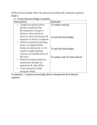
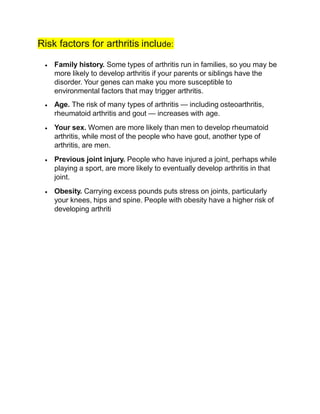
Osteoarthritis
Osteoarthritis is the most common form of arthritis.[3] It can
affect both the larger and the smaller joints of the body,
including the hands, feet, back, hip or knee. The disease is
essentially one acquired from daily wear and tear of the joint,
however, osteoarthritis can also occur as a result of injury.
Osteoarthritis begins in the cartilage and eventually leads to the
two opposing bones eroding into each other. Initially, the
condition starts with minor pain while walking but soon the pain
can be continuous and even occur at night. The pain can be
debilitating and prevent one from doing some activities.
Osteoarthritis typically affects the weight bearing joints such as
the back, spine, and pelvis. Unlike rheumatoid arthritis,
osteoarthritis is most commonly a disease of the elderly. More
than 30 percent of females have some degree of osteoarthritisby
age 65. Risk factors for osteoarthritis include: prior joint trauma,
obesity, sedentary lifestyle.
Osteoarthritis, like rheumatoid arthritis, cannot be cured but one
can prevent the condition from worsening. Weight loss is the
key to improving symptoms and preventing progression.
Physical therapy to strengthen muscles and joints is very helpful.](https://image.slidesharecdn.com/omverma-210129172933/85/Om-verma-18-320.jpg)
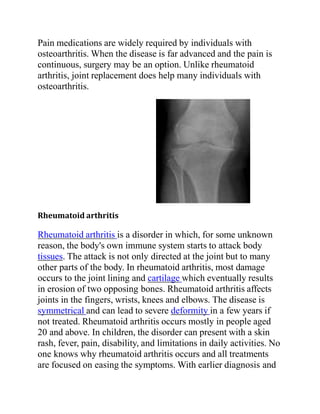
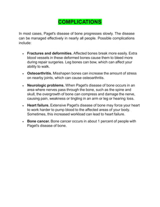
![Gout
Gout is caused by deposition of uric acid crystals in the joint,
causing inflammation. There is also an uncommon form of
gouty arthritis caused by the formation of rhomboid crystals of
calcium pyrophosphate known as pseudogout. In the early
stages, the gouty arthritis usually occur in one joint, but with
time, it can occur in many joints and be quite crippling. The
joints in gout can often become swollen and lose function.[7]
Other
Infectious arthritis is another severe form of arthritis. It presents
with sudden onset of chills, fever and joint pain. The condition
is caused by bacteria elsewhere in the body. Infectious arthritis
must be rapidly diagnosed and treated promptly to prevent
irreversible and permanent joint damage.[8]
Psoriasis is another type of arthritis. With psoriasis, most
individuals develop the skin problem first and then the arthritis.
The typical features are of continuous joint pains, stiffness and
swelling. The disease does recur with periods of remission but
there is no cure for the disorder. A small percentage develop a
severe painful and destructive form of arthritis which destroys
the small joints in the hands and can lead to permanent disability
and loss of hand function. [9]](https://image.slidesharecdn.com/omverma-210129172933/85/Om-verma-21-320.jpg)
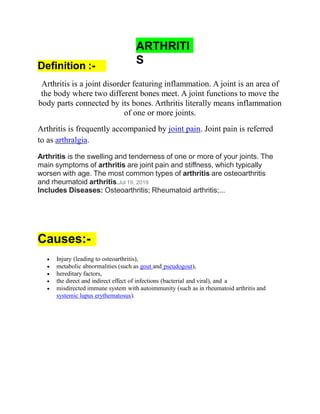
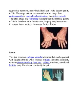
![Signs and symptoms
Regardless of the type of arthritis, the common symptoms for all arthritis
disorders include varied levels of pain, swelling, joint stiffness and
sometimes a constant ache around the joint(s). Arthritic disorders like
lupus and rheumatoid can also affect other organs in the body with a
variety of symptoms.[10]
Inability to use the hand or walk
Malaise and a feeling of tiredness
Fever
Weight loss
Poor sleep
Muscle aches and pains
Tenderness
Difficulty moving the joint
It is common in advanced arthritis for significant secondary changes to
occur. For example, in someone who has limited their physical activity:
Muscle weakness
Loss of flexibility
Decreased aerobic fitness
Assessment & diagnostic findings:-
History and a physical examination.
x-rays,
Bone scans
Bone biopsy.
Blood and urine tests.
Medications:-
NSAIDS(nonsteroidal anti-inflammatory drugs)
COX-2 Inhibitors](https://image.slidesharecdn.com/omverma-210129172933/85/Om-verma-22-320.jpg)


Paget's disease is a chronic bone disorder that causes abnormal bone remodeling, resulting in weakened, deformed bones. It occurs when the normal bone remodeling process shifts out of balance, causing new bone tissue to form improperly. Common symptoms include bone pain, fractures, and arthritis. Diagnosis involves x-rays, blood tests, and bone scans. Treatment focuses on controlling symptoms through medications like bisphosphonates or calcitonin, proper calcium intake, exercise if tolerated, and applying heat or cold for pain relief.


![CASE_PRESENTATION_ON_subdural_hematoma(SDH)[1 FINAL PPT]-1.pptx](https://cdn.slidesharecdn.com/ss_thumbnails/casepresentationonsubduralhematomasdh1finalppt-1-260129172522-d405d375-thumbnail.jpg?width=640&height=640&fit=bounds)


