SlideShare a Scribd company logo
Semi-automated Optical Coherence Tomography in Clinical Practice
Zachary Goodman1, Samantha Lancia1, Gautam Adusumilli1, Courtney Dula1,
Paul Kealey2, Robert T. Naismith1
1Washington University in Saint Louis School of Medicine, 2Optovue
•  Assess feasibility of a portable OCT device (Optovue iScan 500) as a tool for measuring retinal
nerve fiber layer (RNFL) thickness, retinal (macular) thickness and volume, and ganglion cell
complex (GCC) thickness in multiple sclerosis patients with and without self reported ON
•  Assess the relationship of the Pelli-Robson contrast scores and thickness measures obtained
from OCT
•  Recent reports suggest retinal and macular thickness is a biomarker of neurodegenerative
disease progression within the eyes of patients with multiple sclerosis (MS) without optic
neuritis (ON)
•  Optical Coherence Tomography (OCT) can be a useful clinical measure obtained as part of
regular clinical care for monitoring prognosis and disease activity
•  Traditional OCT scanners require considerable technical skill to perform, and are not very
portable
•  We tested a compact, semi-automated OCT scanner for routine use within an MS center
Background
Objectives
Methods
Results
•  Cross-sectional study of 89 MS patients (178 eyes) with and without prior self-reported history
of optic neuritis
•  RNFL thickness, macular thicknesses and volumes, and GCC thicknesses collected using the
Spectral-Domain Optovue iScan 500 OCT scanner
•  15 patients (30 eyes) excluded due to prior ophthalmologic conditions
•  7 eyes excluded due to insufficient scan quality
•  4 eyes excluded due to missing scans
•  RNFL and Macular Volumes were available from 71 MS patients (137 eyes)
•  GCC scans were introduced later within the protocol; pilot data was acquired for 23 patients (46
eyes)
•  4 patients (8 eyes) excluded due to prior ophthalmologic conditions; 19 patients (38 eyes)
analyzed
•  Contrast sensitivity measured for each eye with a Pelli-Robson (PR) contrast chart
•  Mean group differences in Pelli-Robson scores, average RNFL Thickness, average Inner GCC
Thickness, and Macular Volume were assessed by ANOVA
•  Correlations between OCT measurements and Pelli-Robson scores were analyzed using
Spearman correlations
Discussion
A B
A
Subgroups N Spearman’s Correlations
All Eyes (Patients) 137 (71) ρ = 0.38**
Not Affected Eyes (No ON) 84 ρ = 0.33**
Unaffected Eyes (ON-)
•  Inferior
•  Superior
•  Temporal
16 ρ = 0.59*
ρ = 0.55*
ρ = 0.52*
ρ = 0.54*
Affected Eyes (ON+)
•  Inferior
•  Nasal
37 ρ = 0.33*
ρ = 0.42**
ρ = 0.38*
Subgroups N Spearman’s Correlations
All Eyes (Patients) 38 (19) ρ = 0.47**
Not Affected Eyes (No ON) 24 ρ = 0.31
Unaffected Eyes (ON-) 3 ρ = 0.87
Affected Eyes (ON+) 11 ρ = 0.51
Age	
  in	
  
years,	
  
mean	
  ±	
  
SD	
  
(range)
Disease	
  
Duration	
  
in	
  years,	
  
mean	
  ±	
  
SD	
  
(range)
Female,	
  
n	
  (%)
Ethnicity	
  
(Caucasian,	
  
African-­‐
American,	
  
Other),	
  n	
  (%)
Types	
  of	
  MS	
  
(RR/SP/PP/
Unknown),	
  
n	
  (%)
Number	
  of	
  ON	
  
episodes:	
  n	
  =	
  0,	
  
n=	
  1,	
  n	
  >	
  1	


1.	
  Right	
  Eye	


2.	
  Left	
  Eye
ON	
  History	
  
Classification:	
  
No	
  ON/ON+/ON-­‐	
  
1.	
  Right	
  Eye	


2.	
  Left	
  Eye
RNFL	
  n	
  =	
  71	
  
(137	
  eyes)
48	
  ±	
  11	
  
(23	
  -­‐	
  74)
11	
  ±	
  9	
  
(0.2	
  -­‐	
  34)
49	
  
(69%)
57/12/2	
  
(80/17/3)
63/3/4/1	
  
(89/4/6/1)
1.	
  50/14/4	


2.	
  50/16/3
1.	
  41/18/9	


2.	
  43/19/7
GCC	
  n	
  =	
  19	
  
(38	
  eyes)
48	
  ±	
  10	
  
(32	
  -­‐	
  65)
13	
  ±	
  10	
  
(0.6	
  -­‐	
  34)
14	
  
(74%)
15/2/2	
  
(79/11/11)
16/1/2/0	
  
(84/5/11/0)
1.	
  15/3/1	


2.	
  12/6/1
1.	
  12/4/3	


2.	
  12/7/0	
  
1.82 1.86
1.68
1.86
1.75
1.69
1.5
1.6
1.7
1.8
1.9
Not
Affected
Unaffected Affected
Pelli-RobsonScore
Optic Neuritis History
Left Eye
Right Eye
Table 1: Demographic summary statistics of Retinal Nerve Fiber Layer (RNFL) and Ganglion
Cell Complex (GCC) eyes
Figure 1: Optovue OCT iScan 500
Table 2: Means and Standard Deviations of Group differences between MS Patients’Affected Eyes of Pelli Robson
scores, RNFL & GCC stratified by prior ON history Figure 2: Bar graph of mean Pelli-Robson score broken
down by Optic Neuritis History and Eye
Retinal Nerve Fiber Layer (RNFL)
Figure 3: RNFL Thickness stratified by prior ON history and quadrant Table 3: Spearman’s Correlations between RNFL Thickness and Pelli-Robson score
Non-significant correlations not reported.
** Correlation is significant at the 0.01 level (2-tailed)
* Correlation is significant at the 0.05 level (2-tailed)
Figure 4: Correlation scatter plots between Pelli-Robson score and RNFL Thickness
Ganglion Cell Complex (GCC)
Figure 5: RNFL Thickness stratified by prior ON history and quadrant
Table 4: Spearman’s Correlations between GCC Thickness and Pelli-Robson score
Figure 6: Correlation scatter plots between Pelli-Robson score and GCC Thickness
** Correlation is significant at the 0.01 level (2-tailed)
Parafovea Volume (mm3) (mean ± SD) Perifovea Volume (mm3) (mean ± SD)
No ON ON- ON+ Sig No ON ON- ON+ Sig
Inferior 0.47 ± 0.03 0.47 ± 0.04 0.45 ± 0.03 0.03a
0.83 ± 0.05 0.82 ± 0.07 0.80 ± 0.05 <0.01a
Superior 0.47 ± 0.03 0.47 ± 0.04 0.46 ± 0.03 0.04a
0.86 ± 0.05 0.86 ± 0.08 0.84 ± 0.06 NS
Nasal 0.48 ± 0.03 0.48 ± 0.04 0.46 ± 0.03 NS 0.89 ± 0.06 0.89 ± 0.09 0.85 ± 0.06 <0.01a
Temporal 0.46 ± 0.03 0.46 ± 0.04 0.44 ± 0.03 0.02a
0.83 ± 0.05 0.82 ± 0.07 0.80 ± 0.05 0.03a
Results Continued
Macula of Retina
Table 6: Means and Standard Deviations of Full Retinal Volumes and Thicknesses of macular quadrants stratified by prior ON
history
Mean differences evaluated using one-way ANOVA between Not Affected (No ON), Unaffected (ON-), and Affected (ON+) eyes of MS patients
Bold values indicate statistically significant differences by post-hoc analysis with Bonferroni correction.
a = Significant mean difference between No ON and ON+
•  Optovue iScan 500 is a reliable evaluation tool in measuring retinal parameters within an MS
center
•  Capable of stratifying patients based on prior optic neuritis history
•  GCC thickness is available at the time of acquisition, and requires no further analysis or
post-processing
•  Pelli-Robson is feasible to acquire as a single contrast assessment in MS patients’ eyes with and
without optic neuritis
•  Pelli-Robson score, RNFL Thickness, GCC Thickness, and Macular Thickness (Parafovea and
Perifovea) were all significantly reduced in Affected (ON+) eyes in patients with prior ON
•  Unaffected (ON-) eyes were relatively spared in thinning during an ON episode except in
RNFL
•  Optovue is a semi-automated device which can provide detailed information about RNFL,
GCC, and macular volume in approximately 10 minutes
Figure 7: Representative map of
macular regions
Zachary Goodman
Washington University in St. Louis
goodmanz@neuro.wustl.edu
ZG: None, SL: None, GA: None, CD: None, PK: Optovue, RTN: Alkermes, Acorda, Bayer, Biogen, Genentech, Genzyme, EMD Serono, Mallinckrodt, Novartis, Pfiser, Teva
Optovue provided the OCT scanner for evaluation. This investigation was conducted independently from the manufacturer.
No ON ON- ON+ Sig
Macular Volume (mm3
) 5.29 ± 0.30 5.28 ± 0.47 5.10 ± 0.33 0.02
a
•  Parafovea 1.88 ± 0.12 1.89 ± 0.17 1.81 ± 0.12 0.03
a
•  Perifovea 3.41 ± 0.19 3.39 ± 0.30 3.28 ± 0.22 0.01
a
Macular Thickness (µm) 285 ± 22 285 ± 30 275 ± 23 <0.01
a
•  Parafovea 299 ± 18 300 ± 28 289 ± 18 0.03
a
•  Perifovea 271 ± 15 270 ± 24 261 ± 17 <0.01
a
Parafovea Thickness (µm) (mean ± SD) Perifovea Thickness (µm) (mean ± SD)
No ON ON- ON+ Sig No ON ON- ON+ Sig
Inferior 296 ± 18 298 ± 28 286 ± 18 0.02a
265 ± 16 262 ± 23 254 ± 17 <0.01a
Superior 300 ± 19 301 ± 28 290 ± 20 0.04a
272 ± 15 273 ± 25 266 ± 19 NS
Nasal 306 ± 20 309 ± 28 296 ± 19 NS 284 ± 19 284 ± 27 270 ± 20 <0.01a
Temporal 294 ± 18 293 ± 28 283 ± 19 0.02a
264 ± 17 261 ± 23 255 ± 16 0.03a
Table 5: Means±Standard Deviations of Full Retinal Total Volume and Average Thickness
Mean differences evaluated using one-way ANOVA between Not Affected (No ON), Unaffected (ON-), and Affected (ON+) eyes of
MS patients
Bold values indicate statistically significant differences by post-hoc analysis with Bonferroni correction.
a = Significant mean difference between No ON and ON+
Mean differences evaluated using one-way ANOVA between Not Affected (No ON), Unaffected (ON-), and Affected (ON+) eyes of MS patients
Bold values indicate statistically significant differences by post-hoc analysis with Bonferroni correction.
a = Significant mean difference between No ON and ON+; b = Significant mean difference between ON- and ON+
No ON ON- ON+ Sig
Pelli-Robson 1.84	
  +	
  0.20	
   1.80	
  +	
  0.21	
   1.68	
  +	
  0.24	
   0.001
a
RNFL (µm) 92.6	
  +	
  10.6	
   91.8	
  +	
  5.8	
   78.4	
  +	
  15.6	
   <0.001
a
, <0.01
b
GCC (µm) 87.7	
  +	
  9.2	
   81.1	
  +	
  7.4	
   77.2	
  +	
  9.1	
   <0.01
a
Not Affected (No ON) Unaffected (ON-) Affected (ON+)

More Related Content

What's hot

Impact of Dry Eye on Prolonged Reading
Impact of Dry Eye on Prolonged ReadingImpact of Dry Eye on Prolonged Reading
Impact of Dry Eye on Prolonged Reading
Wilmer Eye Institute, Johns Hopkins Medicine
 
dr Oh's article in Korean CLsociety journal
dr Oh's article in Korean CLsociety journaldr Oh's article in Korean CLsociety journal
dr Oh's article in Korean CLsociety journalsomi oh
 
Orthokeratology for Controlling Myopia: Clinical Experiences
Orthokeratology for Controlling Myopia: Clinical ExperiencesOrthokeratology for Controlling Myopia: Clinical Experiences
Orthokeratology for Controlling Myopia: Clinical Experiences
igolenses orthok overnight contact lenses
 
2RT in our Clinic
2RT in our Clinic2RT in our Clinic
Long-term Results of Refractive Lenticule Extraction ReLEx SMILE compared to ...
Long-term Results of Refractive Lenticule Extraction ReLEx SMILE compared to ...Long-term Results of Refractive Lenticule Extraction ReLEx SMILE compared to ...
Long-term Results of Refractive Lenticule Extraction ReLEx SMILE compared to ...
Breyer, Kaymak & Klabe Augenchirurgie
 
MIOL Treatment – Quo Vadis?
MIOL Treatment – Quo Vadis?MIOL Treatment – Quo Vadis?
MIOL Treatment – Quo Vadis?
Breyer, Kaymak & Klabe Augenchirurgie
 
Focus Co Management 2016 Edmison
Focus Co Management 2016 Edmison   Focus Co Management 2016 Edmison
Focus Co Management 2016 Edmison
FocusEye
 
Individualized Patient Care_Comparison of Different Variants of Blended Vision
Individualized Patient Care_Comparison of Different Variants of Blended VisionIndividualized Patient Care_Comparison of Different Variants of Blended Vision
Individualized Patient Care_Comparison of Different Variants of Blended Vision
Breyer, Kaymak & Klabe Augenchirurgie
 
OPHTHALMOLOGY INNOVATION SHOWCASE - Encore Vision
OPHTHALMOLOGY INNOVATION SHOWCASE - Encore VisionOPHTHALMOLOGY INNOVATION SHOWCASE - Encore Vision
OPHTHALMOLOGY INNOVATION SHOWCASE - Encore Vision
Healthegy
 
Excimer Laser for Hyperopia: Wider Limits
Excimer Laser for Hyperopia: Wider LimitsExcimer Laser for Hyperopia: Wider Limits
Excimer Laser for Hyperopia: Wider LimitsLondon Vision Clinic
 
Orthokeratology and Its Effects on Children
Orthokeratology and Its Effects on ChildrenOrthokeratology and Its Effects on Children
Orthokeratology and Its Effects on Children
igolenses orthok overnight contact lenses
 
Ortho - k lenses by Ashith Tripathi
Ortho -  k lenses by Ashith Tripathi Ortho -  k lenses by Ashith Tripathi
Ortho - k lenses by Ashith Tripathi
Ashith Tripathi
 
Blended versus Emmetropic Vision
Blended versus Emmetropic VisionBlended versus Emmetropic Vision
Blended versus Emmetropic Vision
Breyer, Kaymak & Klabe Augenchirurgie
 
IRJET- Segmentation of Optic Disc using Bit Plane Technique for Glaucoma Scre...
IRJET- Segmentation of Optic Disc using Bit Plane Technique for Glaucoma Scre...IRJET- Segmentation of Optic Disc using Bit Plane Technique for Glaucoma Scre...
IRJET- Segmentation of Optic Disc using Bit Plane Technique for Glaucoma Scre...
IRJET Journal
 
IOL Selection- What to Ask and What to Tell Patients
IOL Selection- What to Ask and What to Tell PatientsIOL Selection- What to Ask and What to Tell Patients
IOL Selection- What to Ask and What to Tell Patients
presmedaustralia
 
Strategies for better toric IOL outcomes (Apr 2018)
Strategies for better toric IOL outcomes (Apr 2018)Strategies for better toric IOL outcomes (Apr 2018)
Strategies for better toric IOL outcomes (Apr 2018)
Han Chieh Yu
 
Objective vs subjective outcomes in the latest trifcoal iol Physiol (Finevision)
Objective vs subjective outcomes in the latest trifcoal iol Physiol (Finevision)Objective vs subjective outcomes in the latest trifcoal iol Physiol (Finevision)
Objective vs subjective outcomes in the latest trifcoal iol Physiol (Finevision)
Dr. Ivo Ferreira Rios M.D.
 
IOL power calculation in post-myopic LASIK eyes
IOL power calculation in post-myopic LASIK eyesIOL power calculation in post-myopic LASIK eyes
IOL power calculation in post-myopic LASIK eyes
Han Chieh Yu
 
Orthokeratology Lecture
Orthokeratology LectureOrthokeratology Lecture
Orthokeratology Lecture
Clear View Eye Care
 

What's hot (20)

Impact of Dry Eye on Prolonged Reading
Impact of Dry Eye on Prolonged ReadingImpact of Dry Eye on Prolonged Reading
Impact of Dry Eye on Prolonged Reading
 
dr Oh's article in Korean CLsociety journal
dr Oh's article in Korean CLsociety journaldr Oh's article in Korean CLsociety journal
dr Oh's article in Korean CLsociety journal
 
Orthokeratology for Controlling Myopia: Clinical Experiences
Orthokeratology for Controlling Myopia: Clinical ExperiencesOrthokeratology for Controlling Myopia: Clinical Experiences
Orthokeratology for Controlling Myopia: Clinical Experiences
 
2RT in our Clinic
2RT in our Clinic2RT in our Clinic
2RT in our Clinic
 
Long-term Results of Refractive Lenticule Extraction ReLEx SMILE compared to ...
Long-term Results of Refractive Lenticule Extraction ReLEx SMILE compared to ...Long-term Results of Refractive Lenticule Extraction ReLEx SMILE compared to ...
Long-term Results of Refractive Lenticule Extraction ReLEx SMILE compared to ...
 
MIOL Treatment – Quo Vadis?
MIOL Treatment – Quo Vadis?MIOL Treatment – Quo Vadis?
MIOL Treatment – Quo Vadis?
 
Focus Co Management 2016 Edmison
Focus Co Management 2016 Edmison   Focus Co Management 2016 Edmison
Focus Co Management 2016 Edmison
 
Individualized Patient Care_Comparison of Different Variants of Blended Vision
Individualized Patient Care_Comparison of Different Variants of Blended VisionIndividualized Patient Care_Comparison of Different Variants of Blended Vision
Individualized Patient Care_Comparison of Different Variants of Blended Vision
 
OPHTHALMOLOGY INNOVATION SHOWCASE - Encore Vision
OPHTHALMOLOGY INNOVATION SHOWCASE - Encore VisionOPHTHALMOLOGY INNOVATION SHOWCASE - Encore Vision
OPHTHALMOLOGY INNOVATION SHOWCASE - Encore Vision
 
Excimer Laser for Hyperopia: Wider Limits
Excimer Laser for Hyperopia: Wider LimitsExcimer Laser for Hyperopia: Wider Limits
Excimer Laser for Hyperopia: Wider Limits
 
Orthokeratology and Its Effects on Children
Orthokeratology and Its Effects on ChildrenOrthokeratology and Its Effects on Children
Orthokeratology and Its Effects on Children
 
Ortho - k lenses by Ashith Tripathi
Ortho -  k lenses by Ashith Tripathi Ortho -  k lenses by Ashith Tripathi
Ortho - k lenses by Ashith Tripathi
 
Blended versus Emmetropic Vision
Blended versus Emmetropic VisionBlended versus Emmetropic Vision
Blended versus Emmetropic Vision
 
IRJET- Segmentation of Optic Disc using Bit Plane Technique for Glaucoma Scre...
IRJET- Segmentation of Optic Disc using Bit Plane Technique for Glaucoma Scre...IRJET- Segmentation of Optic Disc using Bit Plane Technique for Glaucoma Scre...
IRJET- Segmentation of Optic Disc using Bit Plane Technique for Glaucoma Scre...
 
IOL Selection- What to Ask and What to Tell Patients
IOL Selection- What to Ask and What to Tell PatientsIOL Selection- What to Ask and What to Tell Patients
IOL Selection- What to Ask and What to Tell Patients
 
Strategies for better toric IOL outcomes (Apr 2018)
Strategies for better toric IOL outcomes (Apr 2018)Strategies for better toric IOL outcomes (Apr 2018)
Strategies for better toric IOL outcomes (Apr 2018)
 
Objective vs subjective outcomes in the latest trifcoal iol Physiol (Finevision)
Objective vs subjective outcomes in the latest trifcoal iol Physiol (Finevision)Objective vs subjective outcomes in the latest trifcoal iol Physiol (Finevision)
Objective vs subjective outcomes in the latest trifcoal iol Physiol (Finevision)
 
IOL power calculation in post-myopic LASIK eyes
IOL power calculation in post-myopic LASIK eyesIOL power calculation in post-myopic LASIK eyes
IOL power calculation in post-myopic LASIK eyes
 
Orthokeratology Lecture
Orthokeratology LectureOrthokeratology Lecture
Orthokeratology Lecture
 
Osa
OsaOsa
Osa
 

Similar to Optovue Poster

Anterior Segment Company Showcase - Encore Vision
Anterior Segment Company Showcase - Encore VisionAnterior Segment Company Showcase - Encore Vision
Anterior Segment Company Showcase - Encore Vision
Healthegy
 
Optical Coherence Tomography in Multiple Sclerosis: a Structural & Functional...
Optical Coherence Tomography in Multiple Sclerosis: a Structural & Functional...Optical Coherence Tomography in Multiple Sclerosis: a Structural & Functional...
Optical Coherence Tomography in Multiple Sclerosis: a Structural & Functional...
Manal AlRomeih
 
Optic Neuritis and OCT in Multiple Sclerosis
Optic Neuritis and OCT in Multiple Sclerosis Optic Neuritis and OCT in Multiple Sclerosis
Optic Neuritis and OCT in Multiple Sclerosis
neurophq8
 
Optic Neuritis and OCT in Multiple Sclerosis
Optic Neuritis and OCT in Multiple Sclerosis Optic Neuritis and OCT in Multiple Sclerosis
Optic Neuritis and OCT in Multiple Sclerosis
neurophq8
 
Optical Coherence Tomography in Multiple Sclerosis
Optical Coherence Tomography in Multiple SclerosisOptical Coherence Tomography in Multiple Sclerosis
Optical Coherence Tomography in Multiple Sclerosis
neurophq8
 
Tomographic fundus features in Pseudoxanthoma Elasticum
Tomographic fundus features in Pseudoxanthoma Elasticum Tomographic fundus features in Pseudoxanthoma Elasticum
Tomographic fundus features in Pseudoxanthoma Elasticum
Abdallah Ellabban
 
Visual Field presentation.nagla.ppt
Visual Field presentation.nagla.pptVisual Field presentation.nagla.ppt
Visual Field presentation.nagla.ppt
nanoAly
 
Interdepartment compilation
Interdepartment compilationInterdepartment compilation
Interdepartment compilation
Panit Cherdchu
 
RESOLVE study ( Journal presentation)
RESOLVE study ( Journal presentation)  RESOLVE study ( Journal presentation)
RESOLVE study ( Journal presentation)
Dr. Mominul Islam, eye consultant
 
Influence-of-dioptric-power-on-retinal-nerve-fiber-layer-thickness-in-myopic-...
Influence-of-dioptric-power-on-retinal-nerve-fiber-layer-thickness-in-myopic-...Influence-of-dioptric-power-on-retinal-nerve-fiber-layer-thickness-in-myopic-...
Influence-of-dioptric-power-on-retinal-nerve-fiber-layer-thickness-in-myopic-...
Iqra Nehal
 
New Versus Former-Generation Diffractive Trifocal Intraocular Lens
New Versus Former-Generation Diffractive Trifocal Intraocular Lens New Versus Former-Generation Diffractive Trifocal Intraocular Lens
New Versus Former-Generation Diffractive Trifocal Intraocular Lens
Breyer, Kaymak & Klabe Augenchirurgie
 
Update on Optic Neuritis and the role of OCT In Multiple Sclerosis
Update on Optic Neuritis and the role of OCT In Multiple Sclerosis Update on Optic Neuritis and the role of OCT In Multiple Sclerosis
Update on Optic Neuritis and the role of OCT In Multiple Sclerosis
neurophq8
 
Oct in glaucoma
Oct in glaucomaOct in glaucoma
Oct in glaucoma
Shagil Khan
 
Electrophysiological assessment of optic neuritis: is there still a role
Electrophysiological assessment of optic neuritis: is there still a roleElectrophysiological assessment of optic neuritis: is there still a role
Electrophysiological assessment of optic neuritis: is there still a role
Clare Fraser
 
Glaucoma.pptx
Glaucoma.pptxGlaucoma.pptx
Glaucoma.pptx
shahilagaz
 
Inferior posterior staphyloma: choroidal maps and macular complications
Inferior posterior staphyloma: choroidal maps and  macular complications Inferior posterior staphyloma: choroidal maps and  macular complications
Inferior posterior staphyloma: choroidal maps and macular complications
Abdallah Ellabban
 
Iop compliance and fluctuations
Iop compliance and fluctuationsIop compliance and fluctuations
Iop compliance and fluctuations
Michael Duplessie
 
The basics of retinal oct ophso.net
The basics of retinal oct ophso.netThe basics of retinal oct ophso.net
The basics of retinal oct ophso.netkebaplik
 
Pwpt eales
Pwpt eales Pwpt eales

Similar to Optovue Poster (20)

Anterior Segment Company Showcase - Encore Vision
Anterior Segment Company Showcase - Encore VisionAnterior Segment Company Showcase - Encore Vision
Anterior Segment Company Showcase - Encore Vision
 
Optical Coherence Tomography in Multiple Sclerosis: a Structural & Functional...
Optical Coherence Tomography in Multiple Sclerosis: a Structural & Functional...Optical Coherence Tomography in Multiple Sclerosis: a Structural & Functional...
Optical Coherence Tomography in Multiple Sclerosis: a Structural & Functional...
 
Optic Neuritis and OCT in Multiple Sclerosis
Optic Neuritis and OCT in Multiple Sclerosis Optic Neuritis and OCT in Multiple Sclerosis
Optic Neuritis and OCT in Multiple Sclerosis
 
pòster-anglès-ok2
pòster-anglès-ok2pòster-anglès-ok2
pòster-anglès-ok2
 
Optic Neuritis and OCT in Multiple Sclerosis
Optic Neuritis and OCT in Multiple Sclerosis Optic Neuritis and OCT in Multiple Sclerosis
Optic Neuritis and OCT in Multiple Sclerosis
 
Optical Coherence Tomography in Multiple Sclerosis
Optical Coherence Tomography in Multiple SclerosisOptical Coherence Tomography in Multiple Sclerosis
Optical Coherence Tomography in Multiple Sclerosis
 
Tomographic fundus features in Pseudoxanthoma Elasticum
Tomographic fundus features in Pseudoxanthoma Elasticum Tomographic fundus features in Pseudoxanthoma Elasticum
Tomographic fundus features in Pseudoxanthoma Elasticum
 
Visual Field presentation.nagla.ppt
Visual Field presentation.nagla.pptVisual Field presentation.nagla.ppt
Visual Field presentation.nagla.ppt
 
Interdepartment compilation
Interdepartment compilationInterdepartment compilation
Interdepartment compilation
 
RESOLVE study ( Journal presentation)
RESOLVE study ( Journal presentation)  RESOLVE study ( Journal presentation)
RESOLVE study ( Journal presentation)
 
Influence-of-dioptric-power-on-retinal-nerve-fiber-layer-thickness-in-myopic-...
Influence-of-dioptric-power-on-retinal-nerve-fiber-layer-thickness-in-myopic-...Influence-of-dioptric-power-on-retinal-nerve-fiber-layer-thickness-in-myopic-...
Influence-of-dioptric-power-on-retinal-nerve-fiber-layer-thickness-in-myopic-...
 
New Versus Former-Generation Diffractive Trifocal Intraocular Lens
New Versus Former-Generation Diffractive Trifocal Intraocular Lens New Versus Former-Generation Diffractive Trifocal Intraocular Lens
New Versus Former-Generation Diffractive Trifocal Intraocular Lens
 
Update on Optic Neuritis and the role of OCT In Multiple Sclerosis
Update on Optic Neuritis and the role of OCT In Multiple Sclerosis Update on Optic Neuritis and the role of OCT In Multiple Sclerosis
Update on Optic Neuritis and the role of OCT In Multiple Sclerosis
 
Oct in glaucoma
Oct in glaucomaOct in glaucoma
Oct in glaucoma
 
Electrophysiological assessment of optic neuritis: is there still a role
Electrophysiological assessment of optic neuritis: is there still a roleElectrophysiological assessment of optic neuritis: is there still a role
Electrophysiological assessment of optic neuritis: is there still a role
 
Glaucoma.pptx
Glaucoma.pptxGlaucoma.pptx
Glaucoma.pptx
 
Inferior posterior staphyloma: choroidal maps and macular complications
Inferior posterior staphyloma: choroidal maps and  macular complications Inferior posterior staphyloma: choroidal maps and  macular complications
Inferior posterior staphyloma: choroidal maps and macular complications
 
Iop compliance and fluctuations
Iop compliance and fluctuationsIop compliance and fluctuations
Iop compliance and fluctuations
 
The basics of retinal oct ophso.net
The basics of retinal oct ophso.netThe basics of retinal oct ophso.net
The basics of retinal oct ophso.net
 
Pwpt eales
Pwpt eales Pwpt eales
Pwpt eales
 

Optovue Poster

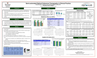
  • 1. Semi-automated Optical Coherence Tomography in Clinical Practice Zachary Goodman1, Samantha Lancia1, Gautam Adusumilli1, Courtney Dula1, Paul Kealey2, Robert T. Naismith1 1Washington University in Saint Louis School of Medicine, 2Optovue •  Assess feasibility of a portable OCT device (Optovue iScan 500) as a tool for measuring retinal nerve fiber layer (RNFL) thickness, retinal (macular) thickness and volume, and ganglion cell complex (GCC) thickness in multiple sclerosis patients with and without self reported ON •  Assess the relationship of the Pelli-Robson contrast scores and thickness measures obtained from OCT •  Recent reports suggest retinal and macular thickness is a biomarker of neurodegenerative disease progression within the eyes of patients with multiple sclerosis (MS) without optic neuritis (ON) •  Optical Coherence Tomography (OCT) can be a useful clinical measure obtained as part of regular clinical care for monitoring prognosis and disease activity •  Traditional OCT scanners require considerable technical skill to perform, and are not very portable •  We tested a compact, semi-automated OCT scanner for routine use within an MS center Background Objectives Methods Results •  Cross-sectional study of 89 MS patients (178 eyes) with and without prior self-reported history of optic neuritis •  RNFL thickness, macular thicknesses and volumes, and GCC thicknesses collected using the Spectral-Domain Optovue iScan 500 OCT scanner •  15 patients (30 eyes) excluded due to prior ophthalmologic conditions •  7 eyes excluded due to insufficient scan quality •  4 eyes excluded due to missing scans •  RNFL and Macular Volumes were available from 71 MS patients (137 eyes) •  GCC scans were introduced later within the protocol; pilot data was acquired for 23 patients (46 eyes) •  4 patients (8 eyes) excluded due to prior ophthalmologic conditions; 19 patients (38 eyes) analyzed •  Contrast sensitivity measured for each eye with a Pelli-Robson (PR) contrast chart •  Mean group differences in Pelli-Robson scores, average RNFL Thickness, average Inner GCC Thickness, and Macular Volume were assessed by ANOVA •  Correlations between OCT measurements and Pelli-Robson scores were analyzed using Spearman correlations Discussion A B A Subgroups N Spearman’s Correlations All Eyes (Patients) 137 (71) ρ = 0.38** Not Affected Eyes (No ON) 84 ρ = 0.33** Unaffected Eyes (ON-) •  Inferior •  Superior •  Temporal 16 ρ = 0.59* ρ = 0.55* ρ = 0.52* ρ = 0.54* Affected Eyes (ON+) •  Inferior •  Nasal 37 ρ = 0.33* ρ = 0.42** ρ = 0.38* Subgroups N Spearman’s Correlations All Eyes (Patients) 38 (19) ρ = 0.47** Not Affected Eyes (No ON) 24 ρ = 0.31 Unaffected Eyes (ON-) 3 ρ = 0.87 Affected Eyes (ON+) 11 ρ = 0.51 Age  in   years,   mean  ±   SD   (range) Disease   Duration   in  years,   mean  ±   SD   (range) Female,   n  (%) Ethnicity   (Caucasian,   African-­‐ American,   Other),  n  (%) Types  of  MS   (RR/SP/PP/ Unknown),   n  (%) Number  of  ON   episodes:  n  =  0,   n=  1,  n  >  1                 1.  Right  Eye               2.  Left  Eye ON  History   Classification:   No  ON/ON+/ON-­‐   1.  Right  Eye               2.  Left  Eye RNFL  n  =  71   (137  eyes) 48  ±  11   (23  -­‐  74) 11  ±  9   (0.2  -­‐  34) 49   (69%) 57/12/2   (80/17/3) 63/3/4/1   (89/4/6/1) 1.  50/14/4                 2.  50/16/3 1.  41/18/9                   2.  43/19/7 GCC  n  =  19   (38  eyes) 48  ±  10   (32  -­‐  65) 13  ±  10   (0.6  -­‐  34) 14   (74%) 15/2/2   (79/11/11) 16/1/2/0   (84/5/11/0) 1.  15/3/1                     2.  12/6/1 1.  12/4/3                       2.  12/7/0   1.82 1.86 1.68 1.86 1.75 1.69 1.5 1.6 1.7 1.8 1.9 Not Affected Unaffected Affected Pelli-RobsonScore Optic Neuritis History Left Eye Right Eye Table 1: Demographic summary statistics of Retinal Nerve Fiber Layer (RNFL) and Ganglion Cell Complex (GCC) eyes Figure 1: Optovue OCT iScan 500 Table 2: Means and Standard Deviations of Group differences between MS Patients’Affected Eyes of Pelli Robson scores, RNFL & GCC stratified by prior ON history Figure 2: Bar graph of mean Pelli-Robson score broken down by Optic Neuritis History and Eye Retinal Nerve Fiber Layer (RNFL) Figure 3: RNFL Thickness stratified by prior ON history and quadrant Table 3: Spearman’s Correlations between RNFL Thickness and Pelli-Robson score Non-significant correlations not reported. ** Correlation is significant at the 0.01 level (2-tailed) * Correlation is significant at the 0.05 level (2-tailed) Figure 4: Correlation scatter plots between Pelli-Robson score and RNFL Thickness Ganglion Cell Complex (GCC) Figure 5: RNFL Thickness stratified by prior ON history and quadrant Table 4: Spearman’s Correlations between GCC Thickness and Pelli-Robson score Figure 6: Correlation scatter plots between Pelli-Robson score and GCC Thickness ** Correlation is significant at the 0.01 level (2-tailed) Parafovea Volume (mm3) (mean ± SD) Perifovea Volume (mm3) (mean ± SD) No ON ON- ON+ Sig No ON ON- ON+ Sig Inferior 0.47 ± 0.03 0.47 ± 0.04 0.45 ± 0.03 0.03a 0.83 ± 0.05 0.82 ± 0.07 0.80 ± 0.05 <0.01a Superior 0.47 ± 0.03 0.47 ± 0.04 0.46 ± 0.03 0.04a 0.86 ± 0.05 0.86 ± 0.08 0.84 ± 0.06 NS Nasal 0.48 ± 0.03 0.48 ± 0.04 0.46 ± 0.03 NS 0.89 ± 0.06 0.89 ± 0.09 0.85 ± 0.06 <0.01a Temporal 0.46 ± 0.03 0.46 ± 0.04 0.44 ± 0.03 0.02a 0.83 ± 0.05 0.82 ± 0.07 0.80 ± 0.05 0.03a Results Continued Macula of Retina Table 6: Means and Standard Deviations of Full Retinal Volumes and Thicknesses of macular quadrants stratified by prior ON history Mean differences evaluated using one-way ANOVA between Not Affected (No ON), Unaffected (ON-), and Affected (ON+) eyes of MS patients Bold values indicate statistically significant differences by post-hoc analysis with Bonferroni correction. a = Significant mean difference between No ON and ON+ •  Optovue iScan 500 is a reliable evaluation tool in measuring retinal parameters within an MS center •  Capable of stratifying patients based on prior optic neuritis history •  GCC thickness is available at the time of acquisition, and requires no further analysis or post-processing •  Pelli-Robson is feasible to acquire as a single contrast assessment in MS patients’ eyes with and without optic neuritis •  Pelli-Robson score, RNFL Thickness, GCC Thickness, and Macular Thickness (Parafovea and Perifovea) were all significantly reduced in Affected (ON+) eyes in patients with prior ON •  Unaffected (ON-) eyes were relatively spared in thinning during an ON episode except in RNFL •  Optovue is a semi-automated device which can provide detailed information about RNFL, GCC, and macular volume in approximately 10 minutes Figure 7: Representative map of macular regions Zachary Goodman Washington University in St. Louis goodmanz@neuro.wustl.edu ZG: None, SL: None, GA: None, CD: None, PK: Optovue, RTN: Alkermes, Acorda, Bayer, Biogen, Genentech, Genzyme, EMD Serono, Mallinckrodt, Novartis, Pfiser, Teva Optovue provided the OCT scanner for evaluation. This investigation was conducted independently from the manufacturer. No ON ON- ON+ Sig Macular Volume (mm3 ) 5.29 ± 0.30 5.28 ± 0.47 5.10 ± 0.33 0.02 a •  Parafovea 1.88 ± 0.12 1.89 ± 0.17 1.81 ± 0.12 0.03 a •  Perifovea 3.41 ± 0.19 3.39 ± 0.30 3.28 ± 0.22 0.01 a Macular Thickness (µm) 285 ± 22 285 ± 30 275 ± 23 <0.01 a •  Parafovea 299 ± 18 300 ± 28 289 ± 18 0.03 a •  Perifovea 271 ± 15 270 ± 24 261 ± 17 <0.01 a Parafovea Thickness (µm) (mean ± SD) Perifovea Thickness (µm) (mean ± SD) No ON ON- ON+ Sig No ON ON- ON+ Sig Inferior 296 ± 18 298 ± 28 286 ± 18 0.02a 265 ± 16 262 ± 23 254 ± 17 <0.01a Superior 300 ± 19 301 ± 28 290 ± 20 0.04a 272 ± 15 273 ± 25 266 ± 19 NS Nasal 306 ± 20 309 ± 28 296 ± 19 NS 284 ± 19 284 ± 27 270 ± 20 <0.01a Temporal 294 ± 18 293 ± 28 283 ± 19 0.02a 264 ± 17 261 ± 23 255 ± 16 0.03a Table 5: Means±Standard Deviations of Full Retinal Total Volume and Average Thickness Mean differences evaluated using one-way ANOVA between Not Affected (No ON), Unaffected (ON-), and Affected (ON+) eyes of MS patients Bold values indicate statistically significant differences by post-hoc analysis with Bonferroni correction. a = Significant mean difference between No ON and ON+ Mean differences evaluated using one-way ANOVA between Not Affected (No ON), Unaffected (ON-), and Affected (ON+) eyes of MS patients Bold values indicate statistically significant differences by post-hoc analysis with Bonferroni correction. a = Significant mean difference between No ON and ON+; b = Significant mean difference between ON- and ON+ No ON ON- ON+ Sig Pelli-Robson 1.84  +  0.20   1.80  +  0.21   1.68  +  0.24   0.001 a RNFL (µm) 92.6  +  10.6   91.8  +  5.8   78.4  +  15.6   <0.001 a , <0.01 b GCC (µm) 87.7  +  9.2   81.1  +  7.4   77.2  +  9.1   <0.01 a Not Affected (No ON) Unaffected (ON-) Affected (ON+) Not Affected (No ON) Unaffected (ON-) Affected (ON+)