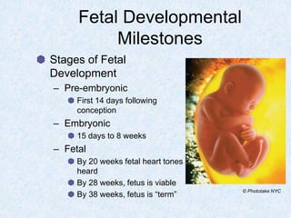
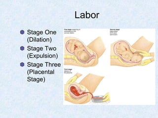
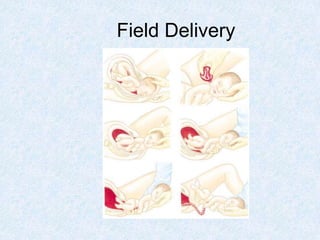
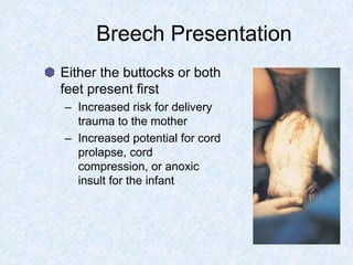
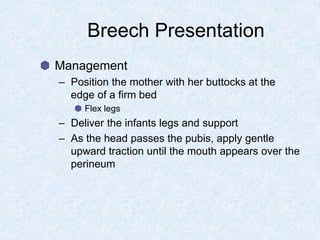
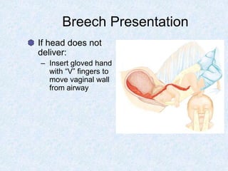
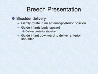
Paramedic Care:
Principles & Practice
Volume 6
Medical Emergencies
Chapter 2
Obstetrics
Topics
The Prenatal Period
General Assessment
General Management
Complications of Pregnancy
Abnormal Delivery Situations
Other Delivery Complications
Maternal Complications of Labor and
Delivery
Introduction
Childbirth occurs daily, usually requiring
only the most basic assistance
Childbirth complications include:
– Preterm labor, multiple births, abnormal
presentations, bleeding, or distressed neonates
Complications of pregnancy are uncommon
– Hypertension, eclampsia, diabetes,
cardiovascular, and trauma are causes
The Prenatal Period
The Prenatal Period
The prenatal period is the time from
conception until delivery of the fetus.
Significant physiological changes occur in
the mother.
– The result of fetal development
Anatomy and Physiology of
the Obstetric Patient
Ovulation
Fertilization
– Normally occurs
in the fallopian
tube
– Blastocyst forms
Implants in the
thickened uterine
lining
Anatomy and Physiology of the
Obstetric Patient
Placenta
– Organ of pregnancy
– Function
Exchanges heat
Exchanges oxygen for carbon dioxide
Delivers nutrients
Removes wastes
– Endocrine function
Anatomy and Physiology of the
Obstetric Patient
Uterus
– Normal capacity 10 mL
– By term
2 pounds
5 Liters
Contains 1/6 of Mom’s blood volume
Anatomy and Physiology of the
Obstetric Patient
Umbilical cord
– Two arteries
and one vein
Vein transports
oxygen rich
blood
Amniotic sac
– “Bag of waters”
– Contains
amniotic fluid
Physiologic Changes
of Pregnancy
Reproductive System
– Uterus increases in size
– Vascular system
– Formation of mucous
plug in cervix
– Estrogen causes vaginal
mucosa to thicken
– Breast enlargement
Physiologic Changes
of Pregnancy
Respiratory System
– Progesterone causes a decrease in airway
resistance
Results in a 20 percent increase in oxygen
consumption and a 40 percent increase in tidal volume
– Slight increase in respiratory rate
Physiologic Changes
of Pregnancy
Cardiovascular System
– Cardiac output increases
– Blood volume increases
Anemia
– Supine hypotension
Physiologic Changes
of Pregnancy
Gastrointestinal System
– Hormone levels
– Peristalsis is slowed
Urinary System
– Urinary frequency is common
Musculoskeletal System
– Loosened pelvic joints
Conception
Normal duration of
pregnancy is 40 weeks
– Estimated date of
confinement (EDC)
– Trimesters
Fetal Developmental
Milestones
2 Weeks Pregnant
Mom has no clue yet
Time of missed menstrual cycle
Positive prego test
4 Weeks Pregnant
Fetal heart beating
8 Weeks
Baby 1.2 inches long
Mom feels like crap
8-12 Weeks
Mom still feels like crap
Baby systems formed
Making urine
13 Weeks
End of 1st trimester
Mom feels better
Less risk of miscarriage
16 Weeks
Baby’s sex determined
2nd ultra sound
20 Weeks
Fetal heart tones present
Baby 8 inches long; 16
ounces
Baby kicking
24 Weeks
Baby breathing fluid
Baby viable
Baby over 1 pound
28 Weeks
Surfactant being formed
Baby 2 -3 pounds
32 Weeks
Baby 3 – 4 pounds
Baby getting some fat
38 – 40 Weeks
Baby term
General Assessment of
the Obstetric Patient
Physical Examination
– Assess fundal height to determine gestation
Fetal Developmental
Milestones
Stages of Fetal
Development
– Pre-embryonic
First 14 days following
conception
– Embryonic
15 days to 8 weeks
– Fetal
By 20 weeks fetal heart tones
heard
By 28 weeks, fetus is viable
By 38 weeks, fetus is “term”
© Phototake NYC
Fetal Circulation
Umbilical vein supplies
blood to fetus
– Connects directly to the
inferior vena cava
Ductus venosus
– Foramen ovale
– Ductus arteriosus
Allows oxygenated blood to
bypass the lungs of fetus
– Changes at birth
General Assessment of
the Obstetric Patient
General Assessment of
the Obstetric Patient
General Information
– Mother’s gravidity and parity
– Length of gestation
– Estimated date of confinement (EDC)
– Complications
– Prenatal care
– State of health
General Assessment of
the Obstetric Patient
Preexisting or Aggravated Conditions
– Diabetes
Diabetics become unstable during pregnancy
Larger baby – shoulder dystocia
Gestational diabetes
Must be treated with insulin
Oral meds cross placenta
– Heart Disease
Congestive heart failure may develop
General Assessment of
the Obstetric Patient
Preexisting or Aggravated Conditions (cont.)
– Hypertension
Common medications may not be used
Pre-eclampsia may develop
– Seizure Disorders
Increased seizures
– Neuromuscular Disorders
Patients may have remission of symptoms during
pregnancy
General Assessment of
the Obstetric Patient
Pain
– Determine when the pain started
Sudden onset or slow
– Duration, location, and radiation
Vaginal Bleeding
– Gain information about the color, amount, and
duration
Assess number of sanitary pads used
– Transport any tissue or clots
General Assessment of
the Obstetric Patient
Active Labor
– Need to push?
– Need to have BM?
– Membranes ruptured?
What did it look like?
General Assessment of
the Obstetric Patient
Physical Examination (cont.)
– Vital signs (lying on left side)
– Evaluate:
vaginal discharge
progression of labor
prolapsed cord
crowning
– Never perform an internal vaginal exam in
the field.
General Management of
the Obstetric Patient
General Management of
the Obstetric Patient
Always remember that you are caring for
two patients, the mother and the fetus.
Maintain the airway, breathing, and
circulation.
– Oxygen administration, IV access
Patient positioning
Transport considerations
Complications of Pregnancy
Trauma
– Pregnant patients are more susceptible to life-
threatening injury
Increased vascularity of the gravid uterus
Transport all trauma patients at 20 weeks or more
gestation
– Anticipate the development of shock
Overt signs of shock are late
– Direct abdominal trauma
Premature separation of the placenta from the uterine
wall, premature labor, abortion, uterine rupture, and
possibly fetal death
Trauma Management
Immobilize as necessary
Administer high-flow oxygen
Initiate two large-bore IVs per protocol
LSB tilted to the left to minimize supine
hypotension
Reassess patient
Monitor the fetus
Medical Conditions
Abdominal pain is a common complaint
– Appendicitis or cholecystitis
Displacement of abdominal organs complicates
assessment
Symptoms of acute cholecystitis may differ from those
in nonpregnant patients
Any pregnant patient with abdominal pain
should be evaluated by a physician.
Bleeding In Pregnancy
Causes
– Abortion
– Ectopic Pregnancy
– Placenta Previa
– Abruptio Placentae
Vaginal bleeding is associated with
potential fetal loss
Abortion
Termination of pregnancy before the 20th
week of gestation
Assessment
– Cramping, abdominal pain, backache, and
vaginal bleeding
– Passage of clots and tissue
Management
– Treat for shock
– Provide emotional support
Ectopic Pregnancy
Abnormal implantation of
the fertilized egg outside
of the uterus
– Most commonly in the
fallopian tube
– Accounts for approximately
10 percent of maternal
mortality
Ectopic Pregnancy
Assessment
– Presence of abdominal pain
– Woman often reports that she missed a period
– The abdomen becomes rigid and the pain
intensifies
– Signs of shock
Management
– Transport this patient immediately
– Treat for shock
Consider use of PASG
Placenta Previa
Abnormal implantation of the placenta on
the lower half of the uterine wall
Contractions pull placenta from uterine wall
– Results in bleeding
Placenta Previa
Placenta Previa
Assessment
– Usually a multigravida in her third trimester of
pregnancy
May have history of placenta previa
– Recent episode of sexual intercourse or vaginal
examination
– Painless bright red vaginal bleeding
Management
– Treat for shock
– Transport to appropriate facility
Abruptio Placentae
Premature separation (abruption) of a
normally implanted placenta from the
uterine wall
Abruptio Placentae
Abruptio Placentae
Predisposing factors
– Multiparity, maternal hypertension, trauma,
cocaine use, increasing maternal age, and
history of abruption in previous pregnancy
20 to 30% fetal mortality
– Increases to 100% when majority of the
placenta has separated
Abruptio Placentae
Assessment
– Varies depending on the extent and character
Partial
Marginal is characterized by vaginal bleeding but no increase in
pain
Central is characterized by sudden sharp, tearing pain and
development of a stiff, board-like abdomen
Complete
Massive vaginal bleeding and profound maternal hypotension
Abruptio Placentae
Management
– Immediate intervention to maintain maternal
oxygenation and perfusion
Oxygenation and intravenous fluid therapy
– Left-lateral recumbent position
– Transport to appropriate facility
Medical Complications
of Pregnancy
Hypertensive Disorders
Supine Hypotensive Syndrome
Gestational Diabetes
Hypertensive Disorders
of Pregnancy
Pre-eclampsia and Eclampsia
– Mild or Severe
– Increase in systolic blood pressure by 30 mmHg
and/or a diastolic increase of 15 mmHg
Two or more occasions
– Most commonly seen in the last 10 weeks of
gestation, during labor, or in the first 48 hours
postpartum
Hypertensive Disorders
of Pregnancy
Preeclampsia and Eclampsia (cont.)
– Mild pre-eclampsia
Hypertension, edema, and protein in the urine
– Severe pre-eclampsia
Maternal blood pressure reaches 160/110 or higher,
generalized edema, large amounts of protein in urine
Other symptoms include headache, visual
disturbances, hyperactive reflexes, and the
development of pulmonary edema
Hypertensive Disorders
of Pregnancy
Pre-eclampsia and Eclampsia (cont.)
– Eclampsia is characterized by SEIZURES
– Patient is usually grossly edematous and has
markedly elevated blood pressure
Hypertensive Disorders
of Pregnancy
Chronic hypertension
– Blood pressure is 140/90 before pregnancy or
prior to the 20th week of gestation
Chronic hypertension superimposed with
pre-eclampsia
Transient hypertension
– A temporary rise in blood pressure which occurs
during labor
Hypertensive Disorders
of Pregnancy
Assessment
– Obtain history
– Physical Exam
Markedly edematous
Pale and apprehensive
Hypereactive reflexes
Elevated blood pressure
Hypertensive Disorders
of Pregnancy
Management
– Hypertension
Closely monitor mother and fetus
– Pre-eclampsia
Keep the patient calm
Dim the lights
Left lateral positioning
IV access
Administration of magnesium sulfate
– Eclampsia
Administer oxygen
Bolus dose of magnesium sulfate
Diazepam as indicated
Supine Hypotensive Syndrome
Gravid uterus compresses the inferior
vena cava
– Mother in the supine position
Assessment
– History of supine positioning
– Patient may complain of dizziness
– Question any recent hemorrhage or fluid loss
Management
– Place the patient in the left lateral recumbent
position or elevate her right hip
– Monitor fetal heart tones and maternal vital
signs
– Transport left-lateral recumbancy
Supine Hypotensive Syndrome
Gestational Diabetes
Hormonal influences cause an increase in
insulin production
– Initially, an increased tissue response to insulin
– During the last 20 weeks placental hormones
cause an increased resistance to insulin and
decreased glucose metabolism
Management requires good prenatal care
Gestational Diabetes
Assessment
– Altered mental status
Always suspect hypoglycemia
– Diaphoresis, tachycardia, N/V, increased
urination
Gestational Diabetes
Management
– If blood glucose less than 60
Obtain blood sample
Administer D50%
Consider oral glucose depending on patient LOC
– If blood glucose greater than 200
Obtain blood sample
Give fluid bolus of 1-2 liters
Medical control may order insulin if delayed or long
transport
Braxton-Hicks Contractions
False labor
Virtually impossible to distinguish false labor
from true labor in the field
Transport patient for evaluation
– Requires examination of the cervix
WHICH WE DO NOT DO
Preterm Labor
Maternal Factors
– Cardiovascular disease, renal disease,
diabetes, uterine and cervical abnormalities,
maternal infection, trauma, contributory factors
Placental Factors
– Placenta previa
– Abruptio placentae
Fetal Factors
– Multiple gestation
– Excessive amniotic fluid
– Fetal infection
Preterm Labor
Assessment
– Determine the approximate gestational age of
the fetus
<38 weeks is pre-term
– Obtain a brief obstetrical history
Ruptured membranes
Need to push or move bowels
Note the intensity and length of the contractions and
contraction interval
– Other common complaints
Preterm Labor
Management
– Preterm labor should be stopped if possible
Tocolysis
Narcotic or barbiturate administration and rest
Fluid bolus administration
Magnesium sulfate or beta agonist administration
– Tocolysis in the field is limited to sedation and
hydration
Labor
Stage One
(Dilation)
Stage Two
(Expulsion)
Stage Three
(Placental
Stage)
Management of
a Patient in Labor
Transport the patient in labor unless delivery
is imminent
Considerations
– Patient’s number of previous pregnancies
– Length of labor during the previous pregnancies
– Frequency of contractions
– Maternal urge to push
– Presence of crowning
Management of
a Patient in Labor
Factors indicating rapid transport despite
imminent delivery
– Prolonged rupture of membranes (> 24 hours)
– Abnormal presentation, such as breech or
transverse
– Prolapsed cord or fetal distress
– Multiple fetuses
Field Delivery
Set up delivery area
Give oxygen to
mother and start IV-
NS TKO
Drape mother with
toweling from OB kit
Monitor fetal heart
rate
As head crowns,
apply gentle
pressure
Suction the mouth
and then the nose
Clamp and cut the
cord
Dry the infant and
keep it warm
Deliver the placenta
and save for
transport with the
mother
Field Delivery
Neonatal Care
Routine neonatal
care
– Maintain warmth
– Clear infant’s
airway by
suctioning mouth
and nose
– Assess the neonate
using APGAR
score
APGAR Scoring
Neonatal Resuscitation
Factors that contribute to the need for
resuscitation
– Prematurity, pregnancy and delivery
complications, maternal health problems, or
inadequate prenatal care
Tactile stimulation
– If respiratory effort insufficient, assist ventilations
with BVM and oxygen
Reassess after 15-30 seconds
Continue as necessary with ventilations
Neonatal Resuscitation
Assess heart rate
– Use a stethoscope to auscultate the apical pulse
If the pulse is 100 or greater with spontaneous
respirations, continue your assessment
If less than 100 - VENTILATE
If less than 60 -
COMPRESSIONS
Neonatal Resuscitation
Transport to appropriate facility
Establish intravenous access
– Umbilical vein
– Peripheral vein
– Intraosseous
– Endotracheal drug administration for cardiac
drugs
Maintain warmth, support ventilations,
oxygenation, and circulation enroute
Abnormal Delivery Situations
Breech
Prolapsed Cord
Limb Presentation
Other Abnormal Presentations
Breech Presentation
Breech Presentation
Either the buttocks or both
feet present first
– Increased risk for delivery
trauma to the mother
– Increased potential for cord
prolapse, cord
compression, or anoxic
insult for the infant
Breech Presentation
Management
– Position the mother with her buttocks at the
edge of a firm bed
Flex legs
– Deliver the infants legs and support
– As the head passes the pubis, apply gentle
upward traction until the mouth appears over the
perineum
Breech Presentation
If head does not
deliver:
– Insert gloved hand
with “V” fingers to
move vaginal wall
from airway
Breech Presentation
Shoulder delivery
– Gently rotate in an anterior-posterior position
– Guide infants body upward
Deliver posterior shoulder
– Guide infant downward to deliver anterior
shoulder
Prolapsed Cord
Occurs when the umbilical cord precedes
the fetal presenting part
– Cord is compressed between the fetus and the
bony pelvis
Impedes circulation
Predisposing factors
– Prematurity
– Multiple births
– Premature rupture of the membranes
Prolapsed Cord
Management
Limb Presentation
Baby is in a transverse lie across the uterus
– Arm or leg is presenting part
Predisposing factors
– Preterm birth and multiple gestation
Management
– Under no circumstance should you attempt a
field delivery
– Assist the mother into a knee-chest position
– Administer oxygen and transport immediately
Other Abnormal Presentations
Occiput-posterior positioning
– Passage through the pelvis is delayed
Most frequent with primigravidas
– Vaginal delivery is impossible in these cases
Management
– Early recognition
– Reassure mother
– Transport with oxygen administration
Amniotic Sac presentation
Other Delivery Complications
Multiple Births
Cephalopelvic Disproportion
Precipitous Delivery
Shoulder Dystocia
Meconium Staining
Multiple Births
Multiple births should also be suspected if
the mother’s abdomen remains large after
delivery of one baby and labor continues
Management
– Normal delivery guidelines
Additional personnel necessary
– Low birth weight increases chance of
hypothermia
Maintain warmth
Cephalopelvic Disproportion
Infant’s head is too big to pass through
pelvis easily
– If not recognized, can cause uterine rupture
Causes include oversized fetus,
hydrocephalus, conjoined twins, or fetal
tumors
Management
– Give oxygen to mother and start IV
– Rapid transport
Precipitous Delivery
Delivery occurs in less than 3 hours of labor
– Usually in patients in grand multipara
– Complications
Fetal trauma, tearing of cord, or maternal lacerations
Management
– Be ready for rapid delivery and attempt to
control the head
– Keep the baby warm
Shoulder Dystocia
Infant’s shoulders are
larger than its head
– Most frequently with
diabetic and obese
mothers and in post-term
pregnancies
During delivery, head
retracts back into
perineum
– Turtle sign
Shoulder Dystocia
Management
– Do not pull on the infant’s head
– McRobert’s Manuever
Flex legs, drop mother’s buttocks off bed
Apply pressure to immediately above symphysis pubis
– If delivery delayed, transport immediately
McRobert’s Maneuver
Meconium Staining
Fetus passes feces into the amniotic fluid
– Indicative of a fetal hypoxic incident
– Risk of aspiration
Management
– Suction the mouth and nose on the perineum
– If staining is light, no further care
– If staining is thick:
Visualize the glottis and suction the hypopharynx and
trachea
Utilize endotracheal tube
Maternal Complications
of Labor and Delivery
Maternal Complications
of Labor and Delivery
Postpartum Hemorrhage
Uterine Rupture
Uterine Inversion
Pulmonary Embolism
Postpartum Hemorrhage
Defined as a loss of more than 500 cc of
blood following delivery
– Most common cause is uterine atony
Multigravida
Precipitous deliveries and prolonged labors
– May occur up to 2 weeks post-delivery
Postpartum Hemorrhage
Assessment
– Rely on the clinical appearance of the patient
and her vital signs
– Assess number of sanitary pads used
– “Boggy” uterus
– Examine the perineum for evidence of traumatic
injury
Postpartum Hemorrhage
Management
– Administer oxygen and begin fundal massage
– Establish IV access
– PASG
– Administration of Pitocin may be indicated
10-20 units in 1 liter of NaCl
Run at 125 cc/hr
May administer 10 units intramuscularly, if necessary
Uterine Rupture
Tearing, or rupture, of the uterus
Patient complains of severe abdominal pain
and will often be in shock. Abdomen is often
tender and rigid.
Fetal heart tones are absent
Management
– Treat for shock
– Give high-flow, high-concentration oxygen and
start two large-bore IVs of normal saline
– Transport patient rapidly
Uterine Inversion
Uterus turns inside out after delivery and
extends through the cervix
– Blood loss ranges from 800 to 1,800 cc
Management
– Place the patient in a supine position and begin
oxygen administration
– Begin fluid resuscitation
– Make one attempt to replace the uterus
– If unsuccessful, transport immediately with moist
towels covering exposed uterus
Pulmonary Embolism
Presents with sudden severe dyspnea and
sharp chest pain
– Tachycardia, tachypnea, jugular vein distention,
and, in severe cases, hypotension
Management
– Administer high-flow, high-concentration oxygen
and support ventilations as needed
– Establish an IV of normal saline
– Transport immediately, monitoring the heart,
vital signs, and oxygen saturation

OB- TCC

  • 1.
    Paramedic Care: Principles &Practice Volume 6 Medical Emergencies
  • 2.
  • 3.
    Topics The Prenatal Period GeneralAssessment General Management Complications of Pregnancy Abnormal Delivery Situations Other Delivery Complications Maternal Complications of Labor and Delivery
  • 4.
    Introduction Childbirth occurs daily,usually requiring only the most basic assistance Childbirth complications include: – Preterm labor, multiple births, abnormal presentations, bleeding, or distressed neonates Complications of pregnancy are uncommon – Hypertension, eclampsia, diabetes, cardiovascular, and trauma are causes
  • 5.
  • 6.
    The Prenatal Period Theprenatal period is the time from conception until delivery of the fetus. Significant physiological changes occur in the mother. – The result of fetal development
  • 7.
    Anatomy and Physiologyof the Obstetric Patient Ovulation Fertilization – Normally occurs in the fallopian tube – Blastocyst forms Implants in the thickened uterine lining
  • 8.
    Anatomy and Physiologyof the Obstetric Patient Placenta – Organ of pregnancy – Function Exchanges heat Exchanges oxygen for carbon dioxide Delivers nutrients Removes wastes – Endocrine function
  • 9.
    Anatomy and Physiologyof the Obstetric Patient Uterus – Normal capacity 10 mL – By term 2 pounds 5 Liters Contains 1/6 of Mom’s blood volume
  • 10.
    Anatomy and Physiologyof the Obstetric Patient Umbilical cord – Two arteries and one vein Vein transports oxygen rich blood Amniotic sac – “Bag of waters” – Contains amniotic fluid
  • 11.
    Physiologic Changes of Pregnancy ReproductiveSystem – Uterus increases in size – Vascular system – Formation of mucous plug in cervix – Estrogen causes vaginal mucosa to thicken – Breast enlargement
  • 12.
    Physiologic Changes of Pregnancy RespiratorySystem – Progesterone causes a decrease in airway resistance Results in a 20 percent increase in oxygen consumption and a 40 percent increase in tidal volume – Slight increase in respiratory rate
  • 13.
    Physiologic Changes of Pregnancy CardiovascularSystem – Cardiac output increases – Blood volume increases Anemia – Supine hypotension
  • 14.
    Physiologic Changes of Pregnancy GastrointestinalSystem – Hormone levels – Peristalsis is slowed Urinary System – Urinary frequency is common Musculoskeletal System – Loosened pelvic joints
  • 15.
    Conception Normal duration of pregnancyis 40 weeks – Estimated date of confinement (EDC) – Trimesters Fetal Developmental Milestones
  • 17.
    2 Weeks Pregnant Momhas no clue yet Time of missed menstrual cycle Positive prego test
  • 18.
  • 19.
    8 Weeks Baby 1.2inches long Mom feels like crap
  • 20.
    8-12 Weeks Mom stillfeels like crap Baby systems formed Making urine
  • 21.
    13 Weeks End of1st trimester Mom feels better Less risk of miscarriage
  • 22.
    16 Weeks Baby’s sexdetermined 2nd ultra sound
  • 23.
    20 Weeks Fetal hearttones present Baby 8 inches long; 16 ounces Baby kicking
  • 24.
    24 Weeks Baby breathingfluid Baby viable Baby over 1 pound
  • 25.
    28 Weeks Surfactant beingformed Baby 2 -3 pounds
  • 26.
    32 Weeks Baby 3– 4 pounds Baby getting some fat
  • 27.
    38 – 40Weeks Baby term
  • 28.
    General Assessment of theObstetric Patient Physical Examination – Assess fundal height to determine gestation
  • 30.
    Fetal Developmental Milestones Stages ofFetal Development – Pre-embryonic First 14 days following conception – Embryonic 15 days to 8 weeks – Fetal By 20 weeks fetal heart tones heard By 28 weeks, fetus is viable By 38 weeks, fetus is “term” © Phototake NYC
  • 31.
    Fetal Circulation Umbilical veinsupplies blood to fetus – Connects directly to the inferior vena cava Ductus venosus – Foramen ovale – Ductus arteriosus Allows oxygenated blood to bypass the lungs of fetus – Changes at birth
  • 33.
    General Assessment of theObstetric Patient
  • 34.
    General Assessment of theObstetric Patient General Information – Mother’s gravidity and parity – Length of gestation – Estimated date of confinement (EDC) – Complications – Prenatal care – State of health
  • 35.
    General Assessment of theObstetric Patient Preexisting or Aggravated Conditions – Diabetes Diabetics become unstable during pregnancy Larger baby – shoulder dystocia Gestational diabetes Must be treated with insulin Oral meds cross placenta – Heart Disease Congestive heart failure may develop
  • 36.
    General Assessment of theObstetric Patient Preexisting or Aggravated Conditions (cont.) – Hypertension Common medications may not be used Pre-eclampsia may develop – Seizure Disorders Increased seizures – Neuromuscular Disorders Patients may have remission of symptoms during pregnancy
  • 37.
    General Assessment of theObstetric Patient Pain – Determine when the pain started Sudden onset or slow – Duration, location, and radiation Vaginal Bleeding – Gain information about the color, amount, and duration Assess number of sanitary pads used – Transport any tissue or clots
  • 38.
    General Assessment of theObstetric Patient Active Labor – Need to push? – Need to have BM? – Membranes ruptured? What did it look like?
  • 39.
    General Assessment of theObstetric Patient Physical Examination (cont.) – Vital signs (lying on left side) – Evaluate: vaginal discharge progression of labor prolapsed cord crowning – Never perform an internal vaginal exam in the field.
  • 40.
    General Management of theObstetric Patient
  • 41.
    General Management of theObstetric Patient Always remember that you are caring for two patients, the mother and the fetus. Maintain the airway, breathing, and circulation. – Oxygen administration, IV access Patient positioning Transport considerations
  • 42.
  • 43.
    Complications of Pregnancy Trauma –Pregnant patients are more susceptible to life- threatening injury Increased vascularity of the gravid uterus Transport all trauma patients at 20 weeks or more gestation – Anticipate the development of shock Overt signs of shock are late – Direct abdominal trauma Premature separation of the placenta from the uterine wall, premature labor, abortion, uterine rupture, and possibly fetal death
  • 44.
    Trauma Management Immobilize asnecessary Administer high-flow oxygen Initiate two large-bore IVs per protocol LSB tilted to the left to minimize supine hypotension Reassess patient Monitor the fetus
  • 45.
    Medical Conditions Abdominal painis a common complaint – Appendicitis or cholecystitis Displacement of abdominal organs complicates assessment Symptoms of acute cholecystitis may differ from those in nonpregnant patients Any pregnant patient with abdominal pain should be evaluated by a physician.
  • 46.
    Bleeding In Pregnancy Causes –Abortion – Ectopic Pregnancy – Placenta Previa – Abruptio Placentae Vaginal bleeding is associated with potential fetal loss
  • 47.
    Abortion Termination of pregnancybefore the 20th week of gestation Assessment – Cramping, abdominal pain, backache, and vaginal bleeding – Passage of clots and tissue Management – Treat for shock – Provide emotional support
  • 48.
    Ectopic Pregnancy Abnormal implantationof the fertilized egg outside of the uterus – Most commonly in the fallopian tube – Accounts for approximately 10 percent of maternal mortality
  • 49.
    Ectopic Pregnancy Assessment – Presenceof abdominal pain – Woman often reports that she missed a period – The abdomen becomes rigid and the pain intensifies – Signs of shock Management – Transport this patient immediately – Treat for shock Consider use of PASG
  • 50.
    Placenta Previa Abnormal implantationof the placenta on the lower half of the uterine wall Contractions pull placenta from uterine wall – Results in bleeding
  • 51.
  • 52.
    Placenta Previa Assessment – Usuallya multigravida in her third trimester of pregnancy May have history of placenta previa – Recent episode of sexual intercourse or vaginal examination – Painless bright red vaginal bleeding Management – Treat for shock – Transport to appropriate facility
  • 53.
    Abruptio Placentae Premature separation(abruption) of a normally implanted placenta from the uterine wall
  • 54.
  • 55.
    Abruptio Placentae Predisposing factors –Multiparity, maternal hypertension, trauma, cocaine use, increasing maternal age, and history of abruption in previous pregnancy 20 to 30% fetal mortality – Increases to 100% when majority of the placenta has separated
  • 56.
    Abruptio Placentae Assessment – Variesdepending on the extent and character Partial Marginal is characterized by vaginal bleeding but no increase in pain Central is characterized by sudden sharp, tearing pain and development of a stiff, board-like abdomen Complete Massive vaginal bleeding and profound maternal hypotension
  • 57.
    Abruptio Placentae Management – Immediateintervention to maintain maternal oxygenation and perfusion Oxygenation and intravenous fluid therapy – Left-lateral recumbent position – Transport to appropriate facility
  • 58.
    Medical Complications of Pregnancy HypertensiveDisorders Supine Hypotensive Syndrome Gestational Diabetes
  • 59.
    Hypertensive Disorders of Pregnancy Pre-eclampsiaand Eclampsia – Mild or Severe – Increase in systolic blood pressure by 30 mmHg and/or a diastolic increase of 15 mmHg Two or more occasions – Most commonly seen in the last 10 weeks of gestation, during labor, or in the first 48 hours postpartum
  • 60.
    Hypertensive Disorders of Pregnancy Preeclampsiaand Eclampsia (cont.) – Mild pre-eclampsia Hypertension, edema, and protein in the urine – Severe pre-eclampsia Maternal blood pressure reaches 160/110 or higher, generalized edema, large amounts of protein in urine Other symptoms include headache, visual disturbances, hyperactive reflexes, and the development of pulmonary edema
  • 61.
    Hypertensive Disorders of Pregnancy Pre-eclampsiaand Eclampsia (cont.) – Eclampsia is characterized by SEIZURES – Patient is usually grossly edematous and has markedly elevated blood pressure
  • 62.
    Hypertensive Disorders of Pregnancy Chronichypertension – Blood pressure is 140/90 before pregnancy or prior to the 20th week of gestation Chronic hypertension superimposed with pre-eclampsia Transient hypertension – A temporary rise in blood pressure which occurs during labor
  • 63.
    Hypertensive Disorders of Pregnancy Assessment –Obtain history – Physical Exam Markedly edematous Pale and apprehensive Hypereactive reflexes Elevated blood pressure
  • 64.
    Hypertensive Disorders of Pregnancy Management –Hypertension Closely monitor mother and fetus – Pre-eclampsia Keep the patient calm Dim the lights Left lateral positioning IV access Administration of magnesium sulfate – Eclampsia Administer oxygen Bolus dose of magnesium sulfate Diazepam as indicated
  • 65.
    Supine Hypotensive Syndrome Graviduterus compresses the inferior vena cava – Mother in the supine position
  • 66.
    Assessment – History ofsupine positioning – Patient may complain of dizziness – Question any recent hemorrhage or fluid loss Management – Place the patient in the left lateral recumbent position or elevate her right hip – Monitor fetal heart tones and maternal vital signs – Transport left-lateral recumbancy Supine Hypotensive Syndrome
  • 67.
    Gestational Diabetes Hormonal influencescause an increase in insulin production – Initially, an increased tissue response to insulin – During the last 20 weeks placental hormones cause an increased resistance to insulin and decreased glucose metabolism Management requires good prenatal care
  • 68.
    Gestational Diabetes Assessment – Alteredmental status Always suspect hypoglycemia – Diaphoresis, tachycardia, N/V, increased urination
  • 69.
    Gestational Diabetes Management – Ifblood glucose less than 60 Obtain blood sample Administer D50% Consider oral glucose depending on patient LOC – If blood glucose greater than 200 Obtain blood sample Give fluid bolus of 1-2 liters Medical control may order insulin if delayed or long transport
  • 70.
    Braxton-Hicks Contractions False labor Virtuallyimpossible to distinguish false labor from true labor in the field Transport patient for evaluation – Requires examination of the cervix WHICH WE DO NOT DO
  • 71.
    Preterm Labor Maternal Factors –Cardiovascular disease, renal disease, diabetes, uterine and cervical abnormalities, maternal infection, trauma, contributory factors Placental Factors – Placenta previa – Abruptio placentae Fetal Factors – Multiple gestation – Excessive amniotic fluid – Fetal infection
  • 72.
    Preterm Labor Assessment – Determinethe approximate gestational age of the fetus <38 weeks is pre-term – Obtain a brief obstetrical history Ruptured membranes Need to push or move bowels Note the intensity and length of the contractions and contraction interval – Other common complaints
  • 73.
    Preterm Labor Management – Pretermlabor should be stopped if possible Tocolysis Narcotic or barbiturate administration and rest Fluid bolus administration Magnesium sulfate or beta agonist administration – Tocolysis in the field is limited to sedation and hydration
  • 74.
  • 75.
    Management of a Patientin Labor Transport the patient in labor unless delivery is imminent Considerations – Patient’s number of previous pregnancies – Length of labor during the previous pregnancies – Frequency of contractions – Maternal urge to push – Presence of crowning
  • 76.
    Management of a Patientin Labor Factors indicating rapid transport despite imminent delivery – Prolonged rupture of membranes (> 24 hours) – Abnormal presentation, such as breech or transverse – Prolapsed cord or fetal distress – Multiple fetuses
  • 77.
    Field Delivery Set updelivery area Give oxygen to mother and start IV- NS TKO Drape mother with toweling from OB kit Monitor fetal heart rate As head crowns, apply gentle pressure Suction the mouth and then the nose Clamp and cut the cord Dry the infant and keep it warm Deliver the placenta and save for transport with the mother
  • 78.
  • 79.
    Neonatal Care Routine neonatal care –Maintain warmth – Clear infant’s airway by suctioning mouth and nose – Assess the neonate using APGAR score
  • 80.
  • 81.
    Neonatal Resuscitation Factors thatcontribute to the need for resuscitation – Prematurity, pregnancy and delivery complications, maternal health problems, or inadequate prenatal care Tactile stimulation – If respiratory effort insufficient, assist ventilations with BVM and oxygen Reassess after 15-30 seconds Continue as necessary with ventilations
  • 82.
    Neonatal Resuscitation Assess heartrate – Use a stethoscope to auscultate the apical pulse If the pulse is 100 or greater with spontaneous respirations, continue your assessment If less than 100 - VENTILATE If less than 60 - COMPRESSIONS
  • 83.
    Neonatal Resuscitation Transport toappropriate facility Establish intravenous access – Umbilical vein – Peripheral vein – Intraosseous – Endotracheal drug administration for cardiac drugs Maintain warmth, support ventilations, oxygenation, and circulation enroute
  • 84.
  • 85.
    Abnormal Delivery Situations Breech ProlapsedCord Limb Presentation Other Abnormal Presentations
  • 86.
  • 87.
    Breech Presentation Either thebuttocks or both feet present first – Increased risk for delivery trauma to the mother – Increased potential for cord prolapse, cord compression, or anoxic insult for the infant
  • 89.
    Breech Presentation Management – Positionthe mother with her buttocks at the edge of a firm bed Flex legs – Deliver the infants legs and support – As the head passes the pubis, apply gentle upward traction until the mouth appears over the perineum
  • 90.
    Breech Presentation If headdoes not deliver: – Insert gloved hand with “V” fingers to move vaginal wall from airway
  • 91.
    Breech Presentation Shoulder delivery –Gently rotate in an anterior-posterior position – Guide infants body upward Deliver posterior shoulder – Guide infant downward to deliver anterior shoulder
  • 92.
    Prolapsed Cord Occurs whenthe umbilical cord precedes the fetal presenting part – Cord is compressed between the fetus and the bony pelvis Impedes circulation Predisposing factors – Prematurity – Multiple births – Premature rupture of the membranes
  • 93.
  • 94.
    Limb Presentation Baby isin a transverse lie across the uterus – Arm or leg is presenting part Predisposing factors – Preterm birth and multiple gestation Management – Under no circumstance should you attempt a field delivery – Assist the mother into a knee-chest position – Administer oxygen and transport immediately
  • 95.
    Other Abnormal Presentations Occiput-posteriorpositioning – Passage through the pelvis is delayed Most frequent with primigravidas – Vaginal delivery is impossible in these cases Management – Early recognition – Reassure mother – Transport with oxygen administration
  • 96.
  • 97.
  • 98.
    Other Delivery Complications MultipleBirths Cephalopelvic Disproportion Precipitous Delivery Shoulder Dystocia Meconium Staining
  • 99.
    Multiple Births Multiple birthsshould also be suspected if the mother’s abdomen remains large after delivery of one baby and labor continues Management – Normal delivery guidelines Additional personnel necessary – Low birth weight increases chance of hypothermia Maintain warmth
  • 100.
    Cephalopelvic Disproportion Infant’s headis too big to pass through pelvis easily – If not recognized, can cause uterine rupture Causes include oversized fetus, hydrocephalus, conjoined twins, or fetal tumors Management – Give oxygen to mother and start IV – Rapid transport
  • 101.
    Precipitous Delivery Delivery occursin less than 3 hours of labor – Usually in patients in grand multipara – Complications Fetal trauma, tearing of cord, or maternal lacerations Management – Be ready for rapid delivery and attempt to control the head – Keep the baby warm
  • 102.
    Shoulder Dystocia Infant’s shouldersare larger than its head – Most frequently with diabetic and obese mothers and in post-term pregnancies During delivery, head retracts back into perineum – Turtle sign
  • 103.
    Shoulder Dystocia Management – Donot pull on the infant’s head – McRobert’s Manuever Flex legs, drop mother’s buttocks off bed Apply pressure to immediately above symphysis pubis – If delivery delayed, transport immediately
  • 104.
  • 105.
    Meconium Staining Fetus passesfeces into the amniotic fluid – Indicative of a fetal hypoxic incident – Risk of aspiration Management – Suction the mouth and nose on the perineum – If staining is light, no further care – If staining is thick: Visualize the glottis and suction the hypopharynx and trachea Utilize endotracheal tube
  • 108.
  • 109.
    Maternal Complications of Laborand Delivery Postpartum Hemorrhage Uterine Rupture Uterine Inversion Pulmonary Embolism
  • 110.
    Postpartum Hemorrhage Defined asa loss of more than 500 cc of blood following delivery – Most common cause is uterine atony Multigravida Precipitous deliveries and prolonged labors – May occur up to 2 weeks post-delivery
  • 111.
    Postpartum Hemorrhage Assessment – Relyon the clinical appearance of the patient and her vital signs – Assess number of sanitary pads used – “Boggy” uterus – Examine the perineum for evidence of traumatic injury
  • 112.
    Postpartum Hemorrhage Management – Administeroxygen and begin fundal massage – Establish IV access – PASG – Administration of Pitocin may be indicated 10-20 units in 1 liter of NaCl Run at 125 cc/hr May administer 10 units intramuscularly, if necessary
  • 113.
    Uterine Rupture Tearing, orrupture, of the uterus Patient complains of severe abdominal pain and will often be in shock. Abdomen is often tender and rigid. Fetal heart tones are absent Management – Treat for shock – Give high-flow, high-concentration oxygen and start two large-bore IVs of normal saline – Transport patient rapidly
  • 115.
    Uterine Inversion Uterus turnsinside out after delivery and extends through the cervix – Blood loss ranges from 800 to 1,800 cc Management – Place the patient in a supine position and begin oxygen administration – Begin fluid resuscitation – Make one attempt to replace the uterus – If unsuccessful, transport immediately with moist towels covering exposed uterus
  • 117.
    Pulmonary Embolism Presents withsudden severe dyspnea and sharp chest pain – Tachycardia, tachypnea, jugular vein distention, and, in severe cases, hypotension Management – Administer high-flow, high-concentration oxygen and support ventilations as needed – Establish an IV of normal saline – Transport immediately, monitoring the heart, vital signs, and oxygen saturation