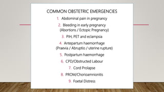
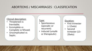
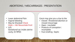
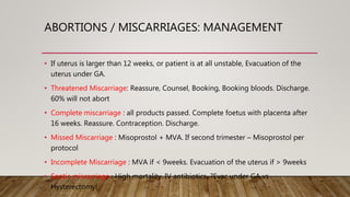
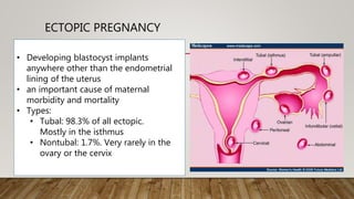
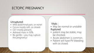
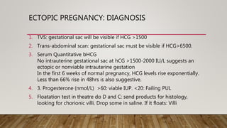
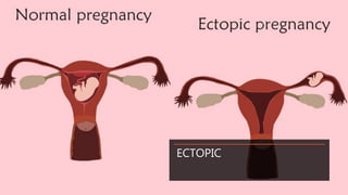
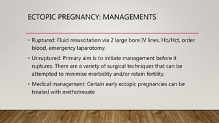
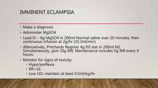
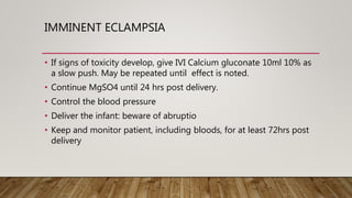
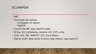
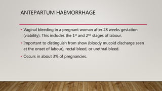
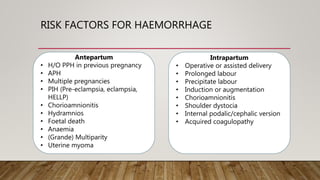
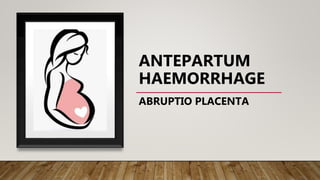
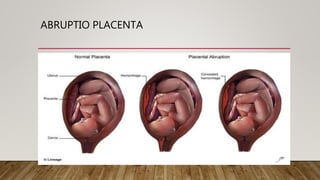
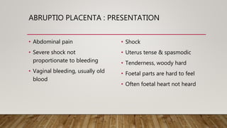
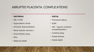
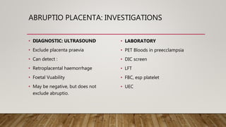
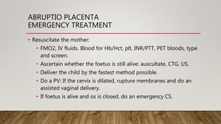
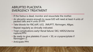
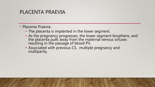
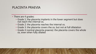
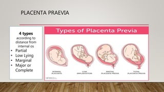
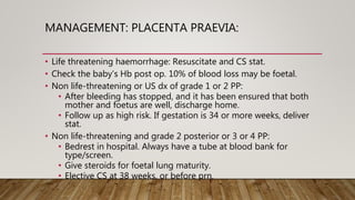
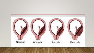
OBS & GYNAE
EMERGENCIES
DR KD DELE IJAGBULU DEPT OF FAMILY MEDICINE
OBSTETRICS EMERGENCIES
• Obstetrical emergencies are
• life threatening medical conditions
• that occur
• in pregnancy or
• during labour and/or
• after delivery.
COMMON OBSTETRIC EMERGENCIES
1. Abdominal pain in pregnancy
2. Bleeding in early pregnancy
(Abortions / Ectopic Pregnancy)
3. PIH, PET and eclampsia
4. Antepartum haemorrhage
(Praevia / Abruptio / uterine rupture)
5. Postpartum haemorrhage
6. CPD/Obstructed Labour
7. Cord Prolapse
8. PROM/Chorioamnionitis
9. Foetal Distress
ABDOMINAL PAIN
IN PREGNANCY
ABDOMINAL PAIN IN PREGNANCY
a. Conditions Incidental To Pregnancy
• These include medical and surgical causes of acute abdominal pain
Acute appendicitis
Acute pancreatitis
Peptic ulcer
Gastroenteritis
Hepatitis
Bowel obstruction
Bowel perforation
Pneumonia
Pulmonary embolus
Intraperitoneal haemorrhage
Splenic rupture
Abdominal trauma
Diabetic ketoacidosis
Sickle Cell Disease (acute)
ABDOMINAL PAIN IN PREGNANCY
b. Conditions Associated With
Pregnancy
Acute cholecystitis
Acute pyelonephritis
Acute cystitis
Rupture of rectus abdominus
muscle
Constipation
Acute Retention of urine
c. Conditions Due To Pregnancy
i.e. Obstetrical Causes
Labour
Miscarriage
Ectopic pregnancy
Rupture corpus luteal cyst
Acute salpingitis
Adnexal torsion
Stretching of round ligament
ABDOMINAL PAIN IN PREGNANCY: MANAGEMENT
• Treat the underlying condition
according to protocol.
BLEEDING IN
EARLY
PREGNANCY
ABORTIONS / MISCARRIAGES
• Definition
• The ending of a pregnancy before it is viable. That is: 26 weeks
after conception or 28 weeks (1000g) after the last menstrual
cycle.
• About 10 to 20 percent of known pregnancies end
in miscarriage.
• But the actual number is likely higher (~50%) because
many miscarriages occur so early in pregnancy that a woman
doesn't realize she's pregnant.
• Major cause of maternal death in South Africa: haemorrhage, sepsis
ABORTIONS / MISCARRIAGES : CLASSIFICATION
Clinical description:
• Threatened vs
Inevitable;
• Incomplete vs
Complete vs Missed;
• Uncomplicated vs
Septic.
Type:
• Spontaneous
(sporadic or
recurrent)
• induced (unsafe
or therapeutic)
Duration:
• First trimester
(<13wks)
• Second
trimester (13-
20wks)
ABORTIONS / MISCARRIAGES : PRESENTATION
• Lower abdominal Pain
• Vaginal bleeding
• May be Shocked! Check
Vitals + Hb! Resuscitate First!
• U/S:
Gestational sac inside uterine
cavity. Or RPOC
Cervix may give you a clue to the
• Closed : threatened abortion or
missed miscarriage
• Open : inevitable
• Products of conception :
incomplete
• Foul smelling : Septic
ABORTIONS / MISCARRIAGES: MANAGEMENT
• If uterus is larger than 12 weeks, or patient is at all unstable, Evacuation of the
uterus under GA.
• Threatened Miscarriage: Reassure, Counsel, Booking, Booking bloods. Discharge.
60% will not abort
• Complete miscarriage : all products passed. Complete foetus with placenta after
16 weeks. Reassure. Contraception. Discharge.
• Missed Miscarriage : Misoprostol + MVA. If second trimester – Misoprostol per
protocol
• Incomplete Miscarriage : MVA if < 9weeks. Evacuation of the uterus if > 9weeks
• Septic miscarriage : High mortality. IV antibiotics. ?Evac under GA vs
Hysterectomy!
BLEEDING IN
EARLY
PREGNANCY
ECTOPIC PREGNANCY
ECTOPIC PREGNANCY
• Developing blastocyst implants
anywhere other than the endometrial
lining of the uterus
• an important cause of maternal
morbidity and mortality
• Types:
• Tubal: 98.3% of all ectopic.
Mostly in the isthmus
• Nontubal: 1.7%. Very rarely in the
ovary or the cervix
ECTOPIC PREGNANCY
PRESENTATION
• Varied.
• From asymptomatic to shock
to acute abdomen
• 50% present with the classic
triad of Pain +++,
Amenorrhea, +/- PV bleeding.
• Pain may be transiently
improved after rupture!
• Others: dizzy, shoulder and
back pain due to peritoneal
irritation.
• When it is too large for the
tube, the following may
occur:
• Tubal Rupture with severe
haemorrhage
• Tubal abortion. May re-
implant leading to intra-
abdominal pregnancy
• Spontaneous resolution
• Chronic ectopic: the
pregnancy ends, but the
patient is left with a chronic
inflammatory mass
ECTOPIC PREGNANCY
Vitals :
• May be normal or unstable
Ruptured:
• patient may be stable, may
be shocked.
• Acute abdomen is common.
• Patient will have PV bleeding
with os closed.
Unruptured:
• Mild abdominal pain, or none!
• Cervix mostly soft, os closed.
• CET mostly present,
• Adnexal mass in 50%.
• Be gentle – you may rupture
the pregnancy!
ECTOPIC PREGNANCY: DIAGNOSIS
1. TVS: gestational sac will be visible if HCG >1500
2. Trans-abdominal scan: gestational sac must be visible if HCG>6500.
3. Serum Quantitative bHCG
No intrauterine gestational sac at hCG >1500-2000 IU/L suggests an
ectopic or nonviable intrauterine gestation
In the first 6 weeks of normal pregnancy, HCG levels rise exponentially.
Less than 66% rise in 48hrs is also suggestive.
4. 3. Progesterone (nmol/L) >60: viable IUP. <20: Failing PUL
5. Floatation test in theatre do D and C: send products for histology,
looking for chorionic villi. Drop some in saline. If it floats: Villi
ECTOPIC
ECTOPIC PREGNANCY: MANAGEMENTS
• Ruptured: Fluid resuscitation via 2 large bore IV lines, Hb/Hct, order
blood, emergency laparotomy.
• Unruptured: Primary aim is to initiate management before it
ruptures. There are a variety of surgical techniques that can be
attempted to minimise morbidity and/or retain fertility.
• Medical management: Certain early ectopic pregnancies can be
treated with methotrexate
CHPT
PIH
PET / SIPET &
ECLAMPSIA
HYPERTENSIVE STATES OF
PREGNANCY
HYPERTENSION IN PREGNANCY
• Hypertension in pregnancy may be defined as:
• Systolic blood pressure of > 140 mmHg
• Diastolics blood pressure of > 90
• (Measured two consecutive times, at least four hours apart)
• Also, Single diastolic > 110mmHg once
• Pathophysiology: poorly understood.
• Abnormal placentation, and inappropriate spiral artery remodelling.
HYPERTENSION IN PREGNANCY
• Risk Factors:
• Primigravida or new father (immunological factors)
• Family Hx of preeclampsia/autoimmune disease
• Multiple pregnancy
• Chronic hypertension
• Chronic renal disease
• DM
• Migraine
• Low SES/malnutririon
HYPERTENSION IN PREGNANCY:
DEFINITION OF TERMS
• Pregnancy Induced Hypertension: Hypertension that develops after
20 weeks in a previously normotensive patient
• Chronic hypertension: preexisting disease, may occur with or
without proteinuria
• Preeclampsia: hypertension that develops after 20 weeks of
pregnancy, with associated organ damage (early pointer –
proteinuria)
• Chronic hypertension with superimposed Preeclampsia (SIPET)
• Eclampsia: seizures that cannot be attributable to other causes in a
woman with preeclampsia.
HYPERTENSION IN PREGNANCY
• Multiple organ systems are affected:
• Liver: periportal necrosis due to
extravasation of blood (EL)
• Kidneys: swelling of glomerular
endothelium with fibrinoid
deposition (proteinuria)
• Brain (cerebral
oedema/haemorrhage)
• Placenta: placental infarcts – leads
to IUGR, high risk of abruptio
HYPERTENSION IN PREGNANCY
• Proteinuria can be measured
on
• a dipstix: 2+ or more is
significant.
• Spot protein/Cr ratio of 0.3 or
more is significant.
• 24-hour urine for DUP (and
creatinine clearance)
HYPERTENSION IN PREGNANCY
• HELLP syndrome:
• H - hemolysis, hemolytic anemia, raised
• EL - liver enzymes: AST > 70 U/l, or LDH > 600 U/l
• LP - low platelets < 100
• May be associated with DIC
• Watch out for 24-48 hrs postpartum
• Preeclampsia may be described as Fulminant if it deteriorates
rapidly over 24-48 hours. May occur with signs of Imminent
Eclampsia
HYPERTENSION IN PREGNANCY
• Imminent Eclampsia: It is characterised by one or more of the
following:
• Restlessness
• Visual disturbances (flashing lights, photophobia, blurred vision,
scotoma)
• Persistent, severe frontal headache
• Itching of the nose (a sign of cerebral irritation)
• Epigastric pain (serious sign, may indicate subcapsular haematoma of
the liver with impending rupture)
• BP>160/110 with proteinuria
• Hyperreflexia/clonus
• Oliguria
• HELLP
HYPERTENSION IN PREGNANCY
• UCE, UA, FBC, Hct, LFT, Abgas, CTG, US
• UCE: signs of renal impairment
• UA>0.35 indicative of poor prognosis
• FBC: low plt, low Hb (HELLP/abruptio), diff - fragments
• Hct: rising Hct indicates severe eclampsia
• LFT: raised transaminases: HELLP; unconj. Bili – haemolysis
• CTG/US: Foetal welfare
• Five most essential: AST, Hb, Plt, Ur, Cr,
HYPERTENSION IN PREGNANCY: MANAGEMENT
Only curative
management is to
terminate the
pregnancy.
Thus very important to
weigh up the risk to the
mother in continuing
the pregnancy versus
the risk to the child if it
is delivered now .
• Make a diagnosis.
• Distinguish it from chronic
HT/renal disease etc.
• Use history, risk profiling,
examination and special
investigation
• Clinically, look for signs of fluid
retention.
• When was HT/proteinuria first
documented
HYPERTENSION IN PREGNANCY: DELIVERY
DELIVERY
• GA 20 – 26 weeks, if severe
or complicated, may need
to be terminated.
• If gestation is between 28
and 34 weeks, give steroids
for lung maturity
• IMI Betamethasone 12mg
stat and 12mg again in 24
hours.
• Continue expectant
management until 34 weeks
INDICATION FOR
IMMEDIATE DELIVERY:
• Mature foetus
• Uncontrollable
hypertension
• Abruptio
• Imminent Eclampsia
• Organ failure/HELLP/DIC
• Foetal distress
HYPERTENSION IN PREGNANCY: PREECLAMPSIA
• Admit
• regular observations,
• intake/output,
• daily weighing.
• Monitor bloods daily.
• Low dose aspirin
• Monitor foetal wellbeing regularly: 4hrly CTG
ASPIRIN 150MG
ASPIRIN 150MG
• Preterm preeclampsia is an important cause of maternal and perinatal death and complications. It is
uncertain whether the intake of low-dose aspirin during pregnancy reduces the risk of preterm preeclampsia.
• METHODS In this multicenter, double-blind, placebo-controlled trial, we randomly assigned 1776 women
with singleton pregnancies who were at high risk for preterm preeclampsia to receive aspirin, at a dose of
150 mg per day, or placebo from 11 to 14 weeks of gestation until 36 weeks of gestation.
• The primary outcome was delivery with preeclampsia before 37 weeks of gestation. The analysis was
performed according to the intention-to-treat principle.
• RESULTS A total of 152 women withdrew consent during the trial, and 4 were lost to follow up, which left 798
participants in the aspirin group and 822 in the placebo group. Preterm preeclampsia occurred in 13
participants (1.6%) in the aspirin group, as compared with 35 (4.3%) in the placebo group (odds ratio in the
aspirin group, 0.38; 95% confidence interval, 0.20 to 0.74; P=0.004).
• CONCLUSIONS Treatment with low-dose aspirin in women at high risk for preterm preeclampsia
resulted in a lower incidence of this diagnosis than placebo.
HYPERTENSION IN PREGNANCY: BP CONTROL
• Aim for BP 140/90. If too low placental circulation is diminished.
• Diastolic <120mmHg:
• Oral alpha methyldopa 250-500mg p o 6 hrly.
• Hydralazine or atenolol may be added
• Diastolic>120mmHg:
• Intravenous antihypertensives eg Labetalol may be used.
• Titrate to effect.
• Aim for diastolic of 110mmHg, or a 15-20% decrease in diastolic over
1 hour.
• Patient needs to be in Hicare.
IMMINENT ECLAMPSIA
• Make a diagnosis
• Administer MgSO4
• Load IV – 6g MgSO4 in 200ml Normal saline over 20 minutes, then
continuous infusion at 2g/hr (33.3ml/min)
• Alternatively, Pritchards Regime: 4g IVI stat in 200ml NS.
Simultaneously, give 10g IMI. Maintenance includes 5g IMI every 4
hours.
• Monitor for signs of toxicity:
• Hypo/areflexia
• RR<16
• Low UO: maintain at least 0.5ml/kg/hr
IMMINENT ECLAMPSIA
• If signs of toxicity develop, give IVI Calcium gluconate 10ml 10% as
a slow push. May be repeated until effect is noted.
• Continue MgSO4 until 24 hrs post delivery.
• Control the blood pressure
• Deliver the infant: beware of abruptio
• Keep and monitor patient, including bloods, for at least 72hrs post
delivery
ECLAMPSIA
• ABC
• Terminate the seizure:
• Lorazepam or valium
• MgSO4
• Control the BP: may need IV meds
• IV line, O2, Catheterise, monitor UO, CVP, a-line
• STAT UCE, FBC, INR/PTT, LFT, Cross Match
• Deliver ASAP. Best within 6 hours. May induce, may need CS.
ANTEPARTUM
HAEMORRHAGE
ANTEPARTUM HAEMORRHAGE
• Vaginal bleeding in a pregnant woman after 28 weeks gestation
(viability). This includes the 1st and 2nd stages of labour.
• Important to distinguish from show (bloody mucoid discharge seen
at the onset of labour), rectal bleed, or urethral bleed.
• Occurs in about 3% of pregnancies.
RISK FACTORS FOR HAEMORRHAGE
Antepartum
• H/O PPH in previous pregnancy
• APH
• Multiple pregnancies
• PIH (Pre-eclampsia, eclampsia,
HELLP)
• Chorioamnionitis
• Hydramnios
• Foetal death
• Anaemia
• (Grande) Multiparity
• Uterine myoma
Intrapartum
• Operative or assisted delivery
• Prolonged labour
• Precipitate labour
• Induction or augmentation
• Chorioamnionitis
• Shoulder dystocia
• Internal podalic/cephalic version
• Acquired coagulopathy
ANTEPARTUM
HAEMORRHAGE
ABRUPTIO PLACENTA
ABRUPTIO PLACENTA
• Abruptio Placenta: Premature separation of a normally situated
placenta in a viable foetus.
• Exact cause is unknown.
• Associated with vascular disease such as HT conditions of
pregnancy
• Also renal disease and connective tissue diseases such as SLE
ABRUPTIO PLACENTA
• Other causes include:
• previous abruptio,
• Blunt abdominal trauma in pregnancy
• poor SES,
• smoking,
• IUGR,
• trauma,
• sudden decrease in uterine volume.
SHER CLASSIFICATION
Grade 1 – mild retroplacental
clot identified after delivery.
Grade 11 – clinical suspicion,
e.g. tense tender abdomen, but
foetus is alive
Grade 111 – foetus is dead.
Grade 111a – without
coagulopathy
Grade 111b – with coagulopathy
ABRUPTIO PLACENTA
ABRUPTIO PLACENTA : PRESENTATION
• Abdominal pain
• Severe shock not
proportionate to bleeding
• Vaginal bleeding, usually old
blood
• Shock
• Uterus tense & spasmodic
• Tenderness, woody hard
• Foetal parts are hard to feel
• Often foetal heart not heard
ABRUPTIO PLACENTA: COMPLICATIONS
• MATERNAL
• DIC 4-10%
• Hypovolemic shock
• Amniotic fluid embolism
• Renal tubular necrosis /
• Acute kidney injury
• PPH
• Maternal death
• FOETAL
• Premature labour
• IUGR
• HIE – hypoxic ischemic
encephalopathy /
• Cerebral palsy
• Foetal distress
• Foetal death
ABRUPTIO PLACENTA: INVESTIGATIONS
• DIAGNOSTIC: ULTRASOUND
• Exclude placenta praevia
• Can detect :
• Retroplacental haemorrhage
• Foetal Vuability
• May be negative, but does not
exclude abruptio.
• LABORATORY
• PET Bloods in preecclampsia
• DIC screen
• LFT
• FBC, esp platelet
• UEC
ABRUPTIO PLACENTA
EMERGENCY TREATMENT
• Treat the shock – large bore IV line, Haemaccel, cross match blood
• Treat DIC – 4FFPs, 4PRBCs
• Deliver the foetus
• Emergency Caesarean if foetus is alive & mature
• Vaginal delivery if cervix is favourable & foetus dead
ABRUPTIO PLACENTA
EMERGENCY TREATMENT
• Resuscitate the mother:
• FMO2, IV fluids. Blood for Hb/Hct, plt, INR/PTT, PET bloods, type
and screen.
• Ascertain whether the foetus is still alive: auscultate, CTG, US.
• Deliver the child by the fastest method possible.
• Do a PV: If the cervix is dilated, rupture membranes and do an
assisted vaginal delivery.
• If foetus is alive and os is closed, do an emergency CS.
ABRUPTIO PLACENTA
EMERGENCY TREATMENT
• If the foetus is dead, monitor and resuscitate the mother.
• An abruptio severe enough to cause IUD will need at least 4 units of
packed cells and 4 units of FFP.
• Take bloods for FBC/diff, UCE, INR/PTT, fibrinogen, ABgas.
• Repeat regularly as clinically indicated.
• Treat complications early! Renal failure/ DIC/ ARDS/Uterine
rupture/PPH
• Be ready to give platelets if count < 50, or cryoprecipitate if
fibrinogen<2.
• Anticipate PPH
ANTEPARTUM
HAEMORRHAGE
PLACENTA PRAEVIA
PLACENTA PRAEVIA
• Placenta Praevia:
• The placenta is implanted in the lower segment.
• As the pregnancy progresses, the lower segment lengthens, and
the placenta pulls away from the maternal venous sinuses
resulting in the passage of blood PV.
• Associated with previous CS, multiple pregnancy and
multiparity.
PLACENTA PRAEVIA
• There are 4 grades:
• Grade 1: the placenta implants in the lower segment but does
not reach the internal os.
• Grade 2: the placenta reaches the internal os.
• Grade 3: the placenta covers the os, but not at full dilatation
• Grade 4 (central placenta praevia): the placenta covers the whole
os, even when fully dilated
PLACENTA PRAEVIA
4 types
according to
distance from
internal os
• Partial
• Low Lying
• Marginal
• Major or
Complete
MANAGEMENT: PLACENTA PRAEVIA:
• Life threatening haemorrhage: Resuscitate and CS stat.
• Check the baby’s Hb post op. 10% of blood loss may be foetal.
• Non life-threatening or US dx of grade 1 or 2 PP:
• After bleeding has stopped, and it has been ensured that both
mother and foetus are well, discharge home.
• Follow up as high risk. If gestation is 34 or more weeks, deliver
stat.
• Non life-threatening and grade 2 posterior or 3 or 4 PP:
• Bedrest in hospital. Always have a tube at blood bank for
type/screen.
• Give steroids for foetal lung maturity.
• Elective CS at 38 weeks, or before prn.
ANTEPARTUM
HAEMORRHAGE
PLACENTA ACRETA
MORBIDLY ADHERENT
PLACENTA
PLACENTA ACCRETA/INCRETA/PERCRETA
• Penetration of the placenta into the uterine myometrium and
beyond
• Placenta accreta – The placenta attaches itself too deeply and too
firmly into the uterus.
• Placenta increta – The placenta attaches itself even more deeply
into the muscle wall of uterus.
• Placenta percreta – The placenta attaches itself and grows through
the uterus, sometimes extending to nearby organs, such as the
bladder.
PLACENTA ACCRETA/INCRETA/PERCRETA
• Often follows precious CS
• Can be caught on U/S but not always and severity questionable
• Can cause: Bleeding and Uterine inversion
• Treatment: Hysterectomy
• True Life threatening emergency if not recognized early
POST PARTUM
HAEMORRHAGE
POST-PARTUM HAEMORRHAGE
• Primary vs Secondary
• Primary PPH is defined as the loss of 500ml or more of blood
from the genital tract during the first 24 hours post delivery.
• Most frequently in the first hour post delivery.
• Secondary PPH is defined as excessive blood loss from genital
tract between 24 hours and 6 weeks post delivery.
POSTPARTUM HAEMORRHAGE: MAIN CAUSES
• Uterine atony
• Retained placenta
• Injuries of the genital tract,
such as cervical laceration and perineal tear
• Uterine rupture
• Other causes: clotting abn.
PPH: RISK FACTORS
• Grande multiparity (5 or
more deliveries)
• Abruptio
• Hx of PPH
• Multiple pregnancy
• Polyhydramnios
• Prolonged labour
• Leiomyomata
• Intrauterine
manipulations
• IUD
• Administration of beta 2
stimulant during delivery
(tocolysis with
salbutamol)
• Halothane anaesthetic
gas
• Oxytocin use during the
first stage of labour
PPH: TREATMENT
• Prevent!
• If a risk factor is present, deliver in a hospital, with an IV line up
• Give oxytocin 10units IMI immediately post delivery. Alternative
is syntometrine 1-amp IMI.
• IV infusion of oxytocin (20-40 units over 8 hours after the
delivery of the placenta) in high risk patients.
PPH: TREATMENT
• Active Mx:
• Aggressive fluid resuscitation, type and screen, Hb, clotting
profile
• If placenta is still in-situ:
• Empty the bladder, massage the uterus, actively deliver the
placenta. If it cannot be delivered, do it under GA. Curette if
necessary
• If placenta is out:
• Empty the bladder.
• Rub uterus up.
• Repeat syntometrine (max 3 amps IMI)
REFERENCES
• American College of Obstetrics and Gynecology (ACOG) with
Beckmann CRB & Ling FW, (2010). Obstetrics and Gynecology.
Lippincott Williams & Wilkins: Philadelphia

Obstetrics Emergencies by Dr KD DELE

  • 1.
    OBS & GYNAE EMERGENCIES DRKD DELE IJAGBULU DEPT OF FAMILY MEDICINE
  • 2.
    OBSTETRICS EMERGENCIES • Obstetricalemergencies are • life threatening medical conditions • that occur • in pregnancy or • during labour and/or • after delivery.
  • 3.
    COMMON OBSTETRIC EMERGENCIES 1.Abdominal pain in pregnancy 2. Bleeding in early pregnancy (Abortions / Ectopic Pregnancy) 3. PIH, PET and eclampsia 4. Antepartum haemorrhage (Praevia / Abruptio / uterine rupture) 5. Postpartum haemorrhage 6. CPD/Obstructed Labour 7. Cord Prolapse 8. PROM/Chorioamnionitis 9. Foetal Distress
  • 4.
  • 5.
    ABDOMINAL PAIN INPREGNANCY a. Conditions Incidental To Pregnancy • These include medical and surgical causes of acute abdominal pain Acute appendicitis Acute pancreatitis Peptic ulcer Gastroenteritis Hepatitis Bowel obstruction Bowel perforation Pneumonia Pulmonary embolus Intraperitoneal haemorrhage Splenic rupture Abdominal trauma Diabetic ketoacidosis Sickle Cell Disease (acute)
  • 6.
    ABDOMINAL PAIN INPREGNANCY b. Conditions Associated With Pregnancy Acute cholecystitis Acute pyelonephritis Acute cystitis Rupture of rectus abdominus muscle Constipation Acute Retention of urine c. Conditions Due To Pregnancy i.e. Obstetrical Causes Labour Miscarriage Ectopic pregnancy Rupture corpus luteal cyst Acute salpingitis Adnexal torsion Stretching of round ligament
  • 7.
    ABDOMINAL PAIN INPREGNANCY: MANAGEMENT • Treat the underlying condition according to protocol.
  • 8.
  • 9.
    ABORTIONS / MISCARRIAGES •Definition • The ending of a pregnancy before it is viable. That is: 26 weeks after conception or 28 weeks (1000g) after the last menstrual cycle. • About 10 to 20 percent of known pregnancies end in miscarriage. • But the actual number is likely higher (~50%) because many miscarriages occur so early in pregnancy that a woman doesn't realize she's pregnant. • Major cause of maternal death in South Africa: haemorrhage, sepsis
  • 10.
    ABORTIONS / MISCARRIAGES: CLASSIFICATION Clinical description: • Threatened vs Inevitable; • Incomplete vs Complete vs Missed; • Uncomplicated vs Septic. Type: • Spontaneous (sporadic or recurrent) • induced (unsafe or therapeutic) Duration: • First trimester (<13wks) • Second trimester (13- 20wks)
  • 11.
    ABORTIONS / MISCARRIAGES: PRESENTATION • Lower abdominal Pain • Vaginal bleeding • May be Shocked! Check Vitals + Hb! Resuscitate First! • U/S: Gestational sac inside uterine cavity. Or RPOC Cervix may give you a clue to the • Closed : threatened abortion or missed miscarriage • Open : inevitable • Products of conception : incomplete • Foul smelling : Septic
  • 12.
    ABORTIONS / MISCARRIAGES:MANAGEMENT • If uterus is larger than 12 weeks, or patient is at all unstable, Evacuation of the uterus under GA. • Threatened Miscarriage: Reassure, Counsel, Booking, Booking bloods. Discharge. 60% will not abort • Complete miscarriage : all products passed. Complete foetus with placenta after 16 weeks. Reassure. Contraception. Discharge. • Missed Miscarriage : Misoprostol + MVA. If second trimester – Misoprostol per protocol • Incomplete Miscarriage : MVA if < 9weeks. Evacuation of the uterus if > 9weeks • Septic miscarriage : High mortality. IV antibiotics. ?Evac under GA vs Hysterectomy!
  • 13.
  • 14.
    ECTOPIC PREGNANCY • Developingblastocyst implants anywhere other than the endometrial lining of the uterus • an important cause of maternal morbidity and mortality • Types: • Tubal: 98.3% of all ectopic. Mostly in the isthmus • Nontubal: 1.7%. Very rarely in the ovary or the cervix
  • 16.
    ECTOPIC PREGNANCY PRESENTATION • Varied. •From asymptomatic to shock to acute abdomen • 50% present with the classic triad of Pain +++, Amenorrhea, +/- PV bleeding. • Pain may be transiently improved after rupture! • Others: dizzy, shoulder and back pain due to peritoneal irritation. • When it is too large for the tube, the following may occur: • Tubal Rupture with severe haemorrhage • Tubal abortion. May re- implant leading to intra- abdominal pregnancy • Spontaneous resolution • Chronic ectopic: the pregnancy ends, but the patient is left with a chronic inflammatory mass
  • 17.
    ECTOPIC PREGNANCY Vitals : •May be normal or unstable Ruptured: • patient may be stable, may be shocked. • Acute abdomen is common. • Patient will have PV bleeding with os closed. Unruptured: • Mild abdominal pain, or none! • Cervix mostly soft, os closed. • CET mostly present, • Adnexal mass in 50%. • Be gentle – you may rupture the pregnancy!
  • 18.
    ECTOPIC PREGNANCY: DIAGNOSIS 1.TVS: gestational sac will be visible if HCG >1500 2. Trans-abdominal scan: gestational sac must be visible if HCG>6500. 3. Serum Quantitative bHCG No intrauterine gestational sac at hCG >1500-2000 IU/L suggests an ectopic or nonviable intrauterine gestation In the first 6 weeks of normal pregnancy, HCG levels rise exponentially. Less than 66% rise in 48hrs is also suggestive. 4. 3. Progesterone (nmol/L) >60: viable IUP. <20: Failing PUL 5. Floatation test in theatre do D and C: send products for histology, looking for chorionic villi. Drop some in saline. If it floats: Villi
  • 19.
  • 20.
    ECTOPIC PREGNANCY: MANAGEMENTS •Ruptured: Fluid resuscitation via 2 large bore IV lines, Hb/Hct, order blood, emergency laparotomy. • Unruptured: Primary aim is to initiate management before it ruptures. There are a variety of surgical techniques that can be attempted to minimise morbidity and/or retain fertility. • Medical management: Certain early ectopic pregnancies can be treated with methotrexate
  • 21.
    CHPT PIH PET / SIPET& ECLAMPSIA HYPERTENSIVE STATES OF PREGNANCY
  • 22.
    HYPERTENSION IN PREGNANCY •Hypertension in pregnancy may be defined as: • Systolic blood pressure of > 140 mmHg • Diastolics blood pressure of > 90 • (Measured two consecutive times, at least four hours apart) • Also, Single diastolic > 110mmHg once • Pathophysiology: poorly understood. • Abnormal placentation, and inappropriate spiral artery remodelling.
  • 23.
    HYPERTENSION IN PREGNANCY •Risk Factors: • Primigravida or new father (immunological factors) • Family Hx of preeclampsia/autoimmune disease • Multiple pregnancy • Chronic hypertension • Chronic renal disease • DM • Migraine • Low SES/malnutririon
  • 24.
    HYPERTENSION IN PREGNANCY: DEFINITIONOF TERMS • Pregnancy Induced Hypertension: Hypertension that develops after 20 weeks in a previously normotensive patient • Chronic hypertension: preexisting disease, may occur with or without proteinuria • Preeclampsia: hypertension that develops after 20 weeks of pregnancy, with associated organ damage (early pointer – proteinuria) • Chronic hypertension with superimposed Preeclampsia (SIPET) • Eclampsia: seizures that cannot be attributable to other causes in a woman with preeclampsia.
  • 25.
    HYPERTENSION IN PREGNANCY •Multiple organ systems are affected: • Liver: periportal necrosis due to extravasation of blood (EL) • Kidneys: swelling of glomerular endothelium with fibrinoid deposition (proteinuria) • Brain (cerebral oedema/haemorrhage) • Placenta: placental infarcts – leads to IUGR, high risk of abruptio
  • 26.
    HYPERTENSION IN PREGNANCY •Proteinuria can be measured on • a dipstix: 2+ or more is significant. • Spot protein/Cr ratio of 0.3 or more is significant. • 24-hour urine for DUP (and creatinine clearance)
  • 27.
    HYPERTENSION IN PREGNANCY •HELLP syndrome: • H - hemolysis, hemolytic anemia, raised • EL - liver enzymes: AST > 70 U/l, or LDH > 600 U/l • LP - low platelets < 100 • May be associated with DIC • Watch out for 24-48 hrs postpartum • Preeclampsia may be described as Fulminant if it deteriorates rapidly over 24-48 hours. May occur with signs of Imminent Eclampsia
  • 28.
    HYPERTENSION IN PREGNANCY •Imminent Eclampsia: It is characterised by one or more of the following: • Restlessness • Visual disturbances (flashing lights, photophobia, blurred vision, scotoma) • Persistent, severe frontal headache • Itching of the nose (a sign of cerebral irritation) • Epigastric pain (serious sign, may indicate subcapsular haematoma of the liver with impending rupture) • BP>160/110 with proteinuria • Hyperreflexia/clonus • Oliguria • HELLP
  • 29.
    HYPERTENSION IN PREGNANCY •UCE, UA, FBC, Hct, LFT, Abgas, CTG, US • UCE: signs of renal impairment • UA>0.35 indicative of poor prognosis • FBC: low plt, low Hb (HELLP/abruptio), diff - fragments • Hct: rising Hct indicates severe eclampsia • LFT: raised transaminases: HELLP; unconj. Bili – haemolysis • CTG/US: Foetal welfare • Five most essential: AST, Hb, Plt, Ur, Cr,
  • 30.
    HYPERTENSION IN PREGNANCY:MANAGEMENT Only curative management is to terminate the pregnancy. Thus very important to weigh up the risk to the mother in continuing the pregnancy versus the risk to the child if it is delivered now . • Make a diagnosis. • Distinguish it from chronic HT/renal disease etc. • Use history, risk profiling, examination and special investigation • Clinically, look for signs of fluid retention. • When was HT/proteinuria first documented
  • 31.
    HYPERTENSION IN PREGNANCY:DELIVERY DELIVERY • GA 20 – 26 weeks, if severe or complicated, may need to be terminated. • If gestation is between 28 and 34 weeks, give steroids for lung maturity • IMI Betamethasone 12mg stat and 12mg again in 24 hours. • Continue expectant management until 34 weeks INDICATION FOR IMMEDIATE DELIVERY: • Mature foetus • Uncontrollable hypertension • Abruptio • Imminent Eclampsia • Organ failure/HELLP/DIC • Foetal distress
  • 32.
    HYPERTENSION IN PREGNANCY:PREECLAMPSIA • Admit • regular observations, • intake/output, • daily weighing. • Monitor bloods daily. • Low dose aspirin • Monitor foetal wellbeing regularly: 4hrly CTG
  • 33.
  • 34.
    ASPIRIN 150MG • Pretermpreeclampsia is an important cause of maternal and perinatal death and complications. It is uncertain whether the intake of low-dose aspirin during pregnancy reduces the risk of preterm preeclampsia. • METHODS In this multicenter, double-blind, placebo-controlled trial, we randomly assigned 1776 women with singleton pregnancies who were at high risk for preterm preeclampsia to receive aspirin, at a dose of 150 mg per day, or placebo from 11 to 14 weeks of gestation until 36 weeks of gestation. • The primary outcome was delivery with preeclampsia before 37 weeks of gestation. The analysis was performed according to the intention-to-treat principle. • RESULTS A total of 152 women withdrew consent during the trial, and 4 were lost to follow up, which left 798 participants in the aspirin group and 822 in the placebo group. Preterm preeclampsia occurred in 13 participants (1.6%) in the aspirin group, as compared with 35 (4.3%) in the placebo group (odds ratio in the aspirin group, 0.38; 95% confidence interval, 0.20 to 0.74; P=0.004). • CONCLUSIONS Treatment with low-dose aspirin in women at high risk for preterm preeclampsia resulted in a lower incidence of this diagnosis than placebo.
  • 35.
    HYPERTENSION IN PREGNANCY:BP CONTROL • Aim for BP 140/90. If too low placental circulation is diminished. • Diastolic <120mmHg: • Oral alpha methyldopa 250-500mg p o 6 hrly. • Hydralazine or atenolol may be added • Diastolic>120mmHg: • Intravenous antihypertensives eg Labetalol may be used. • Titrate to effect. • Aim for diastolic of 110mmHg, or a 15-20% decrease in diastolic over 1 hour. • Patient needs to be in Hicare.
  • 37.
    IMMINENT ECLAMPSIA • Makea diagnosis • Administer MgSO4 • Load IV – 6g MgSO4 in 200ml Normal saline over 20 minutes, then continuous infusion at 2g/hr (33.3ml/min) • Alternatively, Pritchards Regime: 4g IVI stat in 200ml NS. Simultaneously, give 10g IMI. Maintenance includes 5g IMI every 4 hours. • Monitor for signs of toxicity: • Hypo/areflexia • RR<16 • Low UO: maintain at least 0.5ml/kg/hr
  • 38.
    IMMINENT ECLAMPSIA • Ifsigns of toxicity develop, give IVI Calcium gluconate 10ml 10% as a slow push. May be repeated until effect is noted. • Continue MgSO4 until 24 hrs post delivery. • Control the blood pressure • Deliver the infant: beware of abruptio • Keep and monitor patient, including bloods, for at least 72hrs post delivery
  • 39.
    ECLAMPSIA • ABC • Terminatethe seizure: • Lorazepam or valium • MgSO4 • Control the BP: may need IV meds • IV line, O2, Catheterise, monitor UO, CVP, a-line • STAT UCE, FBC, INR/PTT, LFT, Cross Match • Deliver ASAP. Best within 6 hours. May induce, may need CS.
  • 40.
  • 41.
    ANTEPARTUM HAEMORRHAGE • Vaginalbleeding in a pregnant woman after 28 weeks gestation (viability). This includes the 1st and 2nd stages of labour. • Important to distinguish from show (bloody mucoid discharge seen at the onset of labour), rectal bleed, or urethral bleed. • Occurs in about 3% of pregnancies.
  • 42.
    RISK FACTORS FORHAEMORRHAGE Antepartum • H/O PPH in previous pregnancy • APH • Multiple pregnancies • PIH (Pre-eclampsia, eclampsia, HELLP) • Chorioamnionitis • Hydramnios • Foetal death • Anaemia • (Grande) Multiparity • Uterine myoma Intrapartum • Operative or assisted delivery • Prolonged labour • Precipitate labour • Induction or augmentation • Chorioamnionitis • Shoulder dystocia • Internal podalic/cephalic version • Acquired coagulopathy
  • 43.
  • 44.
    ABRUPTIO PLACENTA • AbruptioPlacenta: Premature separation of a normally situated placenta in a viable foetus. • Exact cause is unknown. • Associated with vascular disease such as HT conditions of pregnancy • Also renal disease and connective tissue diseases such as SLE
  • 45.
    ABRUPTIO PLACENTA • Othercauses include: • previous abruptio, • Blunt abdominal trauma in pregnancy • poor SES, • smoking, • IUGR, • trauma, • sudden decrease in uterine volume. SHER CLASSIFICATION Grade 1 – mild retroplacental clot identified after delivery. Grade 11 – clinical suspicion, e.g. tense tender abdomen, but foetus is alive Grade 111 – foetus is dead. Grade 111a – without coagulopathy Grade 111b – with coagulopathy
  • 46.
  • 47.
    ABRUPTIO PLACENTA :PRESENTATION • Abdominal pain • Severe shock not proportionate to bleeding • Vaginal bleeding, usually old blood • Shock • Uterus tense & spasmodic • Tenderness, woody hard • Foetal parts are hard to feel • Often foetal heart not heard
  • 48.
    ABRUPTIO PLACENTA: COMPLICATIONS •MATERNAL • DIC 4-10% • Hypovolemic shock • Amniotic fluid embolism • Renal tubular necrosis / • Acute kidney injury • PPH • Maternal death • FOETAL • Premature labour • IUGR • HIE – hypoxic ischemic encephalopathy / • Cerebral palsy • Foetal distress • Foetal death
  • 49.
    ABRUPTIO PLACENTA: INVESTIGATIONS •DIAGNOSTIC: ULTRASOUND • Exclude placenta praevia • Can detect : • Retroplacental haemorrhage • Foetal Vuability • May be negative, but does not exclude abruptio. • LABORATORY • PET Bloods in preecclampsia • DIC screen • LFT • FBC, esp platelet • UEC
  • 50.
    ABRUPTIO PLACENTA EMERGENCY TREATMENT •Treat the shock – large bore IV line, Haemaccel, cross match blood • Treat DIC – 4FFPs, 4PRBCs • Deliver the foetus • Emergency Caesarean if foetus is alive & mature • Vaginal delivery if cervix is favourable & foetus dead
  • 51.
    ABRUPTIO PLACENTA EMERGENCY TREATMENT •Resuscitate the mother: • FMO2, IV fluids. Blood for Hb/Hct, plt, INR/PTT, PET bloods, type and screen. • Ascertain whether the foetus is still alive: auscultate, CTG, US. • Deliver the child by the fastest method possible. • Do a PV: If the cervix is dilated, rupture membranes and do an assisted vaginal delivery. • If foetus is alive and os is closed, do an emergency CS.
  • 52.
    ABRUPTIO PLACENTA EMERGENCY TREATMENT •If the foetus is dead, monitor and resuscitate the mother. • An abruptio severe enough to cause IUD will need at least 4 units of packed cells and 4 units of FFP. • Take bloods for FBC/diff, UCE, INR/PTT, fibrinogen, ABgas. • Repeat regularly as clinically indicated. • Treat complications early! Renal failure/ DIC/ ARDS/Uterine rupture/PPH • Be ready to give platelets if count < 50, or cryoprecipitate if fibrinogen<2. • Anticipate PPH
  • 53.
  • 54.
    PLACENTA PRAEVIA • PlacentaPraevia: • The placenta is implanted in the lower segment. • As the pregnancy progresses, the lower segment lengthens, and the placenta pulls away from the maternal venous sinuses resulting in the passage of blood PV. • Associated with previous CS, multiple pregnancy and multiparity.
  • 55.
    PLACENTA PRAEVIA • Thereare 4 grades: • Grade 1: the placenta implants in the lower segment but does not reach the internal os. • Grade 2: the placenta reaches the internal os. • Grade 3: the placenta covers the os, but not at full dilatation • Grade 4 (central placenta praevia): the placenta covers the whole os, even when fully dilated
  • 56.
    PLACENTA PRAEVIA 4 types accordingto distance from internal os • Partial • Low Lying • Marginal • Major or Complete
  • 57.
    MANAGEMENT: PLACENTA PRAEVIA: •Life threatening haemorrhage: Resuscitate and CS stat. • Check the baby’s Hb post op. 10% of blood loss may be foetal. • Non life-threatening or US dx of grade 1 or 2 PP: • After bleeding has stopped, and it has been ensured that both mother and foetus are well, discharge home. • Follow up as high risk. If gestation is 34 or more weeks, deliver stat. • Non life-threatening and grade 2 posterior or 3 or 4 PP: • Bedrest in hospital. Always have a tube at blood bank for type/screen. • Give steroids for foetal lung maturity. • Elective CS at 38 weeks, or before prn.
  • 58.
  • 60.
    PLACENTA ACCRETA/INCRETA/PERCRETA • Penetrationof the placenta into the uterine myometrium and beyond • Placenta accreta – The placenta attaches itself too deeply and too firmly into the uterus. • Placenta increta – The placenta attaches itself even more deeply into the muscle wall of uterus. • Placenta percreta – The placenta attaches itself and grows through the uterus, sometimes extending to nearby organs, such as the bladder.
  • 61.
    PLACENTA ACCRETA/INCRETA/PERCRETA • Oftenfollows precious CS • Can be caught on U/S but not always and severity questionable • Can cause: Bleeding and Uterine inversion • Treatment: Hysterectomy • True Life threatening emergency if not recognized early
  • 62.
  • 63.
    POST-PARTUM HAEMORRHAGE • Primaryvs Secondary • Primary PPH is defined as the loss of 500ml or more of blood from the genital tract during the first 24 hours post delivery. • Most frequently in the first hour post delivery. • Secondary PPH is defined as excessive blood loss from genital tract between 24 hours and 6 weeks post delivery.
  • 64.
    POSTPARTUM HAEMORRHAGE: MAINCAUSES • Uterine atony • Retained placenta • Injuries of the genital tract, such as cervical laceration and perineal tear • Uterine rupture • Other causes: clotting abn.
  • 65.
    PPH: RISK FACTORS •Grande multiparity (5 or more deliveries) • Abruptio • Hx of PPH • Multiple pregnancy • Polyhydramnios • Prolonged labour • Leiomyomata • Intrauterine manipulations • IUD • Administration of beta 2 stimulant during delivery (tocolysis with salbutamol) • Halothane anaesthetic gas • Oxytocin use during the first stage of labour
  • 66.
    PPH: TREATMENT • Prevent! •If a risk factor is present, deliver in a hospital, with an IV line up • Give oxytocin 10units IMI immediately post delivery. Alternative is syntometrine 1-amp IMI. • IV infusion of oxytocin (20-40 units over 8 hours after the delivery of the placenta) in high risk patients.
  • 67.
    PPH: TREATMENT • ActiveMx: • Aggressive fluid resuscitation, type and screen, Hb, clotting profile • If placenta is still in-situ: • Empty the bladder, massage the uterus, actively deliver the placenta. If it cannot be delivered, do it under GA. Curette if necessary • If placenta is out: • Empty the bladder. • Rub uterus up. • Repeat syntometrine (max 3 amps IMI)
  • 69.
    REFERENCES • American Collegeof Obstetrics and Gynecology (ACOG) with Beckmann CRB & Ling FW, (2010). Obstetrics and Gynecology. Lippincott Williams & Wilkins: Philadelphia