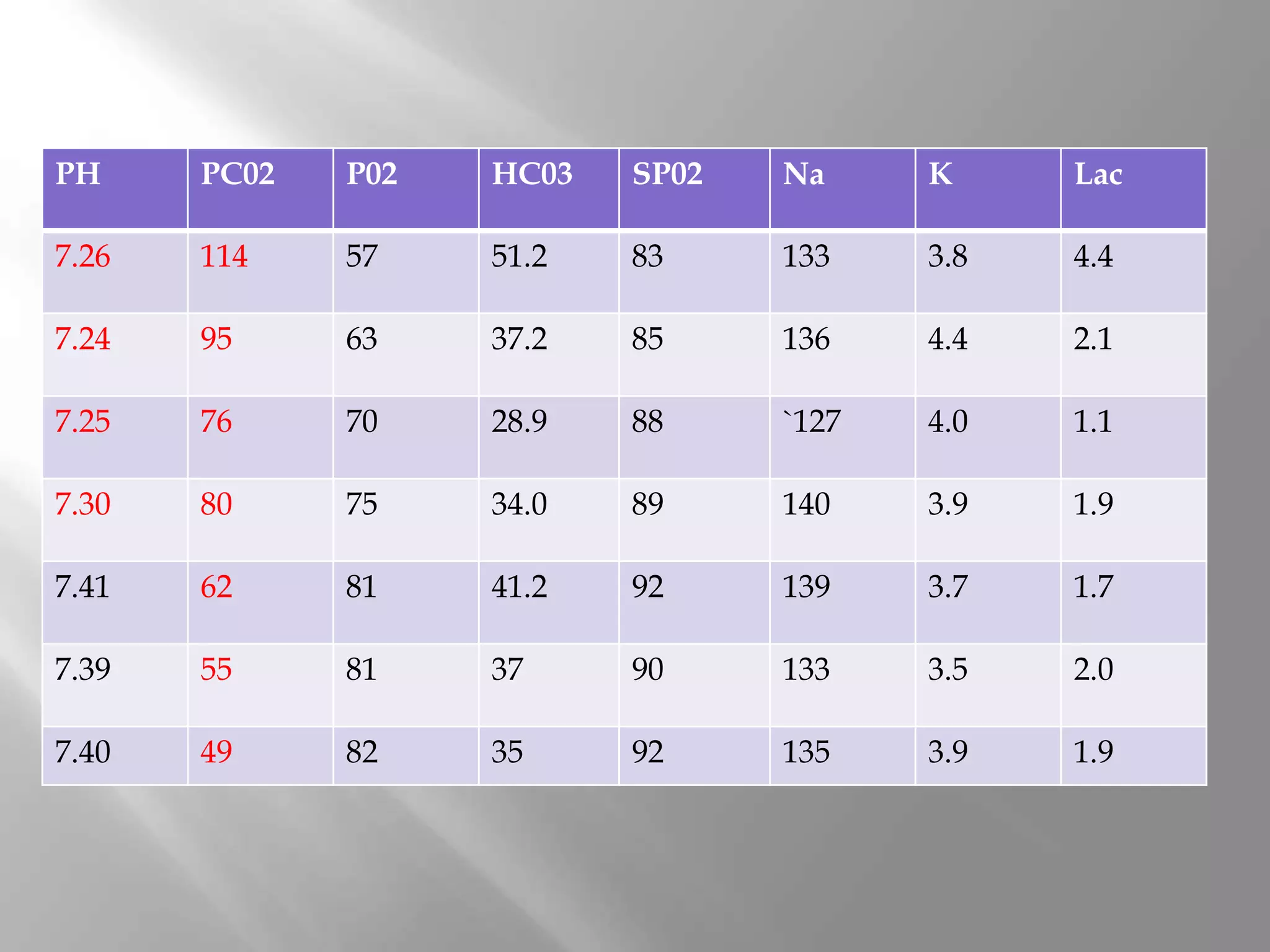
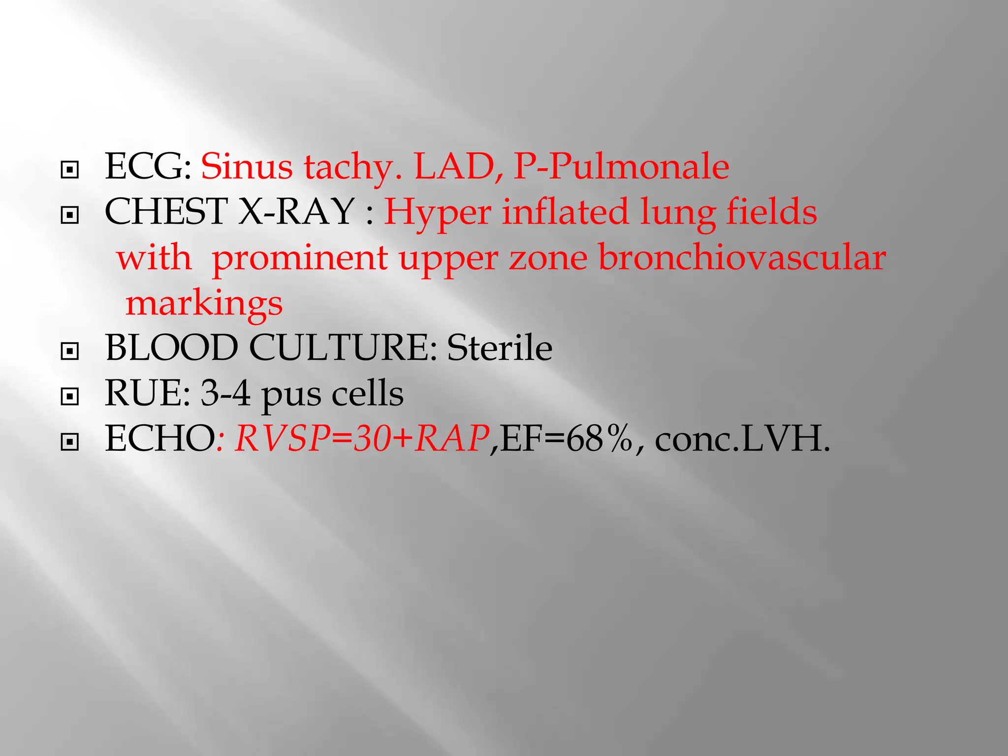
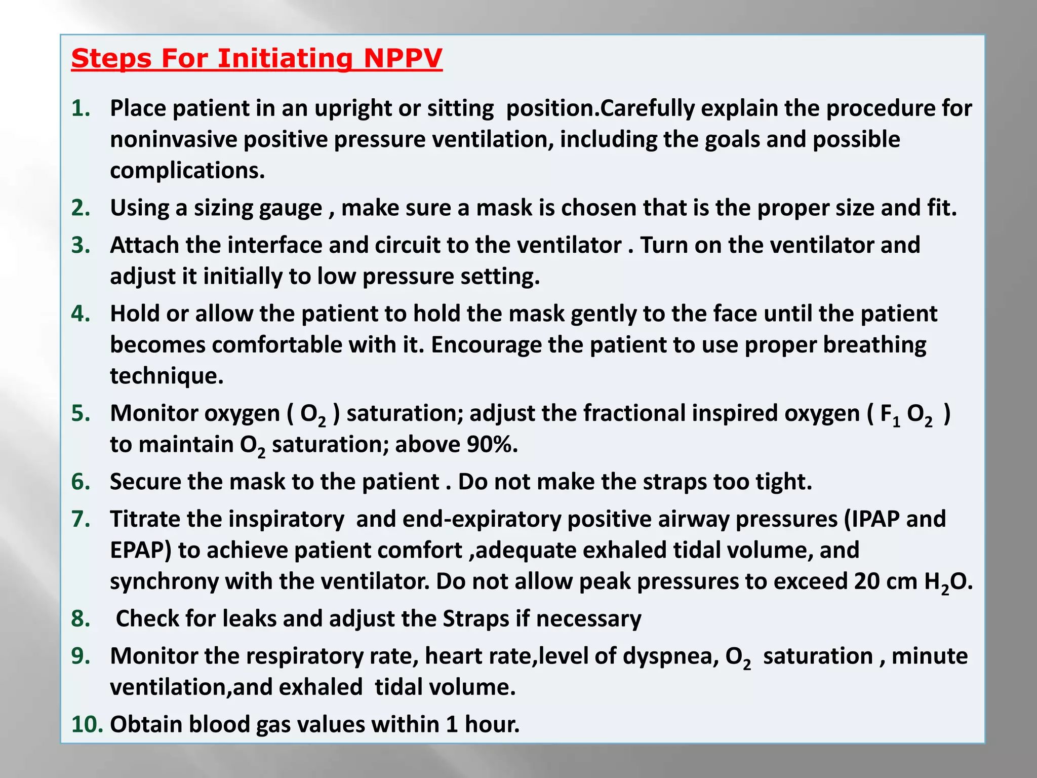
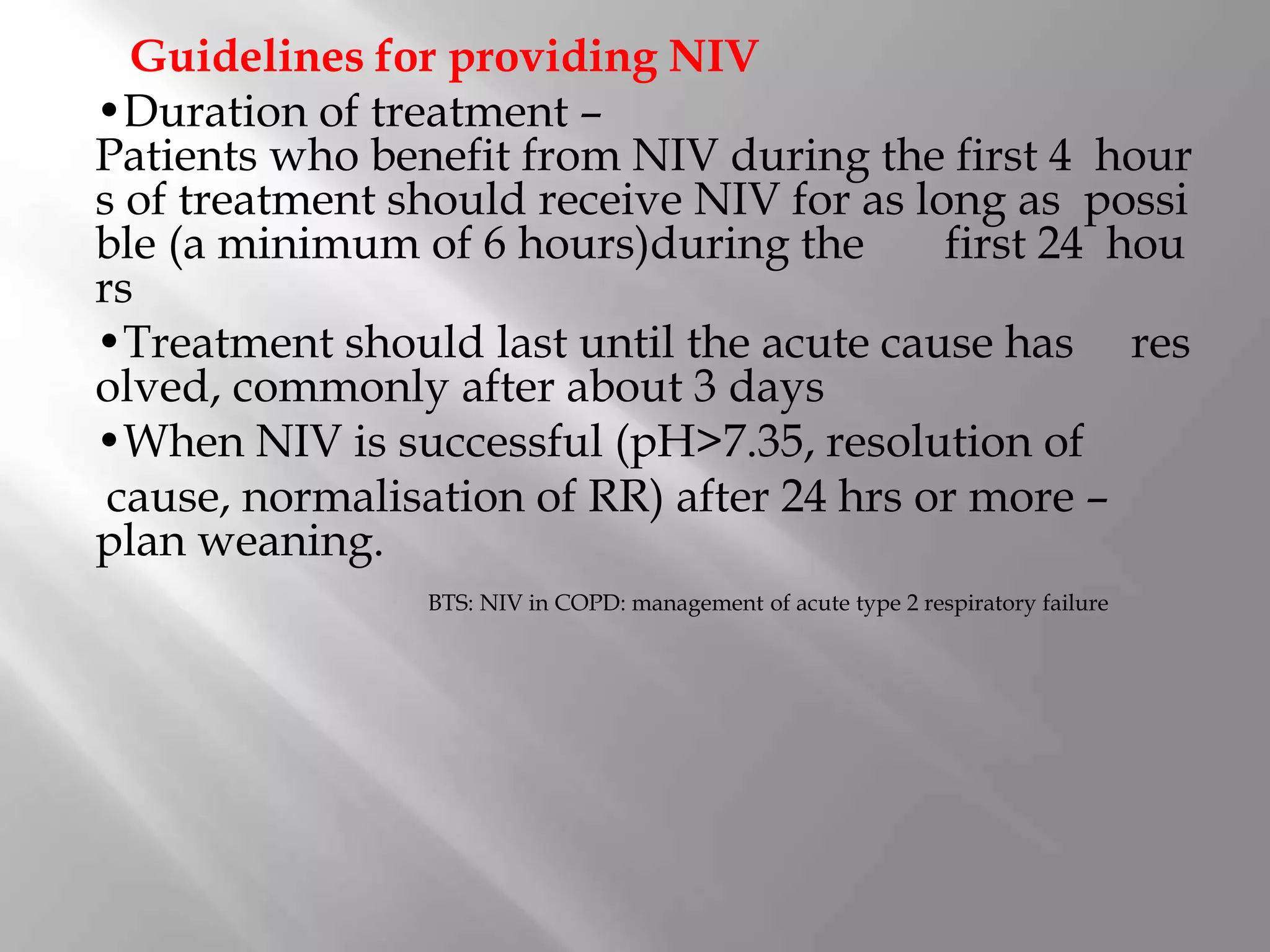
CASE PROFILE
62 year Female k/c/o
HTN/T2DM/COPD on domiciliary 02
C/O: cough with expectoration
worsening of breathlessness 3days
Associated History of :
Orthopnea/PND
No history of:
 Fever
 chest pain
 Haemoptysis
 palpitations.
Drowsy
Vitals:
PR-108/min.
BP-140/80mmhg
RR-28/m
SP02-80%on Room
air
Afebrile to touch
 General Physical
Examination:
 P-
 I- NIL
 C-
 O- B/L LOWER LIMBS
 JVP-RAISED
Chest: B/L Symmetrical
Diffuse Wheeze
Fine Crepts ISA(Bilateral)
CVS: S1,S2 +
P/A: Mild Distention, Mild tenderness RHC
CNS: Drowsy
Moving all limbs Equally.
HTN/T2DM/COPD a/w
 COPD acute exacerbation.
 Congestive cardiac failure .
 PTE.
 ? ? Pneumonia.
HB TLC DLC PLT MCV/MCH HCT
14 12.7 83/12 220 87/25 48
14.5 8.9 77/13 238 88/24 48.2
BIL ALT ALP TP ALB AMY
0.38 58 79 7.7 4.06 88
UREA CREA CPK CA PO4 LDH
38 0.89 70 9.5 2.8 290
CPK GLU PT 1NR D-DIMER
87 132 12.5 1.07 307
PH PC02 P02 HC03 SP02 Na K Lac
7.26 114 57 51.2 83 133 3.8 4.4
7.24 95 63 37.2 85 136 4.4 2.1
7.25 76 70 28.9 88 `127 4.0 1.1
7.30 80 75 34.0 89 140 3.9 1.9
7.41 62 81 41.2 92 139 3.7 1.7
7.39 55 81 37 90 133 3.5 2.0
7.40 49 82 35 92 135 3.9 1.9
 ECG: Sinus tachy. LAD, P-Pulmonale
 CHEST X-RAY : Hyper inflated lung fields
with prominent upper zone bronchiovascular
markings
 BLOOD CULTURE: Sterile
 RUE: 3-4 pus cells
 ECHO: RVSP=30+RAP,EF=68%, conc.LVH.
HTN/T2DM/COPD A/W COPD acute
exacerbation with CCF precipitated by:
? dietary indiscretions and
?non-compliance to medicine
PHARMACOTHERAPY
 O2 INHALATION @4L/min
 Inj ceftriaxone salbactum 1.25g I/V BID
 Tab. Azithromycin
 Inj Methylprednisolone 125mg TID
 Salbutamol nebulisation
 Inj UFH 5000U S/C BD
 Inj Insulin R 4,4,4. Inj Insulin N 6u BT
 Inj Lasix 20mg BD
VENTILATION
 NIV: initially started on IPAP of 10 cm of H2O
& EPAP of 4 cm of H2O
 Subsequently increased to IPAP of 18 & EPAP
of 8 cm of H2O
 NIV given for around 14-16 hrs on D1, 10-12
hrs D2, 8-10hrs D3, 4-6 hrs on D4
DISSCUSSION
Non invasive
ventillation
Is the delivery of ventillatory support
without the need for an invasive airway.
It is provided through a machine
consisting of monitor, flexible tubing, and
mask .
Types of NIV
•Negative pressure NIV
•Positive pressure NIV –
Positive pressure delivered through mask case –
CPAP & BiPAP
Negative Pressure Ventilation (NPV)
•Negative pressure ventilators apply a negative
pressure intermittently around the patient’s body or ch
est wall
•Negative pressure is applied intermittently to the th
oracic area resulting in a pressure drop around the thor
ax •This negative pressure is transmitted to the pleural
space and alveoli creating a pressure gradient be
tween the inside of the lungs and the mouth
CPAP
 In this mode the device delivers a continuous
pressure to the patient at all times. All breaths in
this mode are spontaneous breaths
 CPAP improves pulmonary function by reducing
work of breathing, maintaining inflation of
atelectatic alveoli,improves pulmonary
compliance,improves hemodynamics by
reducing preload & afterload.
 While initiating CPAP pressures 5-15cm of H2o
are most commonly used ,however pressures
greater than 15 cm should be cautiosly used as
they may produce fall in BP
 Used in COPD,asthma ,ARDS,pnemonia.
. Spontaneous mode
. Spontaneous timed mode
. Timed mode
.pressure control mode
. Ventillation control-synchronised intermittent
mandatory ventillation mode
Spontaneous ventillation
 Delivers Bi –level pressure support and
provides spontaneous breaths.
 in this mode ,IPAP is delivered during
inhalation & a lower EPAP is delivered during
exhalation.
Spontaneous /Timed mode
 Delivers Bi- level pressure support .& provides
spontaneous & mandatory breaths. Mandatory
breaths are delivered when the patient does not
breathe spontaneously within a prescribed
breath rate.
Timed Mode
Delivers Bi-level pressure support but it delivers
only mandatory breaths. Means ventilator will not
respond to patient effort .
Pressure Control Mode
The device delivers bi-level pressure support. This
mode delivers assist and mandatory breath. This
mode is identical to S/T mode, except that all
breaths have a fixed inspiratory time.
Pressure controll- synchronised intermittent
mandatory ventillation (PC-SIMV) mode
 The PC-SIMV mode provides
spontaneous,Assist & mandatory breaths.This
mode uses a time window to decide what type
of breaths should be delivered.
 An initial IPAP of 10 cm H2O & EPAP of 4to5 cm H2O
should be used .
 IPAP should be increased by 2to5 cm increments at a
rate of approximately 5 cm H2O every 10 mins, with a usual
IPAP target of 20 cm H2O with EPAP/IPAP=1:2.5
or until a therapeutic response is achieved or patient
tolerability has been reached.
 O2should be entrained into the circuit and the flow
adjusted to SpO2>88–92% .
BTS: NIV in COPD: management of acute type 2 respiratory failure
Indications:
1.Airway obstruction
 –COPD.
 –Facilitation of weaning in COPD.
 –Asthma.
 –Extubation failure in COPD.
 –Cystic Fibrosis .
 –OSA/obesity hypoventilation .
NIV & stable COPD
•NIV increasingly used in stable very severe
COPD
•NIV+O2 therapy–
in selected patients with pronounced daytime
hypercapnia
•Clear benefits in both survival & risk of hospital a
dmission in patients with both COPD & OSA
 At least one of the following
 Respiratory acidosis (pH<7.35 &/or PaCO2>45)
 Severe dyspnea with clinical signs s/o respiratory
muscle fatigue, increased WOB or both
 Use of respiratory accessory muscles
 Paradoxical motion of abdomen
 Intercostal retraction
GOLD update 2013
2)Hypoxemic respiratory failure
–Acute pulmonary edema‐CPAP
–Immunocompromised patients
–Postoperative patients
–ARDS
–Pneumonia
–Trauma or burns
–Restrictive thoracic disorders
 Cardiac/Respiratory arrest.
 Encephalopathy.
 Severe UGI bleed.
 Upper airway obstruction .
 Hemodynamic instability.
 Untreated pneumothorax.
 High risk of aspiration .
 Uncooperative/irritable patient.
 Facial or neurological surgery , trauma or
deformity.
 Less sedation, Non invasive , patient able to
maintain verbalisation,
 Early improvement in hypoxia, acidosis and
hypercapnia
 Shorter hospital stay, decreased rate of
intubation without risks of ET.
 Decreased mortality.
Interface related
.C02 rebreathing
.Discomfort
.Facial skin erythema
.Nasal bridge ulceration
Air pressure/ flow related
.Air leaks
.Nasal or oral dryness or congestion
.Gastric distrension
Patient related
.Aspiration pneumonia
.Barotrauma
.Haemodynamic effects
1. Appropriately monitored location, oximetry, respiratory impedance, vital signs as clinically
indicated
2. Patient in bed or chair at >30 angle
3. Select and fit interface
4. Select ventilator
5. Apply headgear; avoid excessive strap tension (one or two fingers under strap)
6. Connect interface to ventilator tubing and turn on ventilator
7. Start with low pressure in spontaneously triggered mode with backup rate; pressure limited:
8 to 12 cm H2O inspiratory pressure; 3 to 5 cm H2O expiratory pressure
8. Gradually increase inspiratory pressure (10 to 20 cm H2O) as tolerated to achieve alleviation
of dyspnea, decreased respiratory rate, increased tidal volume (if being monitored), and good
patient-ventilator synchrony
9. Provide O2 supplementation as need to keep O2 sat >90 percent
10. Check for air leaks, readjust straps as needed
11. Add humidifier as indicated
12. Consider mild sedation (eg, intravenously administered lorazepam 0.5 mg) in agitated
patients
13. Encouragement, reassurance, and frequent checks and adjustments as needed
14. Monitor occasional blood gases (within 1 to 2 hours) and then as needed
Protocol for initiation of noninvasive positive pressure ventilation
Steps For Initiating NPPV
1. Place patient in an upright or sitting position.Carefully explain the procedure for
noninvasive positive pressure ventilation, including the goals and possible
complications.
2. Using a sizing gauge , make sure a mask is chosen that is the proper size and fit.
3. Attach the interface and circuit to the ventilator . Turn on the ventilator and
adjust it initially to low pressure setting.
4. Hold or allow the patient to hold the mask gently to the face until the patient
becomes comfortable with it. Encourage the patient to use proper breathing
technique.
5. Monitor oxygen ( O2 ) saturation; adjust the fractional inspired oxygen ( F1 O2 )
to maintain O2 saturation; above 90%.
6. Secure the mask to the patient . Do not make the straps too tight.
7. Titrate the inspiratory and end-expiratory positive airway pressures (IPAP and
EPAP) to achieve patient comfort ,adequate exhaled tidal volume, and
synchrony with the ventilator. Do not allow peak pressures to exceed 20 cm H2O.
8. Check for leaks and adjust the Straps if necessary
9. Monitor the respiratory rate, heart rate,level of dyspnea, O2 saturation , minute
ventilation,and exhaled tidal volume.
10. Obtain blood gas values within 1 hour.
ACUTE CARE
•Reduces need for intubation
•Reduces incidence of nosocomial pneumonia
•Shortens stay in intensive care unit
•Shortens hospital stay
•Reduces mortality
•Preserves airway defenses
•Improves patient comfort
•Reduces need for sedation
CHRONIC CARE
•Alleviates symptoms of chronic hypoventilation
•Improves duration and quality of sleep
•Improves functional capacity
•Prolongs survival
clinical Benefits of Noninvasive Positive Pressure Ventilation
Guidelines for providing NIV
•Duration of treatment –
Patients who benefit from NIV during the first 4 hour
s of treatment should receive NIV for as long as possi
ble (a minimum of 6 hours)during the first 24 hou
rs
•Treatment should last until the acute cause has res
olved, commonly after about 3 days
•When NIV is successful (pH>7.35, resolution of
cause, normalisation of RR) after 24 hrs or more –
plan weaning.
BTS: NIV in COPD: management of acute type 2 respiratory failure
Response
Physiological
a) Continuous oximetry.
b) Exhaled tidal volume.
c) ABG at 1 hour or as necessary, at 2 to 6 hour intervals.
Objective
a) Respiratory rate.
b) blood pressure.
c) pulse rate.
Subjective
a) dyspnea.
b) comfort.
c) mental alertness.
Mask
Fit, Comfort, Air leak, Secretions, Skin necrosis.
Respiratory muscle unloading
Accessory muscle activity, paradoxical
abdominal motion.
Abdomen
Gastric distension.
• Most often successful in the critically ill patient
Respironics PerformaTrak® Full Face Mask
Entrainment
valve
Adjustable
Forehead Support
Ball and
Socket Clip
Double-foam
cushion
Pressure
pick-off
port
360
swivel
standard
elbow
Respironics Contour Deluxe™ Mask
Dual flap
cushion
Thin flexible &
bridge
material
Dual density
foam bridge
forehead
support
Nasal Pillows or Nasal Cushions (continued)
• Suitable for patients with
–Claustrophobia
–Skin sensitivities
–Need for visibility
Respironics Comfort Lite Nasal Mask
Advantages of Nasal Masks
Disadvantages of Nasal Masks
• Less risk of aspiration
• Enhanced secretion clearance
• Less claustrophobia
• Easier speech
• Less dead space
• Mouth leak
• Less effectiveness with nasal obstruction
• Nasal irritation and rhinorrhea
• Mouth dryness
Younger age
Lower acuity of illness (APACHE score)
Able to cooperate, better neurologic score
Less air leaking
Moderate hypercarbia (PaCO2 >45 mmHG, <92 mmHG)
Moderate acidemia (pH <7.35, >7.10)
Improvements in gas exchange and heart respiratory rates within first 2 hours
Potential indicators of success in NPPV use
Weaning strategy
•Continue NIV for 16 hours on day 2.
•Continue NIV for 12 hours on day 3 including
6-8 hours overnight use.
•Discontinue NIV day 4, unless continuation
is clinically indicated.
BTS: NIV in COPD: management of acute type 2 respiratory failure
 Inability to tolerate the mask because of
discomfort or pain
 Inability to improve gas exchange or dyspnea
 Need for endotracheal intubation to manage
secretions or protect airway
 Hemodynamic instability
 ECG – ischemia/arrhythmia
 Failure to improve mental status in those with
CO2 narcosis.
 Life threatning Refractory hypoxemia(Pa02<60
mmHg on 100% of inspired oxygen).
 Bronchiectasis with copious secretions.
 Severe Pneumonia.
 Hemodynamic Instability.
THANK YOU

Non invasive ventillation...

  • 2.
    CASE PROFILE 62 yearFemale k/c/o HTN/T2DM/COPD on domiciliary 02 C/O: cough with expectoration worsening of breathlessness 3days Associated History of : Orthopnea/PND No history of:  Fever  chest pain  Haemoptysis  palpitations.
  • 3.
    Drowsy Vitals: PR-108/min. BP-140/80mmhg RR-28/m SP02-80%on Room air Afebrile totouch  General Physical Examination:  P-  I- NIL  C-  O- B/L LOWER LIMBS  JVP-RAISED
  • 4.
    Chest: B/L Symmetrical DiffuseWheeze Fine Crepts ISA(Bilateral) CVS: S1,S2 + P/A: Mild Distention, Mild tenderness RHC CNS: Drowsy Moving all limbs Equally.
  • 5.
    HTN/T2DM/COPD a/w  COPDacute exacerbation.  Congestive cardiac failure .  PTE.  ? ? Pneumonia.
  • 6.
    HB TLC DLCPLT MCV/MCH HCT 14 12.7 83/12 220 87/25 48 14.5 8.9 77/13 238 88/24 48.2 BIL ALT ALP TP ALB AMY 0.38 58 79 7.7 4.06 88 UREA CREA CPK CA PO4 LDH 38 0.89 70 9.5 2.8 290 CPK GLU PT 1NR D-DIMER 87 132 12.5 1.07 307
  • 7.
    PH PC02 P02HC03 SP02 Na K Lac 7.26 114 57 51.2 83 133 3.8 4.4 7.24 95 63 37.2 85 136 4.4 2.1 7.25 76 70 28.9 88 `127 4.0 1.1 7.30 80 75 34.0 89 140 3.9 1.9 7.41 62 81 41.2 92 139 3.7 1.7 7.39 55 81 37 90 133 3.5 2.0 7.40 49 82 35 92 135 3.9 1.9
  • 8.
     ECG: Sinustachy. LAD, P-Pulmonale  CHEST X-RAY : Hyper inflated lung fields with prominent upper zone bronchiovascular markings  BLOOD CULTURE: Sterile  RUE: 3-4 pus cells  ECHO: RVSP=30+RAP,EF=68%, conc.LVH.
  • 9.
    HTN/T2DM/COPD A/W COPDacute exacerbation with CCF precipitated by: ? dietary indiscretions and ?non-compliance to medicine
  • 10.
    PHARMACOTHERAPY  O2 INHALATION@4L/min  Inj ceftriaxone salbactum 1.25g I/V BID  Tab. Azithromycin  Inj Methylprednisolone 125mg TID  Salbutamol nebulisation  Inj UFH 5000U S/C BD  Inj Insulin R 4,4,4. Inj Insulin N 6u BT  Inj Lasix 20mg BD
  • 11.
    VENTILATION  NIV: initiallystarted on IPAP of 10 cm of H2O & EPAP of 4 cm of H2O  Subsequently increased to IPAP of 18 & EPAP of 8 cm of H2O  NIV given for around 14-16 hrs on D1, 10-12 hrs D2, 8-10hrs D3, 4-6 hrs on D4
  • 12.
  • 13.
    Is the deliveryof ventillatory support without the need for an invasive airway. It is provided through a machine consisting of monitor, flexible tubing, and mask .
  • 14.
    Types of NIV •Negativepressure NIV •Positive pressure NIV – Positive pressure delivered through mask case – CPAP & BiPAP Negative Pressure Ventilation (NPV) •Negative pressure ventilators apply a negative pressure intermittently around the patient’s body or ch est wall •Negative pressure is applied intermittently to the th oracic area resulting in a pressure drop around the thor ax •This negative pressure is transmitted to the pleural space and alveoli creating a pressure gradient be tween the inside of the lungs and the mouth
  • 15.
    CPAP  In thismode the device delivers a continuous pressure to the patient at all times. All breaths in this mode are spontaneous breaths  CPAP improves pulmonary function by reducing work of breathing, maintaining inflation of atelectatic alveoli,improves pulmonary compliance,improves hemodynamics by reducing preload & afterload.
  • 16.
     While initiatingCPAP pressures 5-15cm of H2o are most commonly used ,however pressures greater than 15 cm should be cautiosly used as they may produce fall in BP  Used in COPD,asthma ,ARDS,pnemonia.
  • 17.
    . Spontaneous mode .Spontaneous timed mode . Timed mode .pressure control mode . Ventillation control-synchronised intermittent mandatory ventillation mode
  • 18.
    Spontaneous ventillation  DeliversBi –level pressure support and provides spontaneous breaths.  in this mode ,IPAP is delivered during inhalation & a lower EPAP is delivered during exhalation.
  • 19.
    Spontaneous /Timed mode Delivers Bi- level pressure support .& provides spontaneous & mandatory breaths. Mandatory breaths are delivered when the patient does not breathe spontaneously within a prescribed breath rate.
  • 20.
    Timed Mode Delivers Bi-levelpressure support but it delivers only mandatory breaths. Means ventilator will not respond to patient effort . Pressure Control Mode The device delivers bi-level pressure support. This mode delivers assist and mandatory breath. This mode is identical to S/T mode, except that all breaths have a fixed inspiratory time.
  • 21.
    Pressure controll- synchronisedintermittent mandatory ventillation (PC-SIMV) mode  The PC-SIMV mode provides spontaneous,Assist & mandatory breaths.This mode uses a time window to decide what type of breaths should be delivered.
  • 22.
     An initialIPAP of 10 cm H2O & EPAP of 4to5 cm H2O should be used .  IPAP should be increased by 2to5 cm increments at a rate of approximately 5 cm H2O every 10 mins, with a usual IPAP target of 20 cm H2O with EPAP/IPAP=1:2.5 or until a therapeutic response is achieved or patient tolerability has been reached.  O2should be entrained into the circuit and the flow adjusted to SpO2>88–92% . BTS: NIV in COPD: management of acute type 2 respiratory failure
  • 23.
    Indications: 1.Airway obstruction  –COPD. –Facilitation of weaning in COPD.  –Asthma.  –Extubation failure in COPD.  –Cystic Fibrosis .  –OSA/obesity hypoventilation .
  • 24.
    NIV & stableCOPD •NIV increasingly used in stable very severe COPD •NIV+O2 therapy– in selected patients with pronounced daytime hypercapnia •Clear benefits in both survival & risk of hospital a dmission in patients with both COPD & OSA
  • 25.
     At leastone of the following  Respiratory acidosis (pH<7.35 &/or PaCO2>45)  Severe dyspnea with clinical signs s/o respiratory muscle fatigue, increased WOB or both  Use of respiratory accessory muscles  Paradoxical motion of abdomen  Intercostal retraction GOLD update 2013
  • 26.
    2)Hypoxemic respiratory failure –Acutepulmonary edema‐CPAP –Immunocompromised patients –Postoperative patients –ARDS –Pneumonia –Trauma or burns –Restrictive thoracic disorders
  • 27.
     Cardiac/Respiratory arrest. Encephalopathy.  Severe UGI bleed.  Upper airway obstruction .  Hemodynamic instability.  Untreated pneumothorax.  High risk of aspiration .  Uncooperative/irritable patient.  Facial or neurological surgery , trauma or deformity.
  • 28.
     Less sedation,Non invasive , patient able to maintain verbalisation,  Early improvement in hypoxia, acidosis and hypercapnia  Shorter hospital stay, decreased rate of intubation without risks of ET.  Decreased mortality.
  • 29.
    Interface related .C02 rebreathing .Discomfort .Facialskin erythema .Nasal bridge ulceration Air pressure/ flow related .Air leaks .Nasal or oral dryness or congestion .Gastric distrension Patient related .Aspiration pneumonia .Barotrauma .Haemodynamic effects
  • 30.
    1. Appropriately monitoredlocation, oximetry, respiratory impedance, vital signs as clinically indicated 2. Patient in bed or chair at >30 angle 3. Select and fit interface 4. Select ventilator 5. Apply headgear; avoid excessive strap tension (one or two fingers under strap) 6. Connect interface to ventilator tubing and turn on ventilator 7. Start with low pressure in spontaneously triggered mode with backup rate; pressure limited: 8 to 12 cm H2O inspiratory pressure; 3 to 5 cm H2O expiratory pressure 8. Gradually increase inspiratory pressure (10 to 20 cm H2O) as tolerated to achieve alleviation of dyspnea, decreased respiratory rate, increased tidal volume (if being monitored), and good patient-ventilator synchrony 9. Provide O2 supplementation as need to keep O2 sat >90 percent 10. Check for air leaks, readjust straps as needed 11. Add humidifier as indicated 12. Consider mild sedation (eg, intravenously administered lorazepam 0.5 mg) in agitated patients 13. Encouragement, reassurance, and frequent checks and adjustments as needed 14. Monitor occasional blood gases (within 1 to 2 hours) and then as needed Protocol for initiation of noninvasive positive pressure ventilation
  • 31.
    Steps For InitiatingNPPV 1. Place patient in an upright or sitting position.Carefully explain the procedure for noninvasive positive pressure ventilation, including the goals and possible complications. 2. Using a sizing gauge , make sure a mask is chosen that is the proper size and fit. 3. Attach the interface and circuit to the ventilator . Turn on the ventilator and adjust it initially to low pressure setting. 4. Hold or allow the patient to hold the mask gently to the face until the patient becomes comfortable with it. Encourage the patient to use proper breathing technique. 5. Monitor oxygen ( O2 ) saturation; adjust the fractional inspired oxygen ( F1 O2 ) to maintain O2 saturation; above 90%. 6. Secure the mask to the patient . Do not make the straps too tight. 7. Titrate the inspiratory and end-expiratory positive airway pressures (IPAP and EPAP) to achieve patient comfort ,adequate exhaled tidal volume, and synchrony with the ventilator. Do not allow peak pressures to exceed 20 cm H2O. 8. Check for leaks and adjust the Straps if necessary 9. Monitor the respiratory rate, heart rate,level of dyspnea, O2 saturation , minute ventilation,and exhaled tidal volume. 10. Obtain blood gas values within 1 hour.
  • 32.
    ACUTE CARE •Reduces needfor intubation •Reduces incidence of nosocomial pneumonia •Shortens stay in intensive care unit •Shortens hospital stay •Reduces mortality •Preserves airway defenses •Improves patient comfort •Reduces need for sedation CHRONIC CARE •Alleviates symptoms of chronic hypoventilation •Improves duration and quality of sleep •Improves functional capacity •Prolongs survival clinical Benefits of Noninvasive Positive Pressure Ventilation
  • 33.
    Guidelines for providingNIV •Duration of treatment – Patients who benefit from NIV during the first 4 hour s of treatment should receive NIV for as long as possi ble (a minimum of 6 hours)during the first 24 hou rs •Treatment should last until the acute cause has res olved, commonly after about 3 days •When NIV is successful (pH>7.35, resolution of cause, normalisation of RR) after 24 hrs or more – plan weaning. BTS: NIV in COPD: management of acute type 2 respiratory failure
  • 34.
    Response Physiological a) Continuous oximetry. b)Exhaled tidal volume. c) ABG at 1 hour or as necessary, at 2 to 6 hour intervals. Objective a) Respiratory rate. b) blood pressure. c) pulse rate. Subjective a) dyspnea. b) comfort. c) mental alertness.
  • 35.
    Mask Fit, Comfort, Airleak, Secretions, Skin necrosis. Respiratory muscle unloading Accessory muscle activity, paradoxical abdominal motion. Abdomen Gastric distension.
  • 36.
    • Most oftensuccessful in the critically ill patient Respironics PerformaTrak® Full Face Mask Entrainment valve Adjustable Forehead Support Ball and Socket Clip Double-foam cushion Pressure pick-off port
  • 37.
    360 swivel standard elbow Respironics Contour Deluxe™Mask Dual flap cushion Thin flexible & bridge material Dual density foam bridge forehead support
  • 38.
    Nasal Pillows orNasal Cushions (continued) • Suitable for patients with –Claustrophobia –Skin sensitivities –Need for visibility Respironics Comfort Lite Nasal Mask
  • 39.
    Advantages of NasalMasks Disadvantages of Nasal Masks • Less risk of aspiration • Enhanced secretion clearance • Less claustrophobia • Easier speech • Less dead space • Mouth leak • Less effectiveness with nasal obstruction • Nasal irritation and rhinorrhea • Mouth dryness
  • 40.
    Younger age Lower acuityof illness (APACHE score) Able to cooperate, better neurologic score Less air leaking Moderate hypercarbia (PaCO2 >45 mmHG, <92 mmHG) Moderate acidemia (pH <7.35, >7.10) Improvements in gas exchange and heart respiratory rates within first 2 hours Potential indicators of success in NPPV use
  • 41.
    Weaning strategy •Continue NIVfor 16 hours on day 2. •Continue NIV for 12 hours on day 3 including 6-8 hours overnight use. •Discontinue NIV day 4, unless continuation is clinically indicated. BTS: NIV in COPD: management of acute type 2 respiratory failure
  • 42.
     Inability totolerate the mask because of discomfort or pain  Inability to improve gas exchange or dyspnea  Need for endotracheal intubation to manage secretions or protect airway  Hemodynamic instability  ECG – ischemia/arrhythmia  Failure to improve mental status in those with CO2 narcosis.
  • 43.
     Life threatningRefractory hypoxemia(Pa02<60 mmHg on 100% of inspired oxygen).  Bronchiectasis with copious secretions.  Severe Pneumonia.  Hemodynamic Instability.
  • 44.