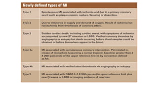
Definition and classification
•Detection of rise or fall of cardiac biomarker with atleast one value
above 99th
percentile above URL along with any one of the following :
1- symptoms of ischemia
2-new or presumed ST-T / LBBB
3- pathological Q wave
4- identification of thrombus by angiography or autopsy
4.
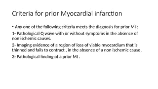
Criteria for priorMyocardial infarction
• Any one of the following criteria meets the diagnosis for prior MI :
1- Pathological Q wave with or without symptoms in the absence of
non ischemic causes.
2- Imaging evidence of a region of loss of viable myocardium that is
thinned and fails to contract , in the absence of a non ischemic cause .
3- Pathological finding of a prior MI .
5.
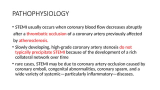
PATHOPHYSIOLOGY
• STEMI usuallyoccurs when coronary blood flow decreases abruptly
after a thrombotic occlusion of a coronary artery previously affected
by atherosclerosis.
• Slowly developing, high-grade coronary artery stenosis do not
typically precipitate STEMI because of the development of a rich
collateral network over time
• rare cases, STEMI may be due to coronary artery occlusion caused by
coronary emboli, congenital abnormalities, coronary spasm, and a
wide variety of systemic—particularly inflammatory—diseases.
6.
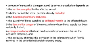
• amount ofmyocardial damage caused by coronary occlusion depends on
1-the territory supplied by the affected vessel,
2-whether or not the vessel becomes totally occluded,
3-the duration of coronary occlusion,
4-the quantity of blood supplied by collateral vessels to the affected tissue,
5-the demand for oxygen of the myocardium whose blood supply has been
suddenly limited,
6-endogenous factors that can produce early spontaneous lysis of the
occlusive thrombus, and
7-the adequacy of myocardial perfusion in the infarct zone when flow is
restored in the occluded epicardial coronary artery.
7.
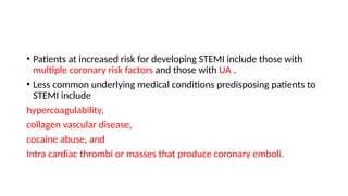
• Patients atincreased risk for developing STEMI include those with
multiple coronary risk factors and those with UA .
• Less common underlying medical conditions predisposing patients to
STEMI include
hypercoagulability,
collagen vascular disease,
cocaine abuse, and
Intra cardiac thrombi or masses that produce coronary emboli.
8.
CLINICAL PRESENTATION
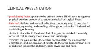
• precipitatingfactor appears to be present before STEMI, such as vigorous
physical exercise, emotional stress, or a medical or surgical illness.
• Pain (m/c) is deep and visceral; adjectives commonly used to describe it
are heavy, squeezing, and crushing; although, occasionally, it is described
as stabbing or burning
• similar in character to the discomfort of angina pectoris but commonly
occurs at rest, is usually more severe, and lasts longer.
• Typically, the pain involves the central portion of the chest and/or the
epigastrium, and, on occasion, it radiates to the arms. Less common sites
of radiation include the abdomen, back, lower jaw, and neck.
9.
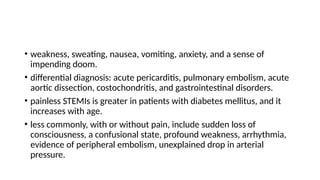
• weakness, sweating,nausea, vomiting, anxiety, and a sense of
impending doom.
• differential diagnosis: acute pericarditis, pulmonary embolism, acute
aortic dissection, costochondritis, and gastrointestinal disorders.
• painless STEMIs is greater in patients with diabetes mellitus, and it
increases with age.
• less commonly, with or without pain, include sudden loss of
consciousness, a confusional state, profound weakness, arrhythmia,
evidence of peripheral embolism, unexplained drop in arterial
pressure.
10.
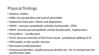
Physical findings
• Anxious,restless
• Pallor a/w perspiration and cool of extremities
• Substernal chest pain >30min and diaphoresis
• AWMI – increase sympathetic activities (tachycardia , HTN)
• IWMI- increase parasympathetic activity (bradycardia , hypotension)
• Precordium – usually quiet
• S3,S4, decrease intensity of first heart sound , paradoxical splitting of S2
• Mid systolic or late systolic murmur
• Decreased carotid pulsation
• transmural infarction, systolic pressure declines by ~10–15 mmHg from the
preinfarction state.
11.
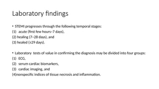
Laboratory findings
• STEMIprogresses through the following temporal stages:
(1) acute (first few hours–7 days),
(2) healing (7–28 days), and
(3) healed (≥29 days).
• Laboratory tests of value in confirming the diagnosis may be divided into four groups:
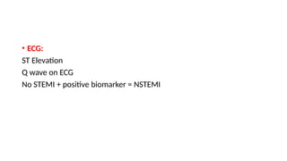
(1) ECG,
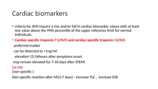
(2) serum cardiac biomarkers,
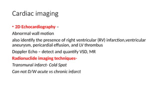
(3) cardiac imaging, and
(4)nonspecific indices of tissue necrosis and inflammation.
Cardiac biomarkers
• criteriafor AMI require a rise and/or fall in cardiac biomarker values with at least
one value above the 99th percentile of the upper reference limit for normal
individuals.
• Cardiac-specific troponin T (cTnT) and cardiac-specific troponin I (cTnI)
preferred marker
can be detected to <1ng/ml
elevation 12-24hours after symptoms onset
may remain elevated for 7-10 days after STEMI
CK-MB
(non specific )
Non specific reaction after MI(3-7 days) - increase TLC , increase ESR
14.
Cardiac imaging
• 2DEchocardiography –
Abnormal wall motion
also identify the presence of right ventricular (RV) infarction,ventricular
aneurysm, pericardial effusion, and LV thrombus
Doppler Echo – detect and quantify VSD, MR
Radionuclide imaging techniques-
Transmural infarct- Cold Spot
Can not D/W acute vs chronic infarct
15.
Management
• prognosis inSTEMI depends on
(1) electrical complications (arrhythmias)
(2) mechanical complications (“pump failure”).
MCC of out of hospital death – Ventrical fibrillation
Goal of initial management –
-control of cardiac discomfort
- Rapid identification of candidates for early perfusion
16.
• Aspirin –buccal absorption of chewed tablet 160-325mg tablet
followed by 75-162mg once daily.
• O2 inhalation via nasal prongs or face mask – 2-4 L/min for first 6-12
hours
• control of discomfort :
• Nitroglycerine : Up to three doses of 0.4 mg should be administered
at about 5-min intervals
• IV nitroglycerine NTG
• Avoid in SBP<90, RV infarction , phosphodiesterase 5 inhibitor
17.
• Morphine –very effective analgesic
Venous pooling – reduce CO and MAP
Vagotomy effect- bradycardia and advanced degree heart block
• Morphine is routinely administered by repetitive (every 5 min)
intravenous injection of small doses (2–4 mg).
• IV Beta blocker
diminishing myocardial O2 demand and hence ischemia.
Reduce the risks of reinfarction and ventricular fibrillation
18.
• metoprolol, 5mg every 2–5 min for a total of three doses, provided the
patient has a heart rate >60 beats/min, systolic pressure >100 mmHg, a PR
interval <0.24 s, and rales that are no higher than 10 cm up from the
diaphragm.
• Fifteen minutes after the last intravenous dose, an oral regimen is initiated of
50 mg every 6 h for 48 h, followed by 100 mg every 12 h.
• Role of CCB in acute phase ? No role
• Glucocorticoids and nonsteroidal anti-inflammatory agents, with the
exception of aspirin, should be avoided in patients with STEMI.
• They can impair infarct healing and increase the risk of myocardial rupture,
and their use may result in a larger infarct scar.
19.
Primary PCI
• PCI,usually angioplasty and/or stenting without preceding
fibrinolysis, referred to as primary PCI, is effective in restoring
perfusion in STEMI when carried out on an emergency basis in the
first few hours of MI.
• Can be done in patients having C/I to fibrinolysis
• more effective than fibrinolysis
• generally preferred when the diagnosis is in doubt, cardiogenic shock
is present, bleeding risk is increased, or symptoms have been present
for at least 2–3 h when the clot is more mature
20.
Fibrinolysis
• If nocontraindications are present (see below), fibrinolytic therapy
should ideally be initiated within 30 min of presentation (i.e., door-to
needle time ≤30 min).
• fibrinolytic agents
• Tissue plasminogen activator (tPA),
• streptokinase, tenecteplase (TNK), and
• reteplase (rPA)
21.
• The currentrecommended regimen of tPA consists of a 15-mg bolus
followed by 50 mg intravenously over the first 30 min, followed by 35
mg over the next 60 min.
• Streptokinase is administered as 1.5 million units (MU) intravenously
over 1 h.
• rPA is administered in a double-bolus regimen consisting of a 10-MU
bolus given over 2–3 min, followed by a second 10-MU bolus 30 min
later.
• TNK is given as a single weight-based intravenous bolus of 0.53 mg/kg
over 10 s.
22.
Contraindication of fibrinolyticagents
• Absolute –
-h/o hemorrhagic CVA at any time,
-non hemorrhagic stroke or other cerebrovascular event within the past
year ,
-marked hypertension (>180/110 mmHg),
-suspicion of aortic dissection, and
-active internal bleeding (excluding menses).
23.
• Relative –
•On anticoagulant INR ≥2,
• recent (<2 weeks) invasive or surgical procedure or
• prolonged (>10 min) cardiopulmonary resuscitation,
• bleeding diathesis,
• pregnancy,
• hemorrhagic ophthalmic condition (e.g., hemorrhagic diabetic retinopathy),
• active peptic ulcer disease,
• history of severe hypertension (Under control).
• STK- used within 5 days to 2 years.
24.
• Cardiac catheterizationand coronary angiography should be carried
out after fibrinolytic therapy if there is evidence of either
• (1) failure of reperfusion (persistent chest pain and ST-segment
elevation >90 min), in which case a rescue PCI should be considered;
or
• (2) coronary artery reocclusion (re-elevation of ST segments and/or
recurrent chest pain) or the development of recurrent ischemia (such
as recurrent angina in the early hospital course or a positive exercise
stress test before discharge),
25.
General management
• Coronarycare units – rhythm and hemodynamic monitoring
• Activity – bed rest for 6-12 hours , ambulation after 2-3 days
• Diet – nil PO or clear fluids in initial 4-12 hrs
• Bowel management- fiber , stool softner
• Sedation – diazepam , oxazepam , or lorazepam
• Many drugs used in the coronary care unit, such as atropine, H2
blockers, and narcotics, can produce delirium, particularly in the
elderly.
26.
Pharmacotherapy
• Antithrombotics
• antiplateletand anticoagulant therapy during the initial phase of
STEMI
• Antiplatelets – Aspirin, P2Y12- Clopidogrel, prasugrel ,ticagrelor ,
cangrelor , GPIIB/IIIA Inhibitors – abciximab, tirofiban
• Anticoagulants – UFH, LMWH , Fondaparinaux , Bivalirudin
• The recommended dose of UFH is an initial bolus of 60 U/kg
(maximum 4000 U) followed by an initial infusion of 12 U/kg per h
(maximum 1000 U/h).