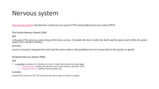
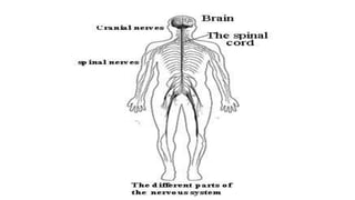
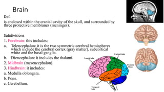
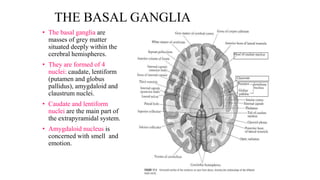
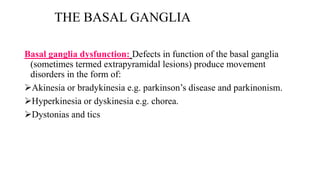
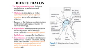
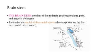
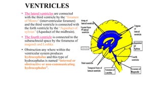
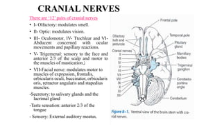
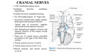
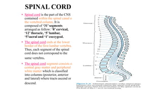
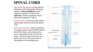
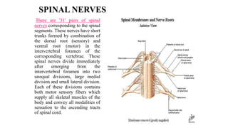
SlideShare a Scribd company logo
Neurological assessment
Dr. Inas Ahmed Al-Amrani
Nervous system
The nervous system is divided into central nervous system (CNS) and peripheral nervous system (PNS).
The Central Nervous System (CNS)
Def.
is the part of the nervous system lying within bony cavities. It includes the brain (within the skull) and the spinal cord (within the spinal
canal of the vertebral column).
Function
receives sensation, integrate them and send the motor orders to the peripheral nervous system then to the muscles or glands.
Peripheral Nervous System (PNS)
Def.
• is classified according to its function or its site of origin from the nervous system into:
• 1. Cranial nerves: emerge from the brain stem except olfactory and optic nerves.
• 2.The spinal nerves: originate from the spinal cord
Function
transmits the sensation to the CNS and carries the motor orders to muscles or glands.
Brain
Def.
is enclosed within the cranial cavity of the skull, and surrounded by
three protective membranes (meninges).
Subdivisions
1. Forebrain: this includes:
a. Telencephalon: it is the two symmetric cerebral hemispheres
which include the cerebral cortex (gray matter), subcortical
white and the basal ganglia.
b. Diencephalon: it includes the thalami.
2. Midbrain (mesencephalon).
3. Hindbrain: it includes:
a. Medulla oblongata.
b. Pons.
c. Cerebellum.
Cerebrum
Def‘.:-It’s the upper and largest part of the brain.
It’s divided incompletely into two hemispheres; Rt.&Lt. cerebral hemispheres by the median
longitudinal cerebral fissure.
At the floor of this fissure a mass of white matter called the corpus callosum unites the 2 cerebral
hemispheres together
• The Cerebral hemispheres occupies the ant., middle and upper part of the post. Cranial fossa
• Covering meningies
The central nervous system is covered by three coverings called meninges. These from inward to outward are pia matter, arachnoid matter and
dura matter.
1.The pia matter fits on very closely to the brain and dips into all sulci.
2.The arachnoid matter is a very thin and delicate membrane and doesn't enter the sulci of the brain. Pia and arachnoid are separated by a
narrow space named the “subarachnoid space" within which the blood vessels of the brain lie.
3. Dura matter is a tough membrane and separated from skull bones by epidural space and from arachnoid matter by subdural space.
Areas of cerebral cortex
• The lobes are separated from each other by sulci (fissures):
The central sulcus separates the frontal from the parietal lobe.
The parieto-occipital sulcus separates the parietal from the occipital
lobe.
The lateral sulcus (sylvian fissure) separates the frontal and parietal
lobes from the temporal lobe.
frontal lobe
comprises the following important areas:
• "Area 4": is the primary motor area. This area controls the voluntary
movements of the skeletal muscles on the opposite side of the body.
• "Area 6": is the premotor area. It takes part in the control of
extrapyramidal system and gives some fibers to the pyramidal tract.
• "Area 8": is concerned with eye movements and papillary changes.
• ''Areas 44 and 45'' (Broca's area) is an important area for speech.
• "Area 9,10,11,12": are the frontal association areas and are concerned
with higher mental function.
Irritative lesion of the frontal lobe leads to:
1-convulsive seizures on the opposite side of the body (focal convulsions).
2-Attacks of conjugate eye deviation to the opposite side of the lesion.
Destructive lesion of the frontal lobe leads to:
1-Contralateral paralysis, contralateral hypertonia of muscles.
2-Paralysis of conjugate eye movement to the opposite side of lesion.
3-Motor aphasia and Agraphia.
4-Mentality, personality and behavioural changes.
5-Incontinence urine and faeces.
parietal lobe
comprises the following important areas:
• "Areas 3,1,2”: are considered the primary sensory areas.
• "Area 5 and 7": are the sensory association areas. They are concerned
with steriognosis and graphoesthesia.
Irritative lesion of the parietal lobe leads to:
1. Contralateral sensory Jaksonian fit.
Destructive lesion of the parietal lobe leads to:
1. Contralateral cortical sensory loss.
2. ALexia, Garone's aphasia (word salad and apraxia.
3. Asteriognosis.
temporal lobe
comprises the following important areas:
• "Area 41and 42": these are the primary auditory areas.
• "Area 22": is the auditory association area and is responsible for knowing the meaning of
sounds.
Irritative lesion of the temporal lobe lead to:
- Auditory hallucination.
Destructive lesion of the temporal lobe lead to:
1-Slight hearing impairment.
2-Auditory agnosia.
3-Temporal seizures.
4-Anterograde amnesia.
5- Perceptive aphasia (in ability of the patient to know the meaning of heard sounds.
occipital lobe
comprises the following important areas:
• "Area 17": it is the primary cortical visual area.
• "Area 18 and 19": are visual association areas and are responsible for
knowledge the meaning of the pictures or words seen.
Irritative lesion of the occipital lobe lead to:
-Visual hallucination sparks, lines, flashes
Destructive lesion of the occipital lobe lead to:
1-Homonymous hemianopia.
2-Visual agnosia.
3-Paralysis of reflex conjugate eye movement.
THE BASAL GANGLIA
• The basal ganglia are
masses of grey matter
situated deeply within the
cerebral hemispheres.
• They are formed of 4
nuclei: caudate, lentiform
(putamen and globus
pallidus), amygdaloid and
claustrum nuclei.
• Caudate and lentiform
nuclei are the main part of
the extrapyramidal system.
• Amygdaloid nucleus is
concerned with smell and
emotion.
THE BASAL GANGLIA
Basal ganglia dysfunction: Defects in function of the basal ganglia
(sometimes termed extrapyramidal lesions) produce movement
disorders in the form of:
Akinesia or bradykinesia e.g. parkinson’s disease and parkinonism.
Hyperkinesia or dyskinesia e.g. chorea.
Dystonias and tics
DIENCEPHALON
The diencephalons includes: thalamus,
subthalamus, hypothalamus and
epithalamus.
• Thalamus is considered to be the
subcortical station for all types of
sensation (especially pain) except
olfaction.
• Lesions of the thalamus produce thalamic
syndrome which is caused mainly by
vascular aetiology.
• Subthalamus: leis between the midbrain
and the thalamus and it is mainly
connected to the extrapyramidal system.
• Epithalamus: concerned with olfaction.
• Hypothalamus: it leis below the thalamus
and has wide varieties functions, among
these functions are: temperature control,
autonomic nervous system control,
endocrine control, emotional stress
reaction, etc.
Brain stem
• THE BRAIN STEM consists of the midbrain (mesencephalon), pons,
and medulla oblongata.
• It contains the nuclei of the cranial nerves (the exceptions are the first
two cranial nerve nuclei),
Brain stem
Midbrain: is the short portion of the
brain stem between the pons (below) and
deincephalon (above).
•The midbrain can be divided into three
main parts:
1.The tectum (quadrigeminal plate).
2. The tegmentum, which is a
continuation of the pons tegmentum.
3.The very large crus cerebri, which
contains the corticofugal fibers.
• The midbrain contains two cranial
nerve nuclei, the oculomotor and
trochlear nuclei and two cerebral
peduncles.
•The most prominent nuclear mass in
the midbrain is the substantia nigra,
Brain stem
• Pons: it leis between the
medulla (below) and mid
brain (above) and in front
of the cerebellum.
• It includes, transverse
pontine fibers and pontine
nuclei, and the trigeminal,
abducent, facial and
vestibule-cochler nerve
nuclei.
Brain stem
• Medulla oblongata: connects the
pons superiorly with the spinal
cord inferiorly.
• The medulla can be divided into
a caudal (closed) portion and a
rostral (open) portion. The
division is based on the absence
or presence of the lower fourth
ventricle.
• It includes: pyramids, olive,
gracile, decussation of pyramids,
cuneate tubercle and cranial
nerve nuclei of the
glossopharyngeal, vagus,
accessory and hypoglossal
nerves.
CEREBELLUM
• The cerebellum is located in
the posterior fossa of the skull
behind the pons and upper
medulla and is separated from
the overlying cerebrum by an
extension of the dura matter
named “tentorium cerebelli”
• It is composed of vermis and
two lateral masses “cerebellar
hemispheres”.
• Lesion of cerebellum produce
trunkal ataxia, swaing,
staggering gait, hypotania,
dysmetria, intention tremors
and inability to perform rapid
alternating movements
(Adiadochokinesia).
VENTRICLES
There are 4 ventricles in the
brain: the two lateral
ventricles, the third ventricle
and the forth ventricle.
• The lateral ventricle is the
cavity of each cerebral
hemisphere.
• The third ventricle is the
cavity of the diencephalon
(between the two thalami).
• The fourth ventricle is the
cavity of the hind brain
(pons, medulla and
cerebellum).
VENTRICLES
• The lateral ventricles are connected
with the third ventricle by the “foramen
of Monro” (interventricular foramen)
and the third ventricle is connected with
the forth ventricle by the “Aqueduct of
sylvius’’ (Aqueduct of the midbrain).
• The fourth ventricle is connected to the
subarachnoid space by the foramena of
magendi and Lushka.
• Obstruction any where within the
ventricular system produces
hydrocephalus and this type of
hydrocephalus is named “internal or
obstructive or non-communicating
hydrocephalus”.
CRANIAL NERVES
There are ‘12’ pairs of cranial nerves
• I- Olfactory: modulates smell.
• II- Optic: modulates vision.
• III- Oculomotor, IV- Ttochlear and VI-
Abducent concerned with ocular
movements and papillary reactions.
• V- Trigemenal: sensory to the face and
anterior 2/3 of the scalp and motor to
the muscles of mastication‫ز‬
• VII-Facial nerve: modulates motor to
muscles of expression, frontalis,
orbicularis oculi, buccinator, orbicularis
oris, retractor angularis and stapedius
muscles.
-Secretory: to salivary glands and the
lacrimal gland
-Taste sensation: anterior 2/3 of the
tongue
- Sensory: External auditory meatus.
CRANIAL NERVES
• VIII- Vestibullocochlear nerve:
-Vestibular division: modulates
equilibrium.
-Cochlear division: modulates hearing.
• IX- Glossopharyngeal, X- Vagus and
- Accessory (cranial part) supply palatal,
pharyngeal and laryngeal muscles.
- Spinal part of accessory: supplies
sternomasoid and trapezius muscles.
• XII- Hypoglossal supplies extrinsic and
intrinsic muscles of the tongue except
palatoglossus.
• There are 12 cranial nerves classified
according to the type of the fibers they
contain.
1- Purely sensory nerves:1,2 and 8.
2- Purely motor nerves:3,4,6,11,12.
3-Mixed (sensory and motor) nerves:
5,7,9,10.
SPINAL CORD
• Spinal cord is the part of the CNS
contained within the spinal canal of
the vertebral column. It is
composed of ‘31’ segments
arranged as follow: ‘8’ cervical,
‘12’ thoracic, ‘5’ lumbar,
‘5’sacral and ‘1’ coccygeal.
• The spinal cord ends at the lower
border of the first lumbar vertebra.
Thus, each segment of the spinal
cord does not correspond to the
same vertebra.
• The spinal cord segment consists of
central gray matter and peripheral
white matter which is classified
into columns (posterior, anterior
and lateral) where tracts ascend or
descend.
SPINAL CORD
• The end of the spinal cord has special
characters and contains the following
regions: conus medullaris (sacral
segments number 3,4 and 5) and
epiconus ( lumbar segments 4 and 5
and sacral segments 1 and 2).
• Cauda equine: are the roots that fill the
spinal canal after the end of the spinal
cord.
• Spinal cord disorders lead to weakness
in a upper motor neuron (UMN) pattern
below the lesion, and weakness in a
nerve root pattern at the level of the
lesion. There are may be sensory loss
below the level of the lesion due to
interruption of ascending tracts.
SPINAL NERVES
There are '31' pairs of spinal
nerves corresponding to the spinal
segments. These nerves have short
trunks formed by combination of
the dorsal root (sensory) and
ventral root (motor) in the
intervertebral foramen of the
corresponding vertebrae. These
spinal nerves divide immediately
after emerging from the
intervertebral foramen into two
unequal divisions, large medial
division and small lateral division.
Each of these divisions contains
both motor sensory fibers which
supply all skeletal muscles of the
body and convey all modalities of
sensation to the ascending tracts
of spinal cord.
investigations
• X-ray
• C.T
• MRI
• EEG
• EMG

More Related Content

Similar to Neurological assessment.pptx

Anatomy and physiology of brain
Anatomy and physiology of brainAnatomy and physiology of brain
Anatomy and physiology of brain
Ravish Yadav
 
The brain
The brainThe brain
Assesment of neurological system
Assesment of neurological systemAssesment of neurological system
Assesment of neurological system
Pinchika S Nair
 
Central and peripheral nervous system
Central and peripheral nervous systemCentral and peripheral nervous system
Central and peripheral nervous system
Fawad Bukhari
 
Biophysio nervous system
Biophysio nervous systemBiophysio nervous system
Biophysio nervous system
Enoch Taclan
 
Nervous system central nervous system peripheral nervous system
Nervous system central nervous system peripheral nervous systemNervous system central nervous system peripheral nervous system
Nervous system central nervous system peripheral nervous system
M Habib
 
Spinal cord
Spinal cordSpinal cord
CNS
CNSCNS
1.Organization of the Nervous System.pptx
1.Organization of the Nervous System.pptx1.Organization of the Nervous System.pptx
1.Organization of the Nervous System.pptx
Sana67616
 
Brain.pptx
Brain.pptxBrain.pptx
Brain.pptx
SaiSai875502
 
The human brain.pptx
The human brain.pptxThe human brain.pptx
The human brain.pptx
Dr. Sarita Sharma
 
Cns Compressed 2 Nd
Cns Compressed 2 NdCns Compressed 2 Nd
Cns Compressed 2 Ndmycomic
 
Primitive,diffused & advance Nervous system in Non chordata.pptx
Primitive,diffused & advance Nervous system in Non chordata.pptxPrimitive,diffused & advance Nervous system in Non chordata.pptx
Primitive,diffused & advance Nervous system in Non chordata.pptx
SoniaBajaj10
 
NEURO PHYSIOLOGY LECTURES.pptx
NEURO PHYSIOLOGY LECTURES.pptxNEURO PHYSIOLOGY LECTURES.pptx
NEURO PHYSIOLOGY LECTURES.pptx
fionagloria1994
 
BRAIN ANATOMY SHOWING THE PARTS OF THE BRAIN
BRAIN ANATOMY SHOWING THE PARTS OF THE BRAINBRAIN ANATOMY SHOWING THE PARTS OF THE BRAIN
BRAIN ANATOMY SHOWING THE PARTS OF THE BRAIN
JyothiK38
 
CNS AND PNS NURSING .pptx1-1.pptx
CNS AND PNS NURSING .pptx1-1.pptxCNS AND PNS NURSING .pptx1-1.pptx
CNS AND PNS NURSING .pptx1-1.pptx
Raghda ALKhateeb
 
Nervous system
Nervous systemNervous system
Nervous system
Sahroz Khan
 
Structure and Functions of cerebrum
Structure and Functions of cerebrumStructure and Functions of cerebrum
Structure and Functions of cerebrum
Maneera Gulzar
 
HUMAN NERVOUS SYSTEM.pptx
HUMAN NERVOUS SYSTEM.pptxHUMAN NERVOUS SYSTEM.pptx
HUMAN NERVOUS SYSTEM.pptx
Arti Sahu
 
Primitive,diffused & advance Nervous system in Non chordata.pptx
Primitive,diffused & advance Nervous system in Non chordata.pptxPrimitive,diffused & advance Nervous system in Non chordata.pptx
Primitive,diffused & advance Nervous system in Non chordata.pptx
SoniaBajaj10
 

Similar to Neurological assessment.pptx (20)

Anatomy and physiology of brain
Anatomy and physiology of brainAnatomy and physiology of brain
Anatomy and physiology of brain
 
The brain
The brainThe brain
The brain
 
Assesment of neurological system
Assesment of neurological systemAssesment of neurological system
Assesment of neurological system
 
Central and peripheral nervous system
Central and peripheral nervous systemCentral and peripheral nervous system
Central and peripheral nervous system
 
Biophysio nervous system
Biophysio nervous systemBiophysio nervous system
Biophysio nervous system
 
Nervous system central nervous system peripheral nervous system
Nervous system central nervous system peripheral nervous systemNervous system central nervous system peripheral nervous system
Nervous system central nervous system peripheral nervous system
 
Spinal cord
Spinal cordSpinal cord
Spinal cord
 
CNS
CNSCNS
CNS
 
1.Organization of the Nervous System.pptx
1.Organization of the Nervous System.pptx1.Organization of the Nervous System.pptx
1.Organization of the Nervous System.pptx
 
Brain.pptx
Brain.pptxBrain.pptx
Brain.pptx
 
The human brain.pptx
The human brain.pptxThe human brain.pptx
The human brain.pptx
 
Cns Compressed 2 Nd
Cns Compressed 2 NdCns Compressed 2 Nd
Cns Compressed 2 Nd
 
Primitive,diffused & advance Nervous system in Non chordata.pptx
Primitive,diffused & advance Nervous system in Non chordata.pptxPrimitive,diffused & advance Nervous system in Non chordata.pptx
Primitive,diffused & advance Nervous system in Non chordata.pptx
 
NEURO PHYSIOLOGY LECTURES.pptx
NEURO PHYSIOLOGY LECTURES.pptxNEURO PHYSIOLOGY LECTURES.pptx
NEURO PHYSIOLOGY LECTURES.pptx
 
BRAIN ANATOMY SHOWING THE PARTS OF THE BRAIN
BRAIN ANATOMY SHOWING THE PARTS OF THE BRAINBRAIN ANATOMY SHOWING THE PARTS OF THE BRAIN
BRAIN ANATOMY SHOWING THE PARTS OF THE BRAIN
 
CNS AND PNS NURSING .pptx1-1.pptx
CNS AND PNS NURSING .pptx1-1.pptxCNS AND PNS NURSING .pptx1-1.pptx
CNS AND PNS NURSING .pptx1-1.pptx
 
Nervous system
Nervous systemNervous system
Nervous system
 
Structure and Functions of cerebrum
Structure and Functions of cerebrumStructure and Functions of cerebrum
Structure and Functions of cerebrum
 
HUMAN NERVOUS SYSTEM.pptx
HUMAN NERVOUS SYSTEM.pptxHUMAN NERVOUS SYSTEM.pptx
HUMAN NERVOUS SYSTEM.pptx
 
Primitive,diffused & advance Nervous system in Non chordata.pptx
Primitive,diffused & advance Nervous system in Non chordata.pptxPrimitive,diffused & advance Nervous system in Non chordata.pptx
Primitive,diffused & advance Nervous system in Non chordata.pptx
 

More from AhmedMufleh1

abdominal surgery.pptx
abdominal surgery.pptxabdominal surgery.pptx
abdominal surgery.pptx
AhmedMufleh1
 
acne.pptx
acne.pptxacne.pptx
acne.pptx
AhmedMufleh1
 
Hip mobilization.pptx
Hip mobilization.pptxHip mobilization.pptx
Hip mobilization.pptx
AhmedMufleh1
 
Physiotherapy in Reconstructive Surgery .pptx
Physiotherapy in Reconstructive Surgery .pptxPhysiotherapy in Reconstructive Surgery .pptx
Physiotherapy in Reconstructive Surgery .pptx
AhmedMufleh1
 
Physiotherapy in Reconstructive Surgery .pptx
Physiotherapy in Reconstructive Surgery .pptxPhysiotherapy in Reconstructive Surgery .pptx
Physiotherapy in Reconstructive Surgery .pptx
AhmedMufleh1
 
Wristand hand-2.pptx
Wristand hand-2.pptxWristand hand-2.pptx
Wristand hand-2.pptx
AhmedMufleh1
 
Orthopedic + erb palsy.pptx
Orthopedic + erb palsy.pptxOrthopedic + erb palsy.pptx
Orthopedic + erb palsy.pptx
AhmedMufleh1
 
elbow injury.pptx
elbow injury.pptxelbow injury.pptx
elbow injury.pptx
AhmedMufleh1
 
Rickets.pptx
Rickets.pptxRickets.pptx
Rickets.pptx
AhmedMufleh1
 
6Neural tube defects.pptx
6Neural tube defects.pptx6Neural tube defects.pptx
6Neural tube defects.pptx
AhmedMufleh1
 
Principles_of_fractures-1.ppt
Principles_of_fractures-1.pptPrinciples_of_fractures-1.ppt
Principles_of_fractures-1.ppt
AhmedMufleh1
 
Shoulder anatomy & examination-2.pptx
Shoulder anatomy & examination-2.pptxShoulder anatomy & examination-2.pptx
Shoulder anatomy & examination-2.pptx
AhmedMufleh1
 
Chromosomal anomalies.ppt
Chromosomal anomalies.pptChromosomal anomalies.ppt
Chromosomal anomalies.ppt
AhmedMufleh1
 
anthropometry lecture-April.pptx
anthropometry lecture-April.pptxanthropometry lecture-April.pptx
anthropometry lecture-April.pptx
AhmedMufleh1
 
Anatomy of skeletal system.ppt
Anatomy of skeletal system.pptAnatomy of skeletal system.ppt
Anatomy of skeletal system.ppt
AhmedMufleh1
 
Massage.pptx
Massage.pptxMassage.pptx
Massage.pptx
AhmedMufleh1
 
orthopedic assessment.pptx
orthopedic assessment.pptxorthopedic assessment.pptx
orthopedic assessment.pptx
AhmedMufleh1
 
Head injury.pptx
Head injury.pptxHead injury.pptx
Head injury.pptx
AhmedMufleh1
 

More from AhmedMufleh1 (18)

abdominal surgery.pptx
abdominal surgery.pptxabdominal surgery.pptx
abdominal surgery.pptx
 
acne.pptx
acne.pptxacne.pptx
acne.pptx
 
Hip mobilization.pptx
Hip mobilization.pptxHip mobilization.pptx
Hip mobilization.pptx
 
Physiotherapy in Reconstructive Surgery .pptx
Physiotherapy in Reconstructive Surgery .pptxPhysiotherapy in Reconstructive Surgery .pptx
Physiotherapy in Reconstructive Surgery .pptx
 
Physiotherapy in Reconstructive Surgery .pptx
Physiotherapy in Reconstructive Surgery .pptxPhysiotherapy in Reconstructive Surgery .pptx
Physiotherapy in Reconstructive Surgery .pptx
 
Wristand hand-2.pptx
Wristand hand-2.pptxWristand hand-2.pptx
Wristand hand-2.pptx
 
Orthopedic + erb palsy.pptx
Orthopedic + erb palsy.pptxOrthopedic + erb palsy.pptx
Orthopedic + erb palsy.pptx
 
elbow injury.pptx
elbow injury.pptxelbow injury.pptx
elbow injury.pptx
 
Rickets.pptx
Rickets.pptxRickets.pptx
Rickets.pptx
 
6Neural tube defects.pptx
6Neural tube defects.pptx6Neural tube defects.pptx
6Neural tube defects.pptx
 
Principles_of_fractures-1.ppt
Principles_of_fractures-1.pptPrinciples_of_fractures-1.ppt
Principles_of_fractures-1.ppt
 
Shoulder anatomy & examination-2.pptx
Shoulder anatomy & examination-2.pptxShoulder anatomy & examination-2.pptx
Shoulder anatomy & examination-2.pptx
 
Chromosomal anomalies.ppt
Chromosomal anomalies.pptChromosomal anomalies.ppt
Chromosomal anomalies.ppt
 
anthropometry lecture-April.pptx
anthropometry lecture-April.pptxanthropometry lecture-April.pptx
anthropometry lecture-April.pptx
 
Anatomy of skeletal system.ppt
Anatomy of skeletal system.pptAnatomy of skeletal system.ppt
Anatomy of skeletal system.ppt
 
Massage.pptx
Massage.pptxMassage.pptx
Massage.pptx
 
orthopedic assessment.pptx
orthopedic assessment.pptxorthopedic assessment.pptx
orthopedic assessment.pptx
 
Head injury.pptx
Head injury.pptxHead injury.pptx
Head injury.pptx
 

Recently uploaded

Pharynx and Clinical Correlations BY Dr.Rabia Inam Gandapore.pptx
Pharynx and Clinical Correlations BY Dr.Rabia Inam Gandapore.pptxPharynx and Clinical Correlations BY Dr.Rabia Inam Gandapore.pptx
Pharynx and Clinical Correlations BY Dr.Rabia Inam Gandapore.pptx
Dr. Rabia Inam Gandapore
 
heat stroke and heat exhaustion in children
heat stroke and heat exhaustion in childrenheat stroke and heat exhaustion in children
heat stroke and heat exhaustion in children
SumeraAhmad5
 
Tom Selleck Health: A Comprehensive Look at the Iconic Actor’s Wellness Journey
Tom Selleck Health: A Comprehensive Look at the Iconic Actor’s Wellness JourneyTom Selleck Health: A Comprehensive Look at the Iconic Actor’s Wellness Journey
Tom Selleck Health: A Comprehensive Look at the Iconic Actor’s Wellness Journey
greendigital
 
Hemodialysis: Chapter 3, Dialysis Water Unit - Dr.Gawad
Hemodialysis: Chapter 3, Dialysis Water Unit - Dr.GawadHemodialysis: Chapter 3, Dialysis Water Unit - Dr.Gawad
Hemodialysis: Chapter 3, Dialysis Water Unit - Dr.Gawad
NephroTube - Dr.Gawad
 
Light House Retreats: Plant Medicine Retreat Europe
Light House Retreats: Plant Medicine Retreat EuropeLight House Retreats: Plant Medicine Retreat Europe
Light House Retreats: Plant Medicine Retreat Europe
Lighthouse Retreat
 
Knee anatomy and clinical tests 2024.pdf
Knee anatomy and clinical tests 2024.pdfKnee anatomy and clinical tests 2024.pdf
Knee anatomy and clinical tests 2024.pdf
vimalpl1234
 
New Directions in Targeted Therapeutic Approaches for Older Adults With Mantl...
New Directions in Targeted Therapeutic Approaches for Older Adults With Mantl...New Directions in Targeted Therapeutic Approaches for Older Adults With Mantl...
New Directions in Targeted Therapeutic Approaches for Older Adults With Mantl...
i3 Health
 
Thyroid Gland- Gross Anatomy by Dr. Rabia Inam Gandapore.pptx
Thyroid Gland- Gross Anatomy by Dr. Rabia Inam Gandapore.pptxThyroid Gland- Gross Anatomy by Dr. Rabia Inam Gandapore.pptx
Thyroid Gland- Gross Anatomy by Dr. Rabia Inam Gandapore.pptx
Dr. Rabia Inam Gandapore
 
ARTIFICIAL INTELLIGENCE IN HEALTHCARE.pdf
ARTIFICIAL INTELLIGENCE IN  HEALTHCARE.pdfARTIFICIAL INTELLIGENCE IN  HEALTHCARE.pdf
ARTIFICIAL INTELLIGENCE IN HEALTHCARE.pdf
Anujkumaranit
 
263778731218 Abortion Clinic /Pills In Harare ,
263778731218 Abortion Clinic /Pills In Harare ,263778731218 Abortion Clinic /Pills In Harare ,
263778731218 Abortion Clinic /Pills In Harare ,
sisternakatoto
 
Sex determination from mandible pelvis and skull
Sex determination from mandible pelvis and skullSex determination from mandible pelvis and skull
Sex determination from mandible pelvis and skull
ShashankRoodkee
 
Pharma Pcd Franchise in Jharkhand - Yodley Lifesciences
Pharma Pcd Franchise in Jharkhand - Yodley LifesciencesPharma Pcd Franchise in Jharkhand - Yodley Lifesciences
Pharma Pcd Franchise in Jharkhand - Yodley Lifesciences
Yodley Lifesciences
 
For Better Surat #ℂall #Girl Service ❤85270-49040❤ Surat #ℂall #Girls
For Better Surat #ℂall #Girl Service ❤85270-49040❤ Surat #ℂall #GirlsFor Better Surat #ℂall #Girl Service ❤85270-49040❤ Surat #ℂall #Girls
For Better Surat #ℂall #Girl Service ❤85270-49040❤ Surat #ℂall #Girls
Savita Shen $i11
 
How to Give Better Lectures: Some Tips for Doctors
How to Give Better Lectures: Some Tips for DoctorsHow to Give Better Lectures: Some Tips for Doctors
How to Give Better Lectures: Some Tips for Doctors
LanceCatedral
 
Lung Cancer: Artificial Intelligence, Synergetics, Complex System Analysis, S...
Lung Cancer: Artificial Intelligence, Synergetics, Complex System Analysis, S...Lung Cancer: Artificial Intelligence, Synergetics, Complex System Analysis, S...
Lung Cancer: Artificial Intelligence, Synergetics, Complex System Analysis, S...
Oleg Kshivets
 
Pictures of Superficial & Deep Fascia.ppt.pdf
Pictures of Superficial & Deep Fascia.ppt.pdfPictures of Superficial & Deep Fascia.ppt.pdf
Pictures of Superficial & Deep Fascia.ppt.pdf
Dr. Rabia Inam Gandapore
 
Evaluation of antidepressant activity of clitoris ternatea in animals
Evaluation of antidepressant activity of clitoris ternatea in animalsEvaluation of antidepressant activity of clitoris ternatea in animals
Evaluation of antidepressant activity of clitoris ternatea in animals
Shweta
 
Triangles of Neck and Clinical Correlation by Dr. RIG.pptx
Triangles of Neck and Clinical Correlation by Dr. RIG.pptxTriangles of Neck and Clinical Correlation by Dr. RIG.pptx
Triangles of Neck and Clinical Correlation by Dr. RIG.pptx
Dr. Rabia Inam Gandapore
 
Are There Any Natural Remedies To Treat Syphilis.pdf
Are There Any Natural Remedies To Treat Syphilis.pdfAre There Any Natural Remedies To Treat Syphilis.pdf
Are There Any Natural Remedies To Treat Syphilis.pdf
Little Cross Family Clinic
 
Ophthalmology Clinical Tests for OSCE exam
Ophthalmology Clinical Tests for OSCE examOphthalmology Clinical Tests for OSCE exam
Ophthalmology Clinical Tests for OSCE exam
KafrELShiekh University
 

Recently uploaded (20)

Pharynx and Clinical Correlations BY Dr.Rabia Inam Gandapore.pptx
Pharynx and Clinical Correlations BY Dr.Rabia Inam Gandapore.pptxPharynx and Clinical Correlations BY Dr.Rabia Inam Gandapore.pptx
Pharynx and Clinical Correlations BY Dr.Rabia Inam Gandapore.pptx
 
heat stroke and heat exhaustion in children
heat stroke and heat exhaustion in childrenheat stroke and heat exhaustion in children
heat stroke and heat exhaustion in children
 
Tom Selleck Health: A Comprehensive Look at the Iconic Actor’s Wellness Journey
Tom Selleck Health: A Comprehensive Look at the Iconic Actor’s Wellness JourneyTom Selleck Health: A Comprehensive Look at the Iconic Actor’s Wellness Journey
Tom Selleck Health: A Comprehensive Look at the Iconic Actor’s Wellness Journey
 
Hemodialysis: Chapter 3, Dialysis Water Unit - Dr.Gawad
Hemodialysis: Chapter 3, Dialysis Water Unit - Dr.GawadHemodialysis: Chapter 3, Dialysis Water Unit - Dr.Gawad
Hemodialysis: Chapter 3, Dialysis Water Unit - Dr.Gawad
 
Light House Retreats: Plant Medicine Retreat Europe
Light House Retreats: Plant Medicine Retreat EuropeLight House Retreats: Plant Medicine Retreat Europe
Light House Retreats: Plant Medicine Retreat Europe
 
Knee anatomy and clinical tests 2024.pdf
Knee anatomy and clinical tests 2024.pdfKnee anatomy and clinical tests 2024.pdf
Knee anatomy and clinical tests 2024.pdf
 
New Directions in Targeted Therapeutic Approaches for Older Adults With Mantl...
New Directions in Targeted Therapeutic Approaches for Older Adults With Mantl...New Directions in Targeted Therapeutic Approaches for Older Adults With Mantl...
New Directions in Targeted Therapeutic Approaches for Older Adults With Mantl...
 
Thyroid Gland- Gross Anatomy by Dr. Rabia Inam Gandapore.pptx
Thyroid Gland- Gross Anatomy by Dr. Rabia Inam Gandapore.pptxThyroid Gland- Gross Anatomy by Dr. Rabia Inam Gandapore.pptx
Thyroid Gland- Gross Anatomy by Dr. Rabia Inam Gandapore.pptx
 
ARTIFICIAL INTELLIGENCE IN HEALTHCARE.pdf
ARTIFICIAL INTELLIGENCE IN  HEALTHCARE.pdfARTIFICIAL INTELLIGENCE IN  HEALTHCARE.pdf
ARTIFICIAL INTELLIGENCE IN HEALTHCARE.pdf
 
263778731218 Abortion Clinic /Pills In Harare ,
263778731218 Abortion Clinic /Pills In Harare ,263778731218 Abortion Clinic /Pills In Harare ,
263778731218 Abortion Clinic /Pills In Harare ,
 
Sex determination from mandible pelvis and skull
Sex determination from mandible pelvis and skullSex determination from mandible pelvis and skull
Sex determination from mandible pelvis and skull
 
Pharma Pcd Franchise in Jharkhand - Yodley Lifesciences
Pharma Pcd Franchise in Jharkhand - Yodley LifesciencesPharma Pcd Franchise in Jharkhand - Yodley Lifesciences
Pharma Pcd Franchise in Jharkhand - Yodley Lifesciences
 
For Better Surat #ℂall #Girl Service ❤85270-49040❤ Surat #ℂall #Girls
For Better Surat #ℂall #Girl Service ❤85270-49040❤ Surat #ℂall #GirlsFor Better Surat #ℂall #Girl Service ❤85270-49040❤ Surat #ℂall #Girls
For Better Surat #ℂall #Girl Service ❤85270-49040❤ Surat #ℂall #Girls
 
How to Give Better Lectures: Some Tips for Doctors
How to Give Better Lectures: Some Tips for DoctorsHow to Give Better Lectures: Some Tips for Doctors
How to Give Better Lectures: Some Tips for Doctors
 
Lung Cancer: Artificial Intelligence, Synergetics, Complex System Analysis, S...
Lung Cancer: Artificial Intelligence, Synergetics, Complex System Analysis, S...Lung Cancer: Artificial Intelligence, Synergetics, Complex System Analysis, S...
Lung Cancer: Artificial Intelligence, Synergetics, Complex System Analysis, S...
 
Pictures of Superficial & Deep Fascia.ppt.pdf
Pictures of Superficial & Deep Fascia.ppt.pdfPictures of Superficial & Deep Fascia.ppt.pdf
Pictures of Superficial & Deep Fascia.ppt.pdf
 
Evaluation of antidepressant activity of clitoris ternatea in animals
Evaluation of antidepressant activity of clitoris ternatea in animalsEvaluation of antidepressant activity of clitoris ternatea in animals
Evaluation of antidepressant activity of clitoris ternatea in animals
 
Triangles of Neck and Clinical Correlation by Dr. RIG.pptx
Triangles of Neck and Clinical Correlation by Dr. RIG.pptxTriangles of Neck and Clinical Correlation by Dr. RIG.pptx
Triangles of Neck and Clinical Correlation by Dr. RIG.pptx
 
Are There Any Natural Remedies To Treat Syphilis.pdf
Are There Any Natural Remedies To Treat Syphilis.pdfAre There Any Natural Remedies To Treat Syphilis.pdf
Are There Any Natural Remedies To Treat Syphilis.pdf
 
Ophthalmology Clinical Tests for OSCE exam
Ophthalmology Clinical Tests for OSCE examOphthalmology Clinical Tests for OSCE exam
Ophthalmology Clinical Tests for OSCE exam
 

Neurological assessment.pptx

  • 2. Nervous system The nervous system is divided into central nervous system (CNS) and peripheral nervous system (PNS). The Central Nervous System (CNS) Def. is the part of the nervous system lying within bony cavities. It includes the brain (within the skull) and the spinal cord (within the spinal canal of the vertebral column). Function receives sensation, integrate them and send the motor orders to the peripheral nervous system then to the muscles or glands. Peripheral Nervous System (PNS) Def. • is classified according to its function or its site of origin from the nervous system into: • 1. Cranial nerves: emerge from the brain stem except olfactory and optic nerves. • 2.The spinal nerves: originate from the spinal cord Function transmits the sensation to the CNS and carries the motor orders to muscles or glands.
  • 3.
  • 4. Brain Def. is enclosed within the cranial cavity of the skull, and surrounded by three protective membranes (meninges). Subdivisions 1. Forebrain: this includes: a. Telencephalon: it is the two symmetric cerebral hemispheres which include the cerebral cortex (gray matter), subcortical white and the basal ganglia. b. Diencephalon: it includes the thalami. 2. Midbrain (mesencephalon). 3. Hindbrain: it includes: a. Medulla oblongata. b. Pons. c. Cerebellum.
  • 5. Cerebrum Def‘.:-It’s the upper and largest part of the brain. It’s divided incompletely into two hemispheres; Rt.&Lt. cerebral hemispheres by the median longitudinal cerebral fissure. At the floor of this fissure a mass of white matter called the corpus callosum unites the 2 cerebral hemispheres together • The Cerebral hemispheres occupies the ant., middle and upper part of the post. Cranial fossa • Covering meningies The central nervous system is covered by three coverings called meninges. These from inward to outward are pia matter, arachnoid matter and dura matter. 1.The pia matter fits on very closely to the brain and dips into all sulci. 2.The arachnoid matter is a very thin and delicate membrane and doesn't enter the sulci of the brain. Pia and arachnoid are separated by a narrow space named the “subarachnoid space" within which the blood vessels of the brain lie. 3. Dura matter is a tough membrane and separated from skull bones by epidural space and from arachnoid matter by subdural space.
  • 6.
  • 7. Areas of cerebral cortex • The lobes are separated from each other by sulci (fissures): The central sulcus separates the frontal from the parietal lobe. The parieto-occipital sulcus separates the parietal from the occipital lobe. The lateral sulcus (sylvian fissure) separates the frontal and parietal lobes from the temporal lobe.
  • 8. frontal lobe comprises the following important areas: • "Area 4": is the primary motor area. This area controls the voluntary movements of the skeletal muscles on the opposite side of the body. • "Area 6": is the premotor area. It takes part in the control of extrapyramidal system and gives some fibers to the pyramidal tract. • "Area 8": is concerned with eye movements and papillary changes. • ''Areas 44 and 45'' (Broca's area) is an important area for speech. • "Area 9,10,11,12": are the frontal association areas and are concerned with higher mental function.
  • 9. Irritative lesion of the frontal lobe leads to: 1-convulsive seizures on the opposite side of the body (focal convulsions). 2-Attacks of conjugate eye deviation to the opposite side of the lesion. Destructive lesion of the frontal lobe leads to: 1-Contralateral paralysis, contralateral hypertonia of muscles. 2-Paralysis of conjugate eye movement to the opposite side of lesion. 3-Motor aphasia and Agraphia. 4-Mentality, personality and behavioural changes. 5-Incontinence urine and faeces.
  • 10. parietal lobe comprises the following important areas: • "Areas 3,1,2”: are considered the primary sensory areas. • "Area 5 and 7": are the sensory association areas. They are concerned with steriognosis and graphoesthesia. Irritative lesion of the parietal lobe leads to: 1. Contralateral sensory Jaksonian fit. Destructive lesion of the parietal lobe leads to: 1. Contralateral cortical sensory loss. 2. ALexia, Garone's aphasia (word salad and apraxia. 3. Asteriognosis.
  • 11. temporal lobe comprises the following important areas: • "Area 41and 42": these are the primary auditory areas. • "Area 22": is the auditory association area and is responsible for knowing the meaning of sounds. Irritative lesion of the temporal lobe lead to: - Auditory hallucination. Destructive lesion of the temporal lobe lead to: 1-Slight hearing impairment. 2-Auditory agnosia. 3-Temporal seizures. 4-Anterograde amnesia. 5- Perceptive aphasia (in ability of the patient to know the meaning of heard sounds.
  • 12. occipital lobe comprises the following important areas: • "Area 17": it is the primary cortical visual area. • "Area 18 and 19": are visual association areas and are responsible for knowledge the meaning of the pictures or words seen. Irritative lesion of the occipital lobe lead to: -Visual hallucination sparks, lines, flashes Destructive lesion of the occipital lobe lead to: 1-Homonymous hemianopia. 2-Visual agnosia. 3-Paralysis of reflex conjugate eye movement.
  • 13. THE BASAL GANGLIA • The basal ganglia are masses of grey matter situated deeply within the cerebral hemispheres. • They are formed of 4 nuclei: caudate, lentiform (putamen and globus pallidus), amygdaloid and claustrum nuclei. • Caudate and lentiform nuclei are the main part of the extrapyramidal system. • Amygdaloid nucleus is concerned with smell and emotion.
  • 14. THE BASAL GANGLIA Basal ganglia dysfunction: Defects in function of the basal ganglia (sometimes termed extrapyramidal lesions) produce movement disorders in the form of: Akinesia or bradykinesia e.g. parkinson’s disease and parkinonism. Hyperkinesia or dyskinesia e.g. chorea. Dystonias and tics
  • 15. DIENCEPHALON The diencephalons includes: thalamus, subthalamus, hypothalamus and epithalamus. • Thalamus is considered to be the subcortical station for all types of sensation (especially pain) except olfaction. • Lesions of the thalamus produce thalamic syndrome which is caused mainly by vascular aetiology. • Subthalamus: leis between the midbrain and the thalamus and it is mainly connected to the extrapyramidal system. • Epithalamus: concerned with olfaction. • Hypothalamus: it leis below the thalamus and has wide varieties functions, among these functions are: temperature control, autonomic nervous system control, endocrine control, emotional stress reaction, etc.
  • 16. Brain stem • THE BRAIN STEM consists of the midbrain (mesencephalon), pons, and medulla oblongata. • It contains the nuclei of the cranial nerves (the exceptions are the first two cranial nerve nuclei),
  • 17. Brain stem Midbrain: is the short portion of the brain stem between the pons (below) and deincephalon (above). •The midbrain can be divided into three main parts: 1.The tectum (quadrigeminal plate). 2. The tegmentum, which is a continuation of the pons tegmentum. 3.The very large crus cerebri, which contains the corticofugal fibers. • The midbrain contains two cranial nerve nuclei, the oculomotor and trochlear nuclei and two cerebral peduncles. •The most prominent nuclear mass in the midbrain is the substantia nigra,
  • 18. Brain stem • Pons: it leis between the medulla (below) and mid brain (above) and in front of the cerebellum. • It includes, transverse pontine fibers and pontine nuclei, and the trigeminal, abducent, facial and vestibule-cochler nerve nuclei.
  • 19. Brain stem • Medulla oblongata: connects the pons superiorly with the spinal cord inferiorly. • The medulla can be divided into a caudal (closed) portion and a rostral (open) portion. The division is based on the absence or presence of the lower fourth ventricle. • It includes: pyramids, olive, gracile, decussation of pyramids, cuneate tubercle and cranial nerve nuclei of the glossopharyngeal, vagus, accessory and hypoglossal nerves.
  • 20. CEREBELLUM • The cerebellum is located in the posterior fossa of the skull behind the pons and upper medulla and is separated from the overlying cerebrum by an extension of the dura matter named “tentorium cerebelli” • It is composed of vermis and two lateral masses “cerebellar hemispheres”. • Lesion of cerebellum produce trunkal ataxia, swaing, staggering gait, hypotania, dysmetria, intention tremors and inability to perform rapid alternating movements (Adiadochokinesia).
  • 21. VENTRICLES There are 4 ventricles in the brain: the two lateral ventricles, the third ventricle and the forth ventricle. • The lateral ventricle is the cavity of each cerebral hemisphere. • The third ventricle is the cavity of the diencephalon (between the two thalami). • The fourth ventricle is the cavity of the hind brain (pons, medulla and cerebellum).
  • 22. VENTRICLES • The lateral ventricles are connected with the third ventricle by the “foramen of Monro” (interventricular foramen) and the third ventricle is connected with the forth ventricle by the “Aqueduct of sylvius’’ (Aqueduct of the midbrain). • The fourth ventricle is connected to the subarachnoid space by the foramena of magendi and Lushka. • Obstruction any where within the ventricular system produces hydrocephalus and this type of hydrocephalus is named “internal or obstructive or non-communicating hydrocephalus”.
  • 23. CRANIAL NERVES There are ‘12’ pairs of cranial nerves • I- Olfactory: modulates smell. • II- Optic: modulates vision. • III- Oculomotor, IV- Ttochlear and VI- Abducent concerned with ocular movements and papillary reactions. • V- Trigemenal: sensory to the face and anterior 2/3 of the scalp and motor to the muscles of mastication‫ز‬ • VII-Facial nerve: modulates motor to muscles of expression, frontalis, orbicularis oculi, buccinator, orbicularis oris, retractor angularis and stapedius muscles. -Secretory: to salivary glands and the lacrimal gland -Taste sensation: anterior 2/3 of the tongue - Sensory: External auditory meatus.
  • 24. CRANIAL NERVES • VIII- Vestibullocochlear nerve: -Vestibular division: modulates equilibrium. -Cochlear division: modulates hearing. • IX- Glossopharyngeal, X- Vagus and - Accessory (cranial part) supply palatal, pharyngeal and laryngeal muscles. - Spinal part of accessory: supplies sternomasoid and trapezius muscles. • XII- Hypoglossal supplies extrinsic and intrinsic muscles of the tongue except palatoglossus. • There are 12 cranial nerves classified according to the type of the fibers they contain. 1- Purely sensory nerves:1,2 and 8. 2- Purely motor nerves:3,4,6,11,12. 3-Mixed (sensory and motor) nerves: 5,7,9,10.
  • 25. SPINAL CORD • Spinal cord is the part of the CNS contained within the spinal canal of the vertebral column. It is composed of ‘31’ segments arranged as follow: ‘8’ cervical, ‘12’ thoracic, ‘5’ lumbar, ‘5’sacral and ‘1’ coccygeal. • The spinal cord ends at the lower border of the first lumbar vertebra. Thus, each segment of the spinal cord does not correspond to the same vertebra. • The spinal cord segment consists of central gray matter and peripheral white matter which is classified into columns (posterior, anterior and lateral) where tracts ascend or descend.
  • 26. SPINAL CORD • The end of the spinal cord has special characters and contains the following regions: conus medullaris (sacral segments number 3,4 and 5) and epiconus ( lumbar segments 4 and 5 and sacral segments 1 and 2). • Cauda equine: are the roots that fill the spinal canal after the end of the spinal cord. • Spinal cord disorders lead to weakness in a upper motor neuron (UMN) pattern below the lesion, and weakness in a nerve root pattern at the level of the lesion. There are may be sensory loss below the level of the lesion due to interruption of ascending tracts.
  • 27. SPINAL NERVES There are '31' pairs of spinal nerves corresponding to the spinal segments. These nerves have short trunks formed by combination of the dorsal root (sensory) and ventral root (motor) in the intervertebral foramen of the corresponding vertebrae. These spinal nerves divide immediately after emerging from the intervertebral foramen into two unequal divisions, large medial division and small lateral division. Each of these divisions contains both motor sensory fibers which supply all skeletal muscles of the body and convey all modalities of sensation to the ascending tracts of spinal cord.