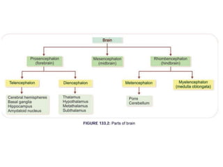
HUMAN NERVOUS SYSTEM
By:
ARTI SAHU
Supervised By:
DR. ANJANA PUROHIT
Department of Psychology
INTRODUCTION
• Nervous system controls all the activities of the body.
• It is divided into 2 major parts:
1. Central Nervous System and
2. Peripheral Nervous System
Central Nervous System (CNS)
• The part of the nervous system, which occupies the central
axis of the body, is called CNS.
• It is formed by neurons and supporting cells called neuroglia.
• It comprises:
1. The Brain
2. The Spinal Cord
The Brain
• Brain weighs about 1.5 kg in adults.
• It is protected by a bony covering called skull and 3
membrane coverings (meninges), namely dura mater,
arachnoid mater and pia mater.
• Cerebrospinal fluid fills the space between arachnoid and
pia, and act as a watery cusion.
The Brain
• It is divided into 3 parts:
1. Fore brain (Prosencephalon)
2. Mid brain (Mesencephalon)
3. Hind brain (Rhombencephalon)
Fore brain (Prosencephalon)
• It subdivided into 2 parts:
1. Telencephalon: It consists of two cerebral hemispheres
(or cerebrum), basal ganglia, hippocampus and
amygdaloid nucleus.
2. Diencephalon: It consists of thalamus, hypothalamus,
metathalamus and subthalamus.
Mid brain (Mesencephalon)
• It is divided by Aqueduct of sylvius (duct that connects
the IIIrd ventricle in the midbrain , with the IV ventricle
in the pons and medulla) into:
1. Cerebral peduncle
2. Tectum
Hind brain (Rhombencephalon)
• It is divided into:
1. Metencephalon, formed by pons and cerebellum
2. Myelencephalon or medulla oblongata
• Midbrain, pons and medulla oblongata are together
called the brainstem.
SPINAL CORD
• The spinal cord is a long (approx 45-50 cm in length),
narrow (about 2 cm in diameter) cylindrical structure.
• It lies outside the skull beginning from its base at the
foramen magnum to terminate at the lower border of 1st
lumbar vertebra.
SPINAL CORD
• Below the 1st lumbar vertebra the spinal cord contains the
lumbar and sacral roots in bands, known as the cauda
equina (or horse tail).
• A cross-section of the spinal cord shows anterior median
fissure, posterior median fissure and central canal.
SPINAL CORD
• Grey matter consisting of nerve cells forms an H-shaped
figure long and narrow dorsal horns; and thick broad
ventral horns.
• Ventral or anterior horn contains the cell bodies of α, γ
and Renshaw motor neurons; axons of α and γ motor
neurons pass out in ventral roots.
SPINAL CORD
• The dorsal horn receives the fibres of the posterior roots
which are purely sensory in function.
• White matter consists of descending and ascending axons
cut across.
• The sensory and motor fibres join o form a mixed nerve
which comes out from the vertebral canal as a peripheral
spinal nerve via the intervertebral foramen.
PERIPHERAL NERVOUS SYSTEM
• This is the part of the nervous system which lies outsude the
CNS.
• It consists of the nerves extending from the brain and Spinal
cord out to all parts of the body.
• It is divided into:
1. Somatic nervous system
2. Autonomic nervous system
SOMATIC NERVOUS SYSTEM
• It is made up of all the nerve fibres going from the CNS to the
skeletal muscle cells.
• It comprises:
1. Spinal nerves: 31 pairs of spinal nerves ( 8 pairs of crania N,
12 pairs of thoracic N, 5 pairs of lumbar N, 5 pairs of sacral N
and 1 pair of coccygeal N)
SOMATIC NERVOUS SYSTEM
2. Cranial nerves: 12 pairs of cranial nerves. They have their cell
bodies in the brain. Some are sensory, some are motor and
some are mixed nerves.
AUTONOMIC NERVOUS SYSTEM
• Also known as vegetative or involuntary nervous system.
• ANS innervates all tissues other than skeletal muscle.
• It is divided into:
1. Sympathetic nervous system
2. Parasympathetic nervous system
AUTONOMIC NERVOUS SYSTEM
• Sympathetic division consists of the thoracic and lumbar
ganglia, therefore, also called thoraco-lumbar division
• Parasympathetic division consists of the cranial ganglia (III,
VII, IX, and X cranial nerves) and sacral ganglia (2nd, 3rd and
4th sacral segments of the spinal nerves), therefore also called
craniosacral division.
Thank You

HUMAN NERVOUS SYSTEM.pptx

  • 1.
    HUMAN NERVOUS SYSTEM By: ARTISAHU Supervised By: DR. ANJANA PUROHIT Department of Psychology
  • 2.
    INTRODUCTION • Nervous systemcontrols all the activities of the body. • It is divided into 2 major parts: 1. Central Nervous System and 2. Peripheral Nervous System
  • 4.
    Central Nervous System(CNS) • The part of the nervous system, which occupies the central axis of the body, is called CNS. • It is formed by neurons and supporting cells called neuroglia. • It comprises: 1. The Brain 2. The Spinal Cord
  • 6.
    The Brain • Brainweighs about 1.5 kg in adults. • It is protected by a bony covering called skull and 3 membrane coverings (meninges), namely dura mater, arachnoid mater and pia mater. • Cerebrospinal fluid fills the space between arachnoid and pia, and act as a watery cusion.
  • 8.
    The Brain • Itis divided into 3 parts: 1. Fore brain (Prosencephalon) 2. Mid brain (Mesencephalon) 3. Hind brain (Rhombencephalon)
  • 10.
    Fore brain (Prosencephalon) •It subdivided into 2 parts: 1. Telencephalon: It consists of two cerebral hemispheres (or cerebrum), basal ganglia, hippocampus and amygdaloid nucleus. 2. Diencephalon: It consists of thalamus, hypothalamus, metathalamus and subthalamus.
  • 11.
    Mid brain (Mesencephalon) •It is divided by Aqueduct of sylvius (duct that connects the IIIrd ventricle in the midbrain , with the IV ventricle in the pons and medulla) into: 1. Cerebral peduncle 2. Tectum
  • 12.
    Hind brain (Rhombencephalon) •It is divided into: 1. Metencephalon, formed by pons and cerebellum 2. Myelencephalon or medulla oblongata • Midbrain, pons and medulla oblongata are together called the brainstem.
  • 13.
    SPINAL CORD • Thespinal cord is a long (approx 45-50 cm in length), narrow (about 2 cm in diameter) cylindrical structure. • It lies outside the skull beginning from its base at the foramen magnum to terminate at the lower border of 1st lumbar vertebra.
  • 14.
    SPINAL CORD • Belowthe 1st lumbar vertebra the spinal cord contains the lumbar and sacral roots in bands, known as the cauda equina (or horse tail). • A cross-section of the spinal cord shows anterior median fissure, posterior median fissure and central canal.
  • 16.
    SPINAL CORD • Greymatter consisting of nerve cells forms an H-shaped figure long and narrow dorsal horns; and thick broad ventral horns. • Ventral or anterior horn contains the cell bodies of α, γ and Renshaw motor neurons; axons of α and γ motor neurons pass out in ventral roots.
  • 17.
    SPINAL CORD • Thedorsal horn receives the fibres of the posterior roots which are purely sensory in function. • White matter consists of descending and ascending axons cut across. • The sensory and motor fibres join o form a mixed nerve which comes out from the vertebral canal as a peripheral spinal nerve via the intervertebral foramen.
  • 18.
    PERIPHERAL NERVOUS SYSTEM •This is the part of the nervous system which lies outsude the CNS. • It consists of the nerves extending from the brain and Spinal cord out to all parts of the body. • It is divided into: 1. Somatic nervous system 2. Autonomic nervous system
  • 19.
    SOMATIC NERVOUS SYSTEM •It is made up of all the nerve fibres going from the CNS to the skeletal muscle cells. • It comprises: 1. Spinal nerves: 31 pairs of spinal nerves ( 8 pairs of crania N, 12 pairs of thoracic N, 5 pairs of lumbar N, 5 pairs of sacral N and 1 pair of coccygeal N)
  • 20.
    SOMATIC NERVOUS SYSTEM 2.Cranial nerves: 12 pairs of cranial nerves. They have their cell bodies in the brain. Some are sensory, some are motor and some are mixed nerves.
  • 21.
    AUTONOMIC NERVOUS SYSTEM •Also known as vegetative or involuntary nervous system. • ANS innervates all tissues other than skeletal muscle. • It is divided into: 1. Sympathetic nervous system 2. Parasympathetic nervous system
  • 22.
    AUTONOMIC NERVOUS SYSTEM •Sympathetic division consists of the thoracic and lumbar ganglia, therefore, also called thoraco-lumbar division • Parasympathetic division consists of the cranial ganglia (III, VII, IX, and X cranial nerves) and sacral ganglia (2nd, 3rd and 4th sacral segments of the spinal nerves), therefore also called craniosacral division.
  • 24.