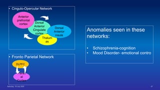
Neural
Circuits
Overview
• Introduction
• History
• Importance
• Basics of Neuro Circuits
• Default mode Network
• Salience Network
• Neuro circuits of psychiatric illness
• Latest Researches
• Summery
• Take Home Message
• References
Introduction
• A neural circuit is a population of neurons interconnected by synapses to carry
out a specific function when activated.*
• Interconnected to form large scale brain network
• Basic kind of connections between neurons are synapses are chemical and
electrical synapses
* Purves, Dale (2011). Neuroscience (5th ed.). Sunderland, Mass.: Sinauer. p. 507. `
Why it was important?
• The practice of human cadaveric dissection during its inception in ancient Greece in 3rd
century BC
• Italy at the beginning of 14th century and subsequent evolution in Europe and the
United States of America over the centuries.
• Microscopic examinations ( Around 1620 Europe ) –Striking resemblance in structural
motifs in animal to animal
• Most of the diseases found to have some molecular / biochemical basis
• For brain treatment is based on complaints/behaviour.
• Equivalent motifs unknown
Challenges
Vast array of cell
types
Knowing about one part is not enough
Challenges
Challanges
•Complex structure and function of nervous system
GENE STRUCTURE FUNCTION
Gene –Structure - Function
Challanges
•Complex structure and function of nervous system
GENE STRUCTURE FUNCTION
Challanges
•Complex structure and function of nervous system
GENE STRUCTURE FUNCTION
Experiences
H Sapiens Iris Ortoria
• Slow development
• Experience based neural
development
• Obligate learner
• Culture based behavior
What we are looking for
Map out the atomic structure at a resolution at which they function
• To determine
• Etiology
• Pathogenesis
• Course
• Treatment
• Prognosis
History
• Golgi's method is a silver
staining technique that is used
to visualize nervous
tissue under light microscopy.
• The method was discovered
by Camillo Golgi,
an Italian physician and scientis
t, who published the first picture
made with the technique in
1873.
History
• Santiago Ramón y
Cajal was a
Spanish neuroscientist, path
ologist,
and histologist specializing
in neuroanatomy .
• Camillo Golgi received
the Nobel Prize in
Physiology or Medicine in
1906.
History (Cajal’s Drawing)
History
• Brodmann areas were originally defined
and numbered by
the German anatomist Korbinian
Brodmann
• Based on
the cytoarchitectural organization
of neurons he observed in the cerebral
cortex using the Nissl method of cell
staining.
• Brodmann published his maps of cortical
areas in humans, monkeys, and other
species in 1909
Concept of Connectomics
• Branch of biotechnology
• Computer assisted image acquisition and analysis
• Structural mapping of sets of neural circuits or complete organism
• Using high speed methods and organizing the results in Database
• Application of dada base in neurology, psychiatry or fundamental
neurosciences
** Kasthuri, N. & Lichtman, J. W. The role of neuronal identity in synaptic competition. Nature 424, 426–430
(2003).
The Pioneer work by Cajal
• Gave the basic
concept
• Sparse network
• Black & White
• Difficult to trace
• Inadequate
information
• Could not explain
the detailed
structure
YFP-16 Transgenic mouse Neural
Structures
Labelling Neural Architecture with
Fluorescent Proteins
Labelling Neural Architecture with
Fluorescent Proteins (BRAIN BOW)
Decreased Convergence with
compensatory synaptic takeover
Concept of Circuit Recognition
Basics Of Circuit
Brain On Tape
Brain On Tape Pro
Electron Microscope Study
In situ Synaptic Achitecture
In situ Synaptic Architecture
1 2 3
Challenges Of the Study
Neuro-imaging
• Historically functional neuro imaging works were
focused on
1. SPECT /PET
2. f MRI
• Indirect estimation of neural activity through the
measurement of
1.Oxygen Metabolism- PET
2.Blood flow- SPECT
3.Blood oxygen level dependent activity- BOLD-f
MRI
F-MRI
studies
Convert Convert measured RF data to image
Receive Receive radio waves re transmitted by subject
Turn off Turn off radio wave transmitter
Transmit Transmit radio waves into subject (2-10 ms)
Put Put subject in big magnetic field
Neuro-imaging
• Primarily TASK BASED studies were done- PET / fMRI
• To assess diffential activation of brain regions during
• Cognitive
• Affective
• Social task
• Ambition was to develop testing hypothesis
• By seeing abnormal BOLD activity
• Identify probable regions effected in different manifestations of mental illness
Neuro-imaging
DLPFC
Amygda
la
Insula
Stiatum
Depression
Cognitive dysfunction in
psychosis
Anxiety Disorder
Addictive Disorder
Later Understanding
• Rarely does one specific activity activity
activates any specific brain region
• Rarely particular psychopathology associated
with abnormal activation only a single brain
region
• Both Input and Output changes the activity of
brain region
Change in the focus of the study
Specific Brain
Region
Neural Circuit
Later Understanding
Normal Relationship between
the brain regions
Successful cognitive and
affective functioning
Not Regions but Connections
DLPFC
SCHIZOPHRENIA
Input
Output
Cognitive Control task ! Abnormality in the DLPFC
Advent Of Studying Functional
Connectivity
• The Main inference of functional connectivity is
• If two regions have high correlated neuronal activity
• Then they are more likely to engage in a common set of processing mechanism
• May provide a tool for understanding the brain regions
• Different areas and circuit for cognitive and effective demands
• Cognitive/emotional /social processing
Resting State Functional Connectivity
• A major breakthrough in 1995 ( Biswal et al)
• Spontaneous activity from regions in the Right &Left motor cortices at resting state
• This was suggestive of functional connectivity between these brain regions
• Spontaneous low frequency fluctuations of the BOLD signal (0.01 to .010 Hz)
• These signals were previously discarded as noise in task-based studies
• This resting state activity consumes a major portion of body’s energy ( aprox 20%)
• Robust studies demonstrated that the fluctuations in brain activity are coherent
Resting State Functional Connectivity
Networks
• Resting state functional connectivity revealed
• Intrinsically organized networks of brain regions that are consistently functionally connected.
• Present even in the absence of task induced perturbations in ongoing brain activity
• Default Mode Network
• Fronto-Parietal Network
• Cingulo-Opercular Network
• Dorsal Attention Network
Resting State Functional Connectivity
Networks
• In a recent study, 356 regions
(Parcels) with homogenous activity,
at resting state were identified
• They grouped into 12 resting state
network that map nicely to networks
identified in previous resting state
study
• These maps closely /partially
matches with the maps identified
task-based studies
Resting State Network
• Mapping of resting state network to known task networks led
to the following hypothesis
• Resting State Network reflect in part organized interactions
that arise from a history of coactivation over development
• Not Dependent on Task Performance
• Present even at rest
• Relatively consistent across tasks and environmental
states
• These are referred as “intrinsic connectivity network”
Human
Connectome
Project
Human Connectome Project
• 5 years Project
• Sponsored by National Institute of Health
• Launched in July 2009
• University of Southern California and 6 other universities participating in the
study
• Goal- Is to build up a “Network Map” (Connectome)
• Which will shed light on anatomical and functional connectivity within
healthy human brain
• Produce a body of DATA which will facilitate research into brain disorders
• Dyslexia
• Autism
• Alzheimer’s Disease
• Schizophrenia
Human Connectome Project
• Wu Minn Oxford Consortium
• Developed improved MRI machine , image acquisition and image
analysis methods for mapping the connectivity
• Collected behavioral and MRI DATA of 1200 healthy individuals
• Twin pairs and their siblings from 300 families
• Used 3 Tesla MRI machine (Also took 184 subjects scan using 7 Tesla
MRI)
• Understanding anatomical & functional connection of brain region
• Related to behavioral DATA / Genetic DATA (From Identical Twins)
Human Connectome Project
• Wu Minn Oxford Consortium
• Understanding relative contribution of Genes and environment in shaping brain
circuitry
• Pinpoint relevant genetic variation
• Methods:
• Resting state fMRI
• Task based f MRI
• MEG
• Diffuse Tensor Imaging
Human Connectome Project
• Wu Minn Oxford Consortium
• Mapping connectomes in macroscale
• Large brain system divided in anatomical and functionally distinct areas
• Not mapping individual neurons
• Data available in public domain as open accessible neuro-formatics platform
Human Connectome Project
• MGH/ Harvard- UCLA Consortium
• Work on spatial resolution of Diffusion MRI for brain mapping
• Diffusion MRI is employed to map brain’s fibrous long-distance connections by tracking
motion of water
• Water diffusion pattern in different types of cells allow detection of different types of tissues
• To understand relationship between brain connectivity and behavior
• Neurological & behavioral functions measured by NIH toolbox
(Cognition/sensation/Motor/Emotions)
Human Connectome Project
• MGH/ Harvard- UCLA Consortium
• The Study fall into 3 categories
• Healthy adult connectomes
• Life span connectome DATA
• Connectomes related to human diseases
Human Connectome Project
• The results of the project confirmed and extended the current
knowledge about
• The critical relationship between individual variations in functional
brain connectivity
• Individual variation in a range of behaviors relevant to mental health
and both negative and positive outcome.
Most Commonly Identified Relevant
Networks
• Cingulo-Opercular Network• Fronto Parietal Network
DLPFC
Parietal
Cortex
Dorsal
Anterior
Cingulate
cortex
Thaluma
s
Anterior
prefrontal
cortex
Dorsal
Anterior
insula
• Cingulo-Opercular Network
• Fronto Parietal Network
DLPFC
Pariet
al
Cortex
Dorsal
Anterior
Cingulate
cortex
Thalum
as
Anterior
prefrontal
cortex
Dorsal
Anterior
insula
Anomalies seen in these
networks:
• Schizophrenia-cognition
• Mood Disorder- emotional contro
Dorsal Attention Network
DLPFC
Parietal Cortex
Eye Field Area
Dorsal
Supplementary
Motor Area
Dorsal Attention Network
DLPFC
Parietal Cortex
Eye Field Area
Dorsal
Supplementary
Motor Area
Dorsal Attention Network
DLPFC
Parietal Cortex
Eye Field Area
Dorsal
Supplementary
Motor Area
Dorsal Attention Network
DLPFC
Parietal Cortex
Eye Field Area
Dorsal
Supplementary
Motor Area
Top-down guided
voluntary allocation of
attention
Ventral Attention Network
Temporo-
Parietal
Junction
VLPFC Salient event in the environment
Dorsal And Ventral Attention
Network on f MRI
• Dorsal fronto parietal system was proposed to
mediate the top down guided voluntary
allocation of attention to location or features
• Ventral fronto parietal system was assumed to
be involved in detecting unattended or
unexpected stimuli triggering shift of attention
**Simone Vossel, Joy J. Geng, Gereon R. Fink,
Neuroscientist. 2014 Apr; 20(2): 150–159
Default Mode Network
Medial
Prefrontal
Cortex
Medial
Posterior
Cingulate
Cortex
Precuneus
• The default mode network (DMN) is
active when a person is not focused on
the outside world
• Brain is at wakeful rest, such as during
daydreaming and mind-wandering.
• Thinking about others, thinking about
themselves, remembering the past, and
planning.
• The network activates “by default” when
a person is not involved in a task.
Default
Mode
Network
Salience Network
• The salience network (SN) is
a large-scale brain network of
the human brain
• Aanterior insula (AI) and
• Dorsal anterior cingulate
cortex (dACC).
• It is involved in detecting and
filtering salient stimuli, as well as in
recruiting relevant functional
networks.
• Contributes to communication, social
behaviour, and self-awareness
through the integration of sensory,
emotional, and cognitive information
Methods For Analysing Resting State
Functional Connectivity
• Region of Interest or a priori network based analysis
• Seed /ROI to whole brain connectivity analysis
• Independent Component analysis
Functional Connectivity Abnormalities in
Schizophrenia
• More than 200 studies done
• Functional connectivity in schizhophrenia spectrum Disorder (as DSM IV)
• Altered thalamic connectivity
• Among multiple hypotheses , Andersen et al , suggested that schizophrenia
involves a disruption in the integration of cortical - sub cortical – cerebellar
circuits
• Hypotheses termed as “Cognitive Dysmetria”
Functional Connectivity Abnormalities in
Schizophrenia
• Large body of studies shown connectivity
• Bilateral thalamic region (Medial Dorsal &Anterior neclei)
• Bilateral DLPFC
• Dorsal Anterior Cingulate Cortex (dACC)
• Parts of striatum
• Bilateral cerebellum
Functional
Connectivity
Abnormalities in
Schizophrenia
• Reduced thalamic
connectivity
(Depicted in Blue)
1. Bilateral prefrontal and
parietal cortex
2. ACC
3. Bilateral Stiatum and
Cerebellum
Functional
Connectivity
Abnormalities in
Schizophrenia
• Increased thalamic
connectivity
(Depicted in Red)
1. Motor Cortex
2. Somatosensory
cortex
3. Visual cortex
Functional Connectivity Abnormalities in Schizophrenia
• Similar pattern of altered connectivity is seen in individuals at clinical and genetic risk
for schizophrenia
• Other disorders characterized by psychosis as Bipolar Disorder
• The pattern of altered thalamic connectivity are most pronounced among those who
subsequently converted to full blown psychosis
• Importantly , reduced connectivity between thalamus and DLPFC predicts cognitive
impairment
• Thalamic connectivity is a potential bio marker for risk of psychosis
• Along with increased presynaptic dopamine availability in the striatum
• Altered glutamate levels in the thalamus
Schizophrenia-Altered Default Mode
Connectivity
• One of the major functions ascribes to the default mode network is attention to
internal emotional states
• A number of researches hypothesized that the attention to internal states must be
dampened when individuals need to engage in specific tasks or attend to external
world
• It was hypothesized that in schizophrenia enhanced attention to internal thoughts ,
feelings and experiences, , due to altered connectivity as well as activity of the
Default Mode Network
• It has been Hypothesized that a failure to suppress default mode network activity
during cognitive task performance leading to disruptive cognitive function in
schizophrenia
Schizophrenia-Altered Default Mode
Connectivity
• The empirical literature on abnormalities in default mode network connectivity is quite
mixed (Narr et al)
• Evidence of Hyper-connectivity and Hypo-connectivity both are present
• One of the challenges in this literature may be that many of the studies focused on
comparing individuals with schizophrenia, regardless of symptom profile to healthy
controls
• Results found to be variable with the same diagnosis and across time in the same
individuals
• It may be critical to examine default mode connectivity in relationship to variation in the
severity of specific symptoms (eg. Hallucination, delusion and cognitive impairment)
Graph Theory Analysis of Functional
Brain Connectivity in Schizophrenia
• Individuals with schizophrenia show reduction in both global and local efficiency,
particularly when examining specific networks
• Most reduced global/local efficiency tend to involve the anterior cingulate and
cingula
• Cingulo-opercular network
• Fronto-parietal network
• These findings are backed by ROI based and ICA based analysis
• Impaired cognition and reduction in global and local connectivity are consistent
finding in patients with schizophrenia.
Graph Theory Analysis of Functional Brain
Connectivity in Schizophrenia
• Individuals with schizophrenia show reductions in both global and local
efficiency
• Specially “Hub” regions in the brain, which are highly interconnected
• Individuals with schizophrenia show significant reductions in the
connections among hubs eg
• insula,
• superior frontal cortex,
• precuneus,
• superior parietal lobe
• They do not show reduction in connection between these hub regions and
non hub regions
Graph Theory Analysis of Functional
Brain Connectivity in Schizophrenia
Graph Theory Analysis of Functional
Brain Connectivity in Schizophrenia
Loss of connectivity in between ” Hubs”
Summery of Schizophrenia Functional
Connectivity Literature
• Reduced thalamic connectivity with frontal, cingulate, striatal and cerebellar
region
• Reduced global and local efficiency in fronto-parietal, cingulo-opercular networks
• Reduced Connectivity in HUB regions.
• Large scale dysconnectivity may result reduced efficiency in information transfer,
causing cognitive impairment
• These impairments are present at the start or even before start of the illness
• More prominent among those who develop full blown psychosis
Functional Connectivity
Abnormalities In Major
Depression
• Amygdala Connectivity and Emotional
Regulation
• Fear Conditioning
• Threat sensitivity
• General response to salient events in the
environment
Amygdala and early life experience
Poverty
Stress
trauma
Amygdal
a
Development
Functioning
Structure
Amygdala Positive And Negative Functional
Connectivity
DMPFC
dACC Amygdala
VMPFC
Hippocampu
s
Amygdala Positive And Negative Functional
Connectivity
DLPFC
dACC
VMPFC
Hippocampu
s
Amygdala
Amygdala Positive And Negative Functional
Connectivity
DLPFC
dACC
Amygdala
VMPFC
Hippocampu
s
PositiveNegetive
Important for effective emotion
regulation
Amygdala Positive And Negative Functional
Connectivity
DLPFC
dACC
VMPFC
Hippocampu
s
Negetive
Important for effective emotion
regulation
Amygdala Positive Functional Connectivity
• Amygdala shows positive and negative
connections with other regions of the
brain
• Positive resting state functional
connectivity
• Hippocampus
• Ventral Medial Prefrontal
cortex
• Amygdala also found to couple with
other regions of salience network
• Ventral anterior cingulate
cortex
• Ventral region Insula
• Anterior Temporal Poles
• Responsible for emotion and stress
response
Amygdala Negative Functional Connectivity
• Negative resting state functional
connectivity (Anti correlations)
• Parietal Region
• Dorsal Prefrontal cortex
• Dorsal Anterior Cingulate
• Amygdala also found to couple with
other regions of salience network
• Ventral anterior cingulate
cortex
• Ventral region Insula
• Anterior Temporal Poles
• Responsible for emotion and stress
response
Implication in Depressive Disorder
• These anti-correlations are thought to indicate top down regulation of emotion and
stress responsivity
• This suggest a critical role for integrity of these connections between amygdala
and fronto-parietal and cingulo-opercular network in mood and effective
functioning
• Early life stress/ maternal deprivation results altered connectivity between
amygdala/ VMPFC/ Anterior Cingulate cortex important for effective emotion
regulation
• Altered amygdala connectivity has been found with these regions and with
components of Salience network /Fronto-Parietal/Cingulo-opercular network in
MDD
Implication in Depressive Disorder
• One hypothesis states that reduced amygdala to prefrontal / Cingulate
connectivity reflects impaired top down regulation of emotion processing
• This contributes to psychopathology
• These pattern of altered connectivity have been found across the life span /at risk
groups
• Connectivity disruption precedes the onset of manifest illness
Default Mode Network Connectivity and
Rumination
• Consistent findings of altered connectivity associated with major depressive
disorder in the domain of Default Mode Network (DMN)
• DMN modulate attention to internal emotional state and govern self referential
processing
• Phenomenologically in MDD the following are seen
• Preoccupation with negative mood
• Negative self evaluation
• Ruminative style of coping with negative events
Increase
connectivity
among Default
Mode Network
regions and
Salience Network
regions
Default Mode Network Connectivity and
MDD
Increase
connectivity
among Default
Mode Network
regions and
Salience Network
regions
Over activity of
DMN+ Salience
Network =
Negative
evaluation of
salient events
Negative Mood
&
Negative self
evaluation
Default Mode Network Connectivity and
MDD
• Increased connectivity between (In MDD)
• Sub Genual Prefrontal Cortex
• Default Mode Network
• Responsible for
• Behavioral withdrawal
• Negative effective appraisal
• Experiencing Guilt
• Promoting safety oriented behaviour
Conclusion
• Brief overview of the current state of field in Human Functional Connectome
• Functional Connection abnormalities relevant for understanding pathophysiology of Major Mental
Illness
• Due to the advancement of imaging technologies multiple Functional Brain Networks has been
identified
• Human connectome Project confirmed individual variations in the intrinsic network connectivity
responsible for positive and negative traits
• Robust evidences demonstrated altered thalamic connectivity connectivity associated with
psychosis
• In MDD altered amygdala to prefrontal –cingulate connectivity relevant for understanding
emotional regulation
• In MDD increased default mode connectivity implicated in increased rumination
References
• Kaplan and Saddock Comprehensive Text Book Of Psychiatry 10th Edition
Neuro circuit

Neuro circuit

  • 1.
  • 2.
    Overview • Introduction • History •Importance • Basics of Neuro Circuits • Default mode Network • Salience Network • Neuro circuits of psychiatric illness • Latest Researches • Summery • Take Home Message • References
  • 3.
    Introduction • A neuralcircuit is a population of neurons interconnected by synapses to carry out a specific function when activated.* • Interconnected to form large scale brain network • Basic kind of connections between neurons are synapses are chemical and electrical synapses * Purves, Dale (2011). Neuroscience (5th ed.). Sunderland, Mass.: Sinauer. p. 507. `
  • 4.
    Why it wasimportant? • The practice of human cadaveric dissection during its inception in ancient Greece in 3rd century BC • Italy at the beginning of 14th century and subsequent evolution in Europe and the United States of America over the centuries. • Microscopic examinations ( Around 1620 Europe ) –Striking resemblance in structural motifs in animal to animal • Most of the diseases found to have some molecular / biochemical basis • For brain treatment is based on complaints/behaviour. • Equivalent motifs unknown
  • 5.
  • 6.
    Knowing about onepart is not enough
  • 7.
  • 8.
    Challanges •Complex structure andfunction of nervous system GENE STRUCTURE FUNCTION
  • 9.
  • 10.
    Challanges •Complex structure andfunction of nervous system GENE STRUCTURE FUNCTION
  • 11.
    Challanges •Complex structure andfunction of nervous system GENE STRUCTURE FUNCTION Experiences
  • 12.
    H Sapiens IrisOrtoria • Slow development • Experience based neural development • Obligate learner • Culture based behavior
  • 13.
    What we arelooking for Map out the atomic structure at a resolution at which they function • To determine • Etiology • Pathogenesis • Course • Treatment • Prognosis
  • 14.
    History • Golgi's methodis a silver staining technique that is used to visualize nervous tissue under light microscopy. • The method was discovered by Camillo Golgi, an Italian physician and scientis t, who published the first picture made with the technique in 1873.
  • 15.
    History • Santiago Ramóny Cajal was a Spanish neuroscientist, path ologist, and histologist specializing in neuroanatomy . • Camillo Golgi received the Nobel Prize in Physiology or Medicine in 1906.
  • 16.
  • 17.
    History • Brodmann areaswere originally defined and numbered by the German anatomist Korbinian Brodmann • Based on the cytoarchitectural organization of neurons he observed in the cerebral cortex using the Nissl method of cell staining. • Brodmann published his maps of cortical areas in humans, monkeys, and other species in 1909
  • 18.
    Concept of Connectomics •Branch of biotechnology • Computer assisted image acquisition and analysis • Structural mapping of sets of neural circuits or complete organism • Using high speed methods and organizing the results in Database • Application of dada base in neurology, psychiatry or fundamental neurosciences ** Kasthuri, N. & Lichtman, J. W. The role of neuronal identity in synaptic competition. Nature 424, 426–430 (2003).
  • 19.
    The Pioneer workby Cajal • Gave the basic concept • Sparse network • Black & White • Difficult to trace • Inadequate information • Could not explain the detailed structure
  • 20.
    YFP-16 Transgenic mouseNeural Structures
  • 21.
    Labelling Neural Architecturewith Fluorescent Proteins
  • 22.
    Labelling Neural Architecturewith Fluorescent Proteins (BRAIN BOW)
  • 24.
  • 25.
    Concept of CircuitRecognition
  • 26.
  • 28.
  • 29.
  • 30.
  • 31.
    In situ SynapticAchitecture
  • 32.
    In situ SynapticArchitecture
  • 33.
    In situ SynapticArchitecture 1 2 3
  • 34.
  • 35.
    Neuro-imaging • Historically functionalneuro imaging works were focused on 1. SPECT /PET 2. f MRI • Indirect estimation of neural activity through the measurement of 1.Oxygen Metabolism- PET 2.Blood flow- SPECT 3.Blood oxygen level dependent activity- BOLD-f MRI
  • 36.
    F-MRI studies Convert Convert measuredRF data to image Receive Receive radio waves re transmitted by subject Turn off Turn off radio wave transmitter Transmit Transmit radio waves into subject (2-10 ms) Put Put subject in big magnetic field
  • 37.
    Neuro-imaging • Primarily TASKBASED studies were done- PET / fMRI • To assess diffential activation of brain regions during • Cognitive • Affective • Social task • Ambition was to develop testing hypothesis • By seeing abnormal BOLD activity • Identify probable regions effected in different manifestations of mental illness
  • 38.
  • 39.
    Later Understanding • Rarelydoes one specific activity activity activates any specific brain region • Rarely particular psychopathology associated with abnormal activation only a single brain region • Both Input and Output changes the activity of brain region
  • 40.
    Change in thefocus of the study Specific Brain Region Neural Circuit
  • 41.
    Later Understanding Normal Relationshipbetween the brain regions Successful cognitive and affective functioning
  • 42.
    Not Regions butConnections DLPFC SCHIZOPHRENIA Input Output Cognitive Control task ! Abnormality in the DLPFC
  • 43.
    Advent Of StudyingFunctional Connectivity • The Main inference of functional connectivity is • If two regions have high correlated neuronal activity • Then they are more likely to engage in a common set of processing mechanism • May provide a tool for understanding the brain regions • Different areas and circuit for cognitive and effective demands • Cognitive/emotional /social processing
  • 44.
    Resting State FunctionalConnectivity • A major breakthrough in 1995 ( Biswal et al) • Spontaneous activity from regions in the Right &Left motor cortices at resting state • This was suggestive of functional connectivity between these brain regions • Spontaneous low frequency fluctuations of the BOLD signal (0.01 to .010 Hz) • These signals were previously discarded as noise in task-based studies • This resting state activity consumes a major portion of body’s energy ( aprox 20%) • Robust studies demonstrated that the fluctuations in brain activity are coherent
  • 45.
    Resting State FunctionalConnectivity Networks • Resting state functional connectivity revealed • Intrinsically organized networks of brain regions that are consistently functionally connected. • Present even in the absence of task induced perturbations in ongoing brain activity • Default Mode Network • Fronto-Parietal Network • Cingulo-Opercular Network • Dorsal Attention Network
  • 46.
    Resting State FunctionalConnectivity Networks • In a recent study, 356 regions (Parcels) with homogenous activity, at resting state were identified • They grouped into 12 resting state network that map nicely to networks identified in previous resting state study • These maps closely /partially matches with the maps identified task-based studies
  • 47.
    Resting State Network •Mapping of resting state network to known task networks led to the following hypothesis • Resting State Network reflect in part organized interactions that arise from a history of coactivation over development • Not Dependent on Task Performance • Present even at rest • Relatively consistent across tasks and environmental states • These are referred as “intrinsic connectivity network”
  • 48.
  • 49.
    Human Connectome Project •5 years Project • Sponsored by National Institute of Health • Launched in July 2009 • University of Southern California and 6 other universities participating in the study • Goal- Is to build up a “Network Map” (Connectome) • Which will shed light on anatomical and functional connectivity within healthy human brain • Produce a body of DATA which will facilitate research into brain disorders • Dyslexia • Autism • Alzheimer’s Disease • Schizophrenia
  • 50.
    Human Connectome Project •Wu Minn Oxford Consortium • Developed improved MRI machine , image acquisition and image analysis methods for mapping the connectivity • Collected behavioral and MRI DATA of 1200 healthy individuals • Twin pairs and their siblings from 300 families • Used 3 Tesla MRI machine (Also took 184 subjects scan using 7 Tesla MRI) • Understanding anatomical & functional connection of brain region • Related to behavioral DATA / Genetic DATA (From Identical Twins)
  • 51.
    Human Connectome Project •Wu Minn Oxford Consortium • Understanding relative contribution of Genes and environment in shaping brain circuitry • Pinpoint relevant genetic variation • Methods: • Resting state fMRI • Task based f MRI • MEG • Diffuse Tensor Imaging
  • 52.
    Human Connectome Project •Wu Minn Oxford Consortium • Mapping connectomes in macroscale • Large brain system divided in anatomical and functionally distinct areas • Not mapping individual neurons • Data available in public domain as open accessible neuro-formatics platform
  • 53.
    Human Connectome Project •MGH/ Harvard- UCLA Consortium • Work on spatial resolution of Diffusion MRI for brain mapping • Diffusion MRI is employed to map brain’s fibrous long-distance connections by tracking motion of water • Water diffusion pattern in different types of cells allow detection of different types of tissues • To understand relationship between brain connectivity and behavior • Neurological & behavioral functions measured by NIH toolbox (Cognition/sensation/Motor/Emotions)
  • 54.
    Human Connectome Project •MGH/ Harvard- UCLA Consortium • The Study fall into 3 categories • Healthy adult connectomes • Life span connectome DATA • Connectomes related to human diseases
  • 55.
    Human Connectome Project •The results of the project confirmed and extended the current knowledge about • The critical relationship between individual variations in functional brain connectivity • Individual variation in a range of behaviors relevant to mental health and both negative and positive outcome.
  • 56.
    Most Commonly IdentifiedRelevant Networks • Cingulo-Opercular Network• Fronto Parietal Network DLPFC Parietal Cortex Dorsal Anterior Cingulate cortex Thaluma s Anterior prefrontal cortex Dorsal Anterior insula
  • 57.
    • Cingulo-Opercular Network •Fronto Parietal Network DLPFC Pariet al Cortex Dorsal Anterior Cingulate cortex Thalum as Anterior prefrontal cortex Dorsal Anterior insula Anomalies seen in these networks: • Schizophrenia-cognition • Mood Disorder- emotional contro
  • 58.
    Dorsal Attention Network DLPFC ParietalCortex Eye Field Area Dorsal Supplementary Motor Area
  • 59.
    Dorsal Attention Network DLPFC ParietalCortex Eye Field Area Dorsal Supplementary Motor Area
  • 60.
    Dorsal Attention Network DLPFC ParietalCortex Eye Field Area Dorsal Supplementary Motor Area
  • 61.
    Dorsal Attention Network DLPFC ParietalCortex Eye Field Area Dorsal Supplementary Motor Area Top-down guided voluntary allocation of attention
  • 62.
  • 63.
    Dorsal And VentralAttention Network on f MRI • Dorsal fronto parietal system was proposed to mediate the top down guided voluntary allocation of attention to location or features • Ventral fronto parietal system was assumed to be involved in detecting unattended or unexpected stimuli triggering shift of attention **Simone Vossel, Joy J. Geng, Gereon R. Fink, Neuroscientist. 2014 Apr; 20(2): 150–159
  • 64.
    Default Mode Network Medial Prefrontal Cortex Medial Posterior Cingulate Cortex Precuneus •The default mode network (DMN) is active when a person is not focused on the outside world • Brain is at wakeful rest, such as during daydreaming and mind-wandering. • Thinking about others, thinking about themselves, remembering the past, and planning. • The network activates “by default” when a person is not involved in a task.
  • 65.
  • 66.
    Salience Network • Thesalience network (SN) is a large-scale brain network of the human brain • Aanterior insula (AI) and • Dorsal anterior cingulate cortex (dACC). • It is involved in detecting and filtering salient stimuli, as well as in recruiting relevant functional networks. • Contributes to communication, social behaviour, and self-awareness through the integration of sensory, emotional, and cognitive information
  • 67.
    Methods For AnalysingResting State Functional Connectivity • Region of Interest or a priori network based analysis • Seed /ROI to whole brain connectivity analysis • Independent Component analysis
  • 68.
    Functional Connectivity Abnormalitiesin Schizophrenia • More than 200 studies done • Functional connectivity in schizhophrenia spectrum Disorder (as DSM IV) • Altered thalamic connectivity • Among multiple hypotheses , Andersen et al , suggested that schizophrenia involves a disruption in the integration of cortical - sub cortical – cerebellar circuits • Hypotheses termed as “Cognitive Dysmetria”
  • 69.
    Functional Connectivity Abnormalitiesin Schizophrenia • Large body of studies shown connectivity • Bilateral thalamic region (Medial Dorsal &Anterior neclei) • Bilateral DLPFC • Dorsal Anterior Cingulate Cortex (dACC) • Parts of striatum • Bilateral cerebellum
  • 70.
    Functional Connectivity Abnormalities in Schizophrenia • Reducedthalamic connectivity (Depicted in Blue) 1. Bilateral prefrontal and parietal cortex 2. ACC 3. Bilateral Stiatum and Cerebellum
  • 71.
    Functional Connectivity Abnormalities in Schizophrenia • Increasedthalamic connectivity (Depicted in Red) 1. Motor Cortex 2. Somatosensory cortex 3. Visual cortex
  • 72.
    Functional Connectivity Abnormalitiesin Schizophrenia • Similar pattern of altered connectivity is seen in individuals at clinical and genetic risk for schizophrenia • Other disorders characterized by psychosis as Bipolar Disorder • The pattern of altered thalamic connectivity are most pronounced among those who subsequently converted to full blown psychosis • Importantly , reduced connectivity between thalamus and DLPFC predicts cognitive impairment • Thalamic connectivity is a potential bio marker for risk of psychosis • Along with increased presynaptic dopamine availability in the striatum • Altered glutamate levels in the thalamus
  • 73.
    Schizophrenia-Altered Default Mode Connectivity •One of the major functions ascribes to the default mode network is attention to internal emotional states • A number of researches hypothesized that the attention to internal states must be dampened when individuals need to engage in specific tasks or attend to external world • It was hypothesized that in schizophrenia enhanced attention to internal thoughts , feelings and experiences, , due to altered connectivity as well as activity of the Default Mode Network • It has been Hypothesized that a failure to suppress default mode network activity during cognitive task performance leading to disruptive cognitive function in schizophrenia
  • 74.
    Schizophrenia-Altered Default Mode Connectivity •The empirical literature on abnormalities in default mode network connectivity is quite mixed (Narr et al) • Evidence of Hyper-connectivity and Hypo-connectivity both are present • One of the challenges in this literature may be that many of the studies focused on comparing individuals with schizophrenia, regardless of symptom profile to healthy controls • Results found to be variable with the same diagnosis and across time in the same individuals • It may be critical to examine default mode connectivity in relationship to variation in the severity of specific symptoms (eg. Hallucination, delusion and cognitive impairment)
  • 75.
    Graph Theory Analysisof Functional Brain Connectivity in Schizophrenia • Individuals with schizophrenia show reduction in both global and local efficiency, particularly when examining specific networks • Most reduced global/local efficiency tend to involve the anterior cingulate and cingula • Cingulo-opercular network • Fronto-parietal network • These findings are backed by ROI based and ICA based analysis • Impaired cognition and reduction in global and local connectivity are consistent finding in patients with schizophrenia.
  • 76.
    Graph Theory Analysisof Functional Brain Connectivity in Schizophrenia • Individuals with schizophrenia show reductions in both global and local efficiency • Specially “Hub” regions in the brain, which are highly interconnected • Individuals with schizophrenia show significant reductions in the connections among hubs eg • insula, • superior frontal cortex, • precuneus, • superior parietal lobe • They do not show reduction in connection between these hub regions and non hub regions
  • 77.
    Graph Theory Analysisof Functional Brain Connectivity in Schizophrenia
  • 78.
    Graph Theory Analysisof Functional Brain Connectivity in Schizophrenia Loss of connectivity in between ” Hubs”
  • 79.
    Summery of SchizophreniaFunctional Connectivity Literature • Reduced thalamic connectivity with frontal, cingulate, striatal and cerebellar region • Reduced global and local efficiency in fronto-parietal, cingulo-opercular networks • Reduced Connectivity in HUB regions. • Large scale dysconnectivity may result reduced efficiency in information transfer, causing cognitive impairment • These impairments are present at the start or even before start of the illness • More prominent among those who develop full blown psychosis
  • 80.
    Functional Connectivity Abnormalities InMajor Depression • Amygdala Connectivity and Emotional Regulation • Fear Conditioning • Threat sensitivity • General response to salient events in the environment
  • 81.
    Amygdala and earlylife experience Poverty Stress trauma Amygdal a Development Functioning Structure
  • 82.
    Amygdala Positive AndNegative Functional Connectivity DMPFC dACC Amygdala VMPFC Hippocampu s
  • 83.
    Amygdala Positive AndNegative Functional Connectivity DLPFC dACC VMPFC Hippocampu s Amygdala
  • 84.
    Amygdala Positive AndNegative Functional Connectivity DLPFC dACC Amygdala VMPFC Hippocampu s PositiveNegetive Important for effective emotion regulation
  • 85.
    Amygdala Positive AndNegative Functional Connectivity DLPFC dACC VMPFC Hippocampu s Negetive Important for effective emotion regulation
  • 86.
    Amygdala Positive FunctionalConnectivity • Amygdala shows positive and negative connections with other regions of the brain • Positive resting state functional connectivity • Hippocampus • Ventral Medial Prefrontal cortex • Amygdala also found to couple with other regions of salience network • Ventral anterior cingulate cortex • Ventral region Insula • Anterior Temporal Poles • Responsible for emotion and stress response
  • 87.
    Amygdala Negative FunctionalConnectivity • Negative resting state functional connectivity (Anti correlations) • Parietal Region • Dorsal Prefrontal cortex • Dorsal Anterior Cingulate • Amygdala also found to couple with other regions of salience network • Ventral anterior cingulate cortex • Ventral region Insula • Anterior Temporal Poles • Responsible for emotion and stress response
  • 88.
    Implication in DepressiveDisorder • These anti-correlations are thought to indicate top down regulation of emotion and stress responsivity • This suggest a critical role for integrity of these connections between amygdala and fronto-parietal and cingulo-opercular network in mood and effective functioning • Early life stress/ maternal deprivation results altered connectivity between amygdala/ VMPFC/ Anterior Cingulate cortex important for effective emotion regulation • Altered amygdala connectivity has been found with these regions and with components of Salience network /Fronto-Parietal/Cingulo-opercular network in MDD
  • 89.
    Implication in DepressiveDisorder • One hypothesis states that reduced amygdala to prefrontal / Cingulate connectivity reflects impaired top down regulation of emotion processing • This contributes to psychopathology • These pattern of altered connectivity have been found across the life span /at risk groups • Connectivity disruption precedes the onset of manifest illness
  • 90.
    Default Mode NetworkConnectivity and Rumination • Consistent findings of altered connectivity associated with major depressive disorder in the domain of Default Mode Network (DMN) • DMN modulate attention to internal emotional state and govern self referential processing • Phenomenologically in MDD the following are seen • Preoccupation with negative mood • Negative self evaluation • Ruminative style of coping with negative events Increase connectivity among Default Mode Network regions and Salience Network regions
  • 91.
    Default Mode NetworkConnectivity and MDD Increase connectivity among Default Mode Network regions and Salience Network regions Over activity of DMN+ Salience Network = Negative evaluation of salient events Negative Mood & Negative self evaluation
  • 92.
    Default Mode NetworkConnectivity and MDD • Increased connectivity between (In MDD) • Sub Genual Prefrontal Cortex • Default Mode Network • Responsible for • Behavioral withdrawal • Negative effective appraisal • Experiencing Guilt • Promoting safety oriented behaviour
  • 93.
    Conclusion • Brief overviewof the current state of field in Human Functional Connectome • Functional Connection abnormalities relevant for understanding pathophysiology of Major Mental Illness • Due to the advancement of imaging technologies multiple Functional Brain Networks has been identified • Human connectome Project confirmed individual variations in the intrinsic network connectivity responsible for positive and negative traits • Robust evidences demonstrated altered thalamic connectivity connectivity associated with psychosis • In MDD altered amygdala to prefrontal –cingulate connectivity relevant for understanding emotional regulation • In MDD increased default mode connectivity implicated in increased rumination
  • 94.
    References • Kaplan andSaddock Comprehensive Text Book Of Psychiatry 10th Edition

Editor's Notes

  • #21 Fluorescent protein from Jelly Fish
  • #23 Promontory Gene Identified (CRE-cause recombination)
  • #49 5 years Project Sponsored by National Institute of Health Launched in July 2009 University of Southern California and 6 other universities participating in the study Goal- Is to build up a “Network Map” (Connectome) Which will shed light on anatomical and functional connectivity within healthy human brain Produce a body of DATA which will facilitate research into brain disorders Dyslexia Autism Alzheimer’s Disease Schizophrenia