Presenter:
Dr. Amrindar JeetKour
JR 2 Psychiatry
AFMC, Pune
Moderator:
Col V S Chauhan
Prof (Psychiatry)
AFMC, Pune
Functional Neural Networks
2.
Overview
• Introduction
• History
•Functional neural networks
• Implication in Psychiatric
disorders
2
• Other networks
• Therapeutic implication
• Future aspects
• Summary
• Take home message
3.
Introduction
Human brain
• 100billion (1011
) neurons
• 100 trillion (1014
) synapses
Brain functions are determined
• Not only by a single neuron or a single brain region independently
• Also by
Clusters of neurons
Neural circuits within a function block
Group of interactions between brain regions
3
F. A. C. Azevedo, L. R. B. Carvalho, L. T. Grinberg et al., “Equal numbers of neuronal and nonneuronal cells make the human brain
an isometrically scaled-up primate brain,” Journal of Comparative Neurology,vol.513, p532,2009.
4.
Introduction
Specific regions ofbrain are specialized for
different functions
Role of particular brain region in production of
specific behaviors cannot be viewed in isolation
Speech a complex faculty
Role to be considered within context of neural
circuits connecting neurons with other brain regions
4
Smitha KA, Akhil Raja K, Arun KM, Rajesh PG, Thomas B, Kapilamoorthy TR, Kesavadas C. Resting state fMRI: A review on methods in
resting state connectivity analysis and resting state networks. The neuroradiology journal. 2017 Aug;30(4):305-17.
5.
History
1943 ,Warren Mcculloch and Walter Pitts describe
how neurons in the brain work
Modeled a simple neural network using electrical circuits
1949, Donald Hebb wrote
The Organization of Behavior
Neural pathways are strengthened each time
they are used
1950's, simulation of hypothetical neural network by
Nathanial Rochester
5
7
Functional neural networks
GordonEM, Laumann TO, Adeyemo B, Huckins JF, Kelley WM, Petersen SE. Generation and evaluation of a cortical area parcellation
from resting-state correlations. Cereb Cortex. 2016;26(1):288–303
8.
Default mode network
8
McCormick,E.M., Telzer, E.H. Contributions of default mode network stability and deactivation to adolescent task engagement. Sci
Rep 8, 18049 (2018).
Default Mode Network:Functions
Neurological basis for the self:
• Autobiographical information: Memories of collection of events and
facts about one's self
• Self-reference: Referring to traits and descriptions of one's self
Remembering the past and thinking about the future:
• Remembering the past: Recalling events that happened in the past
• Imagining the future: Envisioning events that might happen in the future
10
Andrews-Hanna JR. The brain’s default network and its adaptive role in internal mentation. The Neuroscientist. 2012 Jun;18(3):251-
70.
11.
Default Mode Network:Functions
Thinking about others:
Theory of mind: Thinking about the thoughts of others and what they
might or might not know
Emotions of other: Understanding the emotions of other people and
empathizing with their feelings
Moral reasoning: Determining just and unjust result of an action
Social evaluations: Good-bad attitude judgments about social concepts
11
Andrews-Hanna JR. The brain’s default network and its adaptive role in internal mentation. The Neuroscientist. 2012
Jun;18(3):251-70.
12.
Default mode Network:Task related
behavior
Down-regulated in order to perform goal-directed behaviours.
Default Mode Network shows marked suppression during
task engagement
visual attention or
cognitive tasks
Thus network labelled as task negative network
Activation in DMN regions : inverse relationship with “task-active” regions
12
Andrews-Hanna JR. The brain’s default network and its adaptive role in internal mentation. The Neuroscientist. 2012
Jun;18(3):251-70.
13.
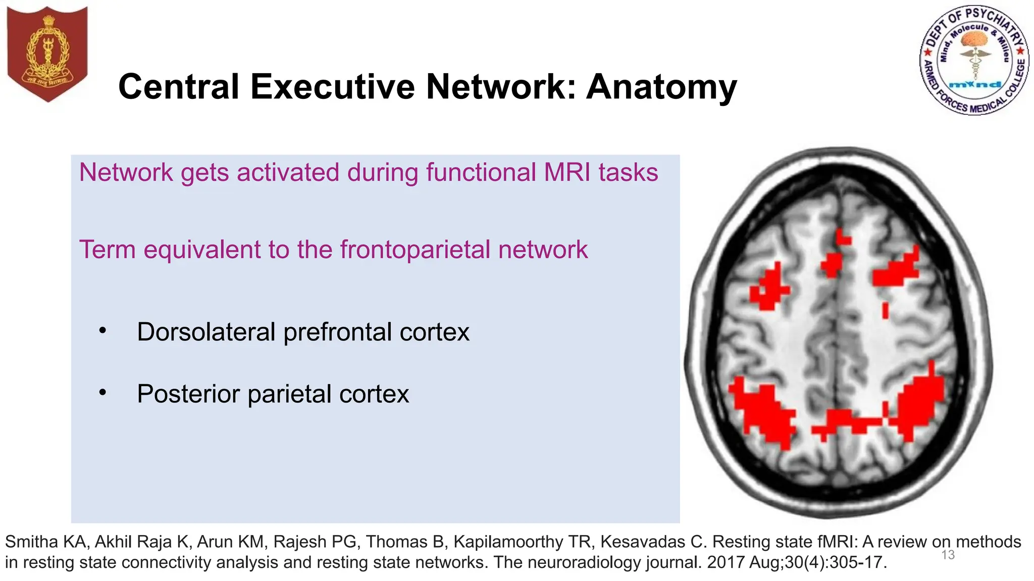
Central Executive Network:Anatomy
Network gets activated during functional MRI tasks
Term equivalent to the frontoparietal network
13
• Dorsolateral prefrontal cortex
• Posterior parietal cortex
Smitha KA, Akhil Raja K, Arun KM, Rajesh PG, Thomas B, Kapilamoorthy TR, Kesavadas C. Resting state fMRI: A review on methods
in resting state connectivity analysis and resting state networks. The neuroradiology journal. 2017 Aug;30(4):305-17.
14.
Executive functions
Tasks whichneed cognitive control and working memory
Target-directed activities
Control of intellectual activities
Active during task condition
Exhibits anticorrelated network during resting condition
14
Central Executive Network: Function
Smitha KA, Akhil Raja K, Arun KM, Rajesh PG, Thomas B, Kapilamoorthy TR, Kesavadas C. Resting state fMRI: A review on
methods in resting state connectivity analysis and resting state networks. The neuroradiology journal. 2017 Aug;30(4):305-17.
15.
Salience network
Anatomy
Dorsal anteriorcingulate cortex
Bilateral insula
Presupplementary motor area
15
Smitha KA, Akhil Raja K, Arun KM, Rajesh PG, Thomas B, Kapilamoorthy TR, Kesavadas C. Resting state fMRI: A review on
methods in resting state connectivity analysis and resting state networks. The neuroradiology journal. 2017 Aug;30(4):305-17.
16.
Salience network: Functions
Switchingbetween other large-scale networks to facilitate access to
attention and working memory resources when a salient event occurs
Strong functional coupling with Anterior Cingulate cortex to facilitate rapid
access to motor system
16
Smitha KA, Akhil Raja K, Arun KM, Rajesh PG, Thomas B, Kapilamoorthy TR, Kesavadas C. Resting state fMRI: A review
on methods in resting state connectivity analysis and resting state networks. The neuroradiology journal. 2017
Aug;30(4):305-17.
17.
Switching in Functionalnetworks
17
Bressler SL, Menon V. Large-scale brain networks in cognition: emerging methods and principles. Trends in cognitive sciences.
2018 Jun 1;14(6):277-90.
Substance use disorder
19
ZhangR, Volkow ND. Brain defaultmode network dysfunction in addiction. Neuroimage. 2019 Oct 15;200:313-31.
Substance use disorder
Aberrant patterns of brain functional
connectivity in Default Mode Network
Resting State Functional Connectivity
anterior region decreased
posterior region increased
Associated with craving and relapse
Prominence decreased during
intoxication phases
Disruption of the Default Mode Network in
addiction reflects in changes in
dopaminergic, glutamatergic and others
Neuro transmitters
20.
Schizophrenia
Significant hypoconnectivities wereobserved between seed regions and
Areas in auditory network (left insula)
Core network (right superior temporal cortex)
Default mode network
Self-referential network (right superior temporal cortex)
Somatomotor network (right precentral gyrus)
20
Lewis DA, Curley AA, Glausier JR, Volk DW. Cortical parvalbumin interneurons and cognitive dysfunction in schizophrenia. Trends
Neurosci. 2012;35;57–67
21.
Obsessive Compulsive Disorder
Intrinsicfunctional connectivity in Salience Network
Decreased intrinsic functional connectivity strength within Salience
Network subregions
Decreased intrinsic functional connectivity strength between left thalamus
and left cerebellum
Between left insula and right thalamus
Between right cerebellum and bilateral insula, and right Ant Cingulate
Cortex
21
Yun JY, Boedhoe PS, Vriend C, Jahanshad N, Abe Y, Ameis SH, Anticevic A, Arnold PD, Batistuzzo MC, Benedetti F,
Beucke JC. Brain structural covariance networks in obsessive-compulsive disorder: a graph analysis from the
ENIGMA Consortium. Brain. 2020 Feb 1;143(2):684-700.
22.
Obsessive Compulsive Disorder
FunctionalConnectivity Between the SN and Other Networks
Exhibited decreased intrinsic Functional Connectivity strength between
the Salience Network and Default Mode Network
Intrinsic Functional Connectivity strength between Salience Network and
Central Executive Network was significantly decreased in patients with
OCD
Specifically between Salience Network subregions (left insula) and ventral
lateral prefrontal cortex (VLPFC)
22
Yun JY, Boedhoe PS, Vriend C, Jahanshad N, Abe Y, Ameis SH, Anticevic A, Arnold PD, Batistuzzo MC, Benedetti F, Beucke
JC. Brain structural covariance networks in obsessive-compulsive disorder: a graph analysis from the ENIGMA Consortium.
Brain. 2020 Feb 1;143(2):684-700.
23.
23
Depression
Rive MM, vanRooijen G, Veltman DJ, Phillips ML, Schene AH, Ruhé HG. Neural correlates of dysfunctional emotion regulation in
major depressive disorder. A systematic review of neuroimaging studies. Neuroscience & Biobehavioral Reviews. 2013 Dec
1;37(10):2529-53
Amygdala connectivity and emotion regulation
Positive functional connectivity with amygdala
Hippocampus
Ventro Medial Pre Frontal Cortex
Anterior temporal poles
Negative functional connectivity with amygdala
DorsoLateral Prefrontal Cortex
Parietal regions
Dorsal Anterior Cingulate
• Also seen in early life stress, or at risk for depression (eg, family
history)
24.
24
Depression
Rive MM, vanRooijen G, Veltman DJ, Phillips ML, Schene AH, Ruhé HG. Neural correlates of dysfunctional emotion regulation in
major depressive disorder. A systematic review of neuroimaging studies. Neuroscience & Biobehavioral Reviews. 2013 Dec
1;37(10):2529-53.
Default Mode Network connectivity and rumination
Increase in Default Mode Network connectivity (Intra-network)
Preoccupation with negative mood, negative self-evaluation and
rumination
Increased connectivity of Default Mode Network with salience network
Dominance of Default Mode Network over Salience Network
Negative evaluation bias to salient information in environment
Degree of hyperconnectivity between sub genual Prefrontal Cortex and
Default Mode Network predicts the level of rumination
25.
Stroke
Damage to theattention network and symptoms of spatial neglect
Functional connectivity correlates with severity of symptoms
Alterations in default-mode network associated with post stroke
depression and episodic memory dysfunction
25
Ovadia-Caro S, Margulies DS, Villringer A. The value of resting-state functional magnetic resonance imaging in stroke. Stroke. 2014
Sep;45(9):2818-24.
26.
Autism spectrum disorder
SalienceNetwork is decisive for identifying salient stimuli
Builds central interface between sensory information processing,
attention, motor function and cognition
Alterations in social cognition is characteristic
Alterations in the Salience Network is characteristically seen
26
Neufeld J, Kuja-Halkola R, Mevel K, Cauvet É, Fransson P, Bölte S. Alterations in resting state connectivity along the
autism trait continuum: a twin study. Molecular psychiatry. 2018 Jul;23(7):1659-65.
27.
Autism spectrum disorder
Earlyhyperconnectivity followed by decreased connectivity in adulthood
Negative within-pair correlation between autistic traits and connectivity
between rt Ant Insula and ventromedial Prefrontal Cortex only in adults
Default Mode Network under-active owing to dysfunctional regulatory
mechanisms depending on other networks
Reduced connectivity of Default Mode Network at different ages
27
Neufeld J, Kuja-Halkola R, Mevel K, Cauvet É, Fransson P, Bölte S. Alterations in resting state connectivity along the autism
trait continuum: a twin study. Molecular psychiatry. 2018 Jul;23(7):1659-65.
28.
Attention Deficit HyperactivityDisorder
Higher local efficiency and decreased global efficiency
Indicating a developmental delay of whole-brain functional networks
Medial prefrontal, temporal, and occipital cortices
Regional loss of efficiency
Inferior frontal gyrus
Increased nodal efficiency
Default mode network connectivity
Delayed maturation
Decreased anterior-posterior connectivity
28
29.
Stress
Impact of earlylife stress associated
Decreased default network connectivity
Changes in structure and function in medial prefrontal cortex
Increases in connectivity between amygdala and medial prefrontal cortex
29
Philip NS, Sweet LH, Tyrka AR, Price LH, Bloom RF, Carpenter LL. Decreased default network connectivity is
associated with early life stress in medication-free healthy adults. European Neuropsychopharmacology. 2013 Jan
1;23(1):24-32.
Dorsal and VentralAttention Networks
24
Vossel S, Geng JJ, Fink GR. Dorsal and ventral attention systems: distinct neural circuits but collaborative roles. The
Neuroscientist. 2014 Apr;20(2):150-9.
32.
Dorsal and VentralAttention Networks:
Function
Top-down, goal-driven attention
Orienting after symbolic predictive cues
Visual search
Visual short-term maintenance
Stimulus-driven attention to salient behaviorally relevant events
Orienting to exogenous cues
Reorienting to unexpected events
Response to contextual cues
32
Vossel S, Geng JJ, Fink GR. Dorsal and ventral attention systems: distinct neural circuits but collaborative roles. The
Neuroscientist. 2014 Apr;20(2):150-9.
33.
Post Traumatic StressDisorder
Greater cross-network connectivity involving Salience Network (dorsal Ant Cingulate
Cortex) and default mode network (ventromedial Prefrontal cortex) associated with
impairments in disengagement and orienting of attention, processes
Both attentional orienting processes and balance observed to be disrupted in
veterans with Post Traumatic Stress Disorder
Interventions that utilize attention training, such as mindfulness might be useful for
alleviating attentional impairments in Post Traumatic Stress Disorder
33
Block SR, King AP, Sripada RK, Weissman DH, Welsh R, Liberzon I. Behavioral and neural correlates of disrupted
orienting attention in posttraumatic stress disorder. Cognitive, Affective, & Behavioral Neuroscience. 2017
Apr;17(2):422-36.
34.
Cingulo-Opercular Network: Function
Functionhas been particularly difficult to characterize
Network's pervasive activity
Frequent co-activation with other control-related networks
Functions
Sustaining alertness
Cognitive control functions
34
Coste CP, Kleinschmidt A. Cingulo-opercular network activity maintains alertness. Neuroimage. 2016 Mar 1;128:264-72.
35.
Precuneus network
Core ofdefault mode network
High metabolic rate compared to other networks
Part of superior Parietal lobule
Located in front of Cuneus
Functions
Autobiographical memory retrieval,
Emotional stimulus processing
Reward outcome monitoring
Assists behavioral function
35
Smitha KA, Akhil Raja K, Arun KM, Rajesh PG, Thomas B, Kapilamoorthy TR, Kesavadas C. Resting state fMRI: A
review on methods in resting state connectivity analysis and resting state networks. The neuroradiology journal.
2017 Aug;30(4):305-17.
36.
Visual Network: Anatomy& Function
Primary visual areas
Lateral occipital gyrus
Superior occipital gyrus
Function
Perception of visual stimuli
36
Smitha KA, Akhil Raja K, Arun KM, Rajesh PG, Thomas B, Kapilamoorthy TR, Kesavadas C. Resting state fMRI: A review on
methods in resting state connectivity analysis and resting state networks. The neuroradiology journal. 2017 Aug;30(4):305-17.
37.
Visuo-spatial Network: Anatomy& Function
Posterior parietal cortex of the
occipitoparietal junction
Midline of the precuneus
Posterior cingulate cortex
Frontal pole
37
Smitha KA, Akhil Raja K, Arun KM, Rajesh PG, Thomas B, Kapilamoorthy TR, Kesavadas C. Resting state fMRI: A review on methods
in resting state connectivity analysis and resting state networks. The neuroradiology journal. 2017 Aug;30(4):305-17.
Functions
Spatial attention
Orienting to salient
visuospatial cues
38.
Language Network: Anatomy
38
Broca’sareas
Wernicke’s areas
Additional regions
Prefrontal
Temporo-parietal
Subcortical regions
Wernick
area
Broca s
area
Auditory
cortex
Smitha KA, Akhil Raja K, Arun KM, Rajesh PG, Thomas B, Kapilamoorthy TR, Kesavadas C. Resting state fMRI: A review on
methods in resting state connectivity analysis and resting state networks. The neuroradiology journal. 2017 Aug;30(4):305-17.
Sensorimotor Network: Anatomy& Functions
40
Smitha KA, Akhil Raja K, Arun KM, Rajesh PG, Thomas B, Kapilamoorthy TR, Kesavadas C. Resting state fMRI: A review on
methods in resting state connectivity analysis and resting state networks. The neuroradiology journal. 2017 Aug;30(4):305-17.
Primary motor cortex
Pre-central gyrus
Somatosensory motor cortex
Post-central gyrus
Function
Performing and co-ordinating
motor tasks
Mindfulness
Associated with brainactivity in areas overlapping with default mode,
salience, and central executive networks (DMN, SN, CEN)
Four different mental states during meditation
Focus on the present experience was most strongly related to dorso-lateral
prefrontal cortex activation of central executive network (CEN)
Mind wandering was associated with default mode network (DMN),
Awareness of mind wandering linked with activation salience network (SN)
Shift of attention back towards focus on the present experience was again
linked with of central executive network (CEN)
43
Doll A, Hölzel BK, Boucard CC, Wohlschläger AM, Sorg C. Mindfulness is associated with intrinsic functional
connectivity between default mode and salience networks. Frontiers in human neuroscience. 2015 Aug 25;9:461.
44.
Mindfulness
Mindfulness is correlatedwith inter-intrinsic Functional Connectivity of
subnetworks of Default Mode network
Show decreased correlation between anterior and posterior-dorsal
Default Mode Network
Stronger negative correlation between the insula Salience Network and
posterior ventral Default Mode Network
44
Repetitive Trans cranialMagnetic Stimulation
Applied over left dorso-lateral prefrontal cortex (DLPFC)
Treatment response mechanism based on modulations in functional
networks, particularly the meso-cortico-limbic reward circuit.
Stimulation of left Dorsol-ateral Prefrontal Cortex modulates anterior
cingulate cortex connectivity in one specific meso-cortico-limbic network
46
Tik M, Hoffmann A, Sladky R, Tomova L, Hummer A, de Lara LN, Bukowski H, Pripfl J, Biswal B, Lamm C, Windischberger C.
Towards understanding rTMS mechanism of action: stimulation of the DLPFC causes network-specific increase in functional
connectivity. N
euroimage. 2017 Nov 15;162:289-96.
47.
Transcranial Direct CurrentStimulation
• Psychiatric disorders demonstrate disrupted functional and structural
neural networks,
• tDCS application modulate functional connectivity and induce
synchronization changes
• Active tDCS enhanced functional connectivity in frontal and fronto-parietal
regions regions known to play important underlying role in depression
• tDCS results in increase in cortical excitability within DLPFC leads to a
subsequent disturbance of the integrity of DMN
• tDCS-induced deactivations of Default Mode N facilitate reallocation of
cerebral resources to support task performance
47
Baeken C, Brunelin J, Duprat R, Vanderhasselt MA. The application of tDCS in psychiatric disorders: a brain imaging view.
Socioaffective neuroscience & psychology. 2016 Jan 1;6(1):29588.
48.
Future Aspects
Recent advancesshould focus on persistent needs to identify neural
networks involved in biologic process of psychiatric illness and aid in drug
discovery process
Non invasive Interventions targeting various networks can be utilized for
benefits in whom drugs are not indicated
Networks can act as potential biomarker for predicting clinical outcomes
and intervention at appropriate time
48
#4 Regions of the brain are specialized for different functions. . However, speech is a complex faculty that depends not only on the integrity of Broca area but also on the distributed processing of information across numerous brain regions through various interconnections. Thus, the role of any particular brain region or group of neurons in the production of specific behaviors or in the pathophysiology of a given neuropsychiatric disorder cannot be viewed in isolation, but must be considered within the context of the neural circuits connecting the neurons with other brain regions.
#7 Over the last 20 years neuroimaging studies have vastly expanded our knowledge of corticocortical connectivity in humans. However, it is important to note that most of these neuroimaging studies have identified functional connectivity (i.e., brain areas that are coactivated during a specific task) and not physical connections between regions. Evidence of direct anatomical corticocortical connections in the human brain is sparse, although newer imaging techniques such as Diffusion Tensor Imaging do provide some structural detail. Large-scale cortical networks linking distributed areas that subserve particular functions, such as attention or memory, have long been suggested and are now more readily studied with the neuroimaging techniques.
Numerous networks DMN VIS, FRP,CING OPER,Dorsal attention, ventral attention have been described, but three particular networks have received the greatest focus. They are dmn cen sn
#8 Regions of the DMN include posterior cingulate or precuneus ,angular gyrus medial-prefrontal, hippocampal, and lateral temporal areas
Regions showing strong functional connectivity with the posterior cingulate during rest
#10 The default mode network is active during passive rest and mind-wandering[4] which usually involves thinking about others, thinking about one's self, remembering the past, and envisioning the future rather than the task being performed
Functional hubs:[32] Information regarding the self
Posterior cingulate cortex (PCC) & precuneus: Combines bottom-up (not controlled) attention with information from memory and perception. The ventral (lower) part of PCC activates in all tasks which involve the DMN including those related to the self, related to others, remembering the past, thinking about future,
Medial prefrontal cortex (mPFC): Decisions about self processing such as personal information, autobiographical memories, future goals and events, and decision making regarding those personally very close such as family
#12 Additionally, during attention demanding tasks, sufficient deactivation of the default mode network at the time of memory encoding has been shown to result in more successful long-term memory consolidation.
In particular, the DMN shows marked suppression during task engagement, and need to be down-regulated in order to perform goal-directed behaviors such as visual attention or cognitive tasks thus this network labelled as the task-negative network.[6]
#13 Network which gets activated during Fmri tasks involving executive functions
he term central executive network (CEN) is generally equivalent to the frontoparietal network in literature,[
It constitutes
#14 CEN nodes that show strong intrinsic functional coupling also show strong coactivation during cognitively challenging tasks. In particular, the CEN is critical for actively maintaining and manipulating information in working memory, and for judgment and decision making in the context of goal-directed behavior
#15 Salience network (red) (b) Salience sub-divisions as defined showing ventral anterior insula network (red), dorsal anterior insula network (blue) and the overlap between the two (purple)The salience network constitutes the dorsal anterior cingulate cortex, bilateral insula and presupplementary motor area. The dysfunction of the network will disrupt the functioning of other networks, because it has a key role in regulating the dynamic changes in other networks. Moreover, the network is indispensable during the rapid change of behaviour. That is, what to do next or not to do is decided by the appropriate functioning of the network. For these reasons the salient network’s proper functioning is inevitable for the commencement of control of cognition processes
#16 It is involved in detecting and filtering salient stimuli, as well as in recruiting relevant functional networks.[3][4] Together with its interconnected brain networks, the SN contributes to a variety of complex functions, including communication, social behavior, and self-awareness through the integration of sensory, emotional, and cognitive information.[5]
Switching between other large-scale networks to facilitate access to attention and working memory resources when a salient event occurs
Interaction of the anterior and posterior insula to modulate autonomic reactivity to salient stimuli
Strong functional coupling with the ACC to facilitate rapid access to the motor system
Key role in regulating dynamic changes in other networks
Network indispensable in rapid change of behavior
Commencement of control of cognition processes
#19 ant dmn self-referential mental thought and post dmn engages in episodic memory retrieval and scene construction
Imbalance between anterior and posteriorDMN caused by altered DA-related reward function inaddiction. During the binge-intoxication stage, the rewarding effects of drug (red) are mediated by acti-vation of the DA reward system projecting from theVTA into the VS (and to a lesser extent the DS) and the PFC including regions from the anterior DMN and regionsfrom the ECN while suppressing the function of pos-terior DMN shifting attention to external stimuli.
Chronic drug use however decreases the activity ofthe brain DA reward system (grey) of addicted in-dividuals resulting in opposite effects than during acute drug intoxication. Reduced emotional control(mediated by anterior DMN) and increased self-referential processing (mediated by posterior DMN)can enhance ruminatory behaviors and negative affect(mediated by amygdala and habenula and decreased dopaminergic signaling in VS) during withdrawal and contribute to negative reinforcement of drug use. Lower baseline activity of anterior DMN exaggerates the reactivity to drug cues. Along with compromised cognitive control (mediated by ECN), it facilitates craving and relapsing behavior in the preoccupation stage of the addiction cycle.
#20 Default mode network (right medial prefrontal cortex, and left precuneus
and anterior cingulate cortex )
No hyperconnectivity between seed regions and any other areas within the networks was detected in patients
Altered Default Mode Connectivity
Altered perceptual experiences that individuals with schizophrenia reflect enhanced attention to internal thoughts, feelings, and experiences,
Failure to suppress default mode network activity during cognitive task performance leading to disrupted cognitive function
Presence of auditory verbal hallucinations tends to be associated with reduced connectivity among these default mode structures.
#22 Functional Connectivity Between the SN and Other Networks
Exhibited decreased intrinsic Functional Connectivity strength between the Salience Network and Default Mode Network mainly found in Salience Network subregions (left insula) and the MCC
Patients with OCD exhibited decreased iFC strength between the SN and the DMN compared to the HCs, which mainly foun
#25 Damage to the attention network and corresponding symptoms of spatial neglect have been associated with decreased interhemispheric connectivity in structurally intact areas that are part of the attention network.Importantly, functional connectivity correlates with the severity of symptoms.24 rs-fMRI has also bee
Another network that has been explored after stroke is the default-mode network. The default-mode network is a network of regions including the posterior cingulate and precuneus, the temporoparietal junction, and the medial prefrontal cortex. It has been widely implicated in various neurological and mental disorders and has been linked to tasks such as autobiographical memory retrieval and theory of mind functions.26 After stroke, alterations in default-mode network functional connectivity have been associated with poststroke depression27 and episodic memory dysfunction
#26 1Alterations in social cognition is characteristic of ASD
2SN has been hypothesized to be decisive for identifying salient stimuli, building a central interface between bottom-up sensory information processing, attention guidance, emotion processing, motor function and cognition
It has been suggested that might be related to alterations in the SN since these functions depend on the perception of what is salient.56 In line with this notion, a meta-analysis found consistently reduced activation of the insula in ASD compared to typically developing individuals during social processing.57 Altered salience processing might further be related to sensory issues such as sensory hyper- or hypo-sensitivity56 as well as enhanced attention to detail characteristic of ASD.58
#27 connectivity between these networks in adults with higher autistic traits could indicate that this modulatory function is weaker. In line with this notion, it has been hypothesized that the DMN might be under-active in ASD owing to dysfunctional regulatory mechanisms depending on other networks.11 Current developmental models of ASD suggest early hyperconnectivity followed by decreased connectivity in adulthood.10,55,
Reduced connectivity of DMN in ASD at different ages
Early hyperconnectivity followed by decreased connectivity in adulthood
Negative within-pair correlation between autistic traits and connectivity between rAI and vmPFC only in adults
Reduced RS connectivity between networks in adults with higher autistic traits indicate weak modulatory function
DMN under-active in ASD owing to dysfunctional regulatory mechanisms depending on other networks
#28 Weaker interconnectivity in the DMN, dorsal attention network, and cerebellum
Stronger short-range connectivity within reward network
#29 In the paper entitled ‘‘Decreased Default Network Connectivity is Associated with Early Life Stress in Medication-Free Healthy Adults’’ by Philip et al. (in this issue) the impact of early life stress was associated with changes in structure and function in the medial prefrontal cortex, as part of the default network. Early life stress was associated with decreased default network connectivity and trend-level increases in connectivity between the amygdala and the medial prefrontal cortex, suggesting an altered resting state connectivity is a correlate of stress exposure, rather than a product of medication or psychiatric morbidity. In the paper
#31 .IT HAS BEEN proposed tht neither of the two networks controls attentional processes in isolation and that the flexible interaction between both systems enables the dynamic control of attention in relation to top-down goals and bottom-up sensory stimulation
The dorsal network (Fig. 1, blue) is supposed to be organized bilaterally and comprises the intraparietal sulcus (IPS) and the frontal eye fields (FEF) of each hemisphere. These areas are active when attention is overtly or covertly oriented in space
The ventral network comprises the temporoparietal junction (TPJ) and the ventral frontal cortex (VFC) (Fig. 1, orange) and typically responds when behaviorally relevant stimuli occur unexpectedly (e.g., when they appear outside the cued focus of spatial attention). I
#33 PTSD group exhibited greater rsFC between the salience network and (a) the default mode network, (b) the dorsal attention network, and (c) the ventral attention network. Moreover, problems with disengaging spatial attention increased the rsFC between the networks above in the control group, but not in the PTSD group. The present findings link PTSD to both altered orienting of spatial attention and altered relationships between spatial orienting and functional connectivity involving the salience network. Interventions that target orienting and disengaging spatial attention may be a new avenue for PTSD research.
ePTSD group exhibited greater resting-state functional connectivity between the SN (dACC) and the default mode network (vmPFC) independent of attentional performance (Supplementary materials), replicating our previous findings (Sripada et al. 2012a). The control group did not exhibit greater rsFC than the PTSD group between the SN and any other regions.
#37 Ghazi-Saidi L. Visuospatial and Executive Deficits in Parkinson’s Disease: A Review. Acta Scientific Neurology (ISSN: 2582-1121). 2020 Apr;3(4).
Visuospatial perception permits the recognition and interpretation of information regarding many aspects of the visual world and is distinct from visuospatial attention, which is specific to directing attention to a particular location in space. Visuospatial deficit has been associated with dysfunction of several brain regions including occipital lobe, parietal lobe, frontal lobe, thalamus, and the superior longitudinal fasciculus
Application
Studies of visuospatial function in PD have largely focused on visual hallucination, perception, attention, and neglect given the common occurrence of related symptoms in the disease
#39 Speech
Comprehension
Reading
Interpreting
Mimicking
Broca’s area is the location of mirror neurons
Observing similar movements of others
Imitate motor activities
#40 neuroscience, the sensorimotor network (SMN) is a large-scale brain network . The SMN includes somatosensory (post-central gyrus) and motor (pre-central gyrus) regions and extends to the supplementary motor areas.[1] Studies have shown that this network is activated during motor tasks such as finger tapping[2] indicating that these regions may involve a pre-mediated state that ready the brain when performing and coordinating a motor task.[1] Dysfunction in the SMN has been implicated in various neuropsychiatric disorders.
Pathophysiology[edit]
Bipolar Disorder: The psychomotor disturbances that characterize the depressive and manic phases of bipolar disorder may be related to dysfunction in the sensorimotor network (SMN) and its balance with other large-scale networks such as the default mode network.[3][4]
Amyotrophic Lateral Sclerosis: Altered functional connectivity patterns in the SMN may contribute to various symptoms in the neurodegenerative disease .[
#42 Needless to say, the hand is the principal instrument for writing and writing is one of the most fundamental things we do with the hand. It is therefore worthwhile to consider in some detail the hand in a book dealing with several aspects of writing. In this chapter I do not claim to present a complete and detailed review of the functions of the hand, but I will try to describe some fundamental sensory and motor mechanisms in an attempt to construct an overview of hand functioning. The hand is both a sensory and a motor organ, so that the sensory functions cannot be completely separated from the motor ones. When we explore the world with our hands, the tactile information arriving at our brain plays an essential role in the ongoing motor behavior. This continuous sensory inflow and motor outflow represents what has been called “active touch” (Gibson, 1962) or “haptics” (Revesz, 1950; Kennedy, 1978). Thus these terms mean the combined action of skin, joints and muscles for acquiring information from the external world. On the other hand, the movement
#43 Mindfulness is attention to present moment experience without judgment. Mindfulness practice is associated with brain activity in areas overlapping with the default mode, salience, and central executive networks (DMN, SN, CEN). Intrinsic functional connectivity (iFC; i.e., synchronized ongoing activity) across these networks is associated with mindfulness scores.
four different mental states during meditation with each state being preferentially related to activity in different intrinsic brain networks: focus on the present experience was most strongly related to dorso-lateral prefrontal cortex activation of the central executive network (CEN),
mind wandering was associated with the default mode network (DMN),
awareness of mind wandering was linked with activation in the salience network (SN),
shift of attention back towards focus on the present experience was again linked with the right dorsolateral prefrontal cortex (DLPFC) and right posterior parietal cortex with both regions being part of the CEN.
. These networks are believed to subserve specific cognitive functions like attentional control or core affect (Fox and Raichle, 2007) as their patterns of coherent ongoing activity overlap and reflect the activation patterns observed during goal directed
. These networks are believed to subserve specific cognitive functions like attentional control or core affect as their patterns of coherent ongoing activity overlap and reflect the activation patterns observed during goal directed
#44 A decreased connectivity of the aDMN and pDMN might indicate that more mindful individuals interpret the affective relevance of a given stimulus as less self-related. This is also supported by the association of the MAAS questionnaire with this connectivity. The MAAS focuses on measuring the ability to consciously perceive the present moment (Brown and Ryan, 2003). This present moment experience has been associated with a deactivation of the PCC/Precuneus (Garrison et al., 2013) area and with activations in dorso medial (Hölzel et al., 2007; Dickenson et al., 2013) and lateral prefrontal regions (Brefczynski-Lewis et al., 2007). Thus, this would speak for a reduced synchrony of the antDMN and the pDMN regions during this experience, which could transition into a stronger decoupling of these parts of the DMN network in more mindful individuals during rest. Instead, these regions may be coupled more strongly to either lateral parietal or DLPFC in expert meditators. e.g., Brewer et al. (2011) found increased connectivity between the PCC, dorsal ACC and DLPFC in participants with more meditation experience both during rest and during different kinds of meditation. The authors interpreted these results as an at baseline increased connectivity and activity of task positive control regions together with reduced activation of the DMN in experienced meditators regardless of condition. Other authors have argued for a coactivation of the aDMN together with inferior parietal regions during rest, which might reduce distractibility by mind wandering in experienced meditators (Hasenkamp and Barsalou, 2012). Our data are more in accordance with the model by Hasenkamp and Barsalou (2012), which suggests a critical interplay between medial DMN and lateral CEN for engaging attention on present experience. Instead of being engaged in mind-wandering which results in activation of the anterior and posterior DMN (Mason et al., 2007), regions in the dmPFC might be responsible for focusing attention back to present experience likely reflected by stronger anti-correlated coupling between CEN and DMN. Future studies will have to further clarify the directionality of connectivity between the anterior DMN, posterior DMN, and attention-relevant regions of frontal and parietal CEN and how this is related to meditation experience
#45 Abbreviations: aDMN, anterior default mode network; pvDMN, posterior ventral default mode network; pdDMN, posterior dorsal default mode network; accSN, cingular salience network; insSN, insula salience network; lCEN, left central executive network; rCEN, right central executive network.
#46 Transcranial magnetic stimulation (TMS) is a powerful non-invasive technique for the modulation of brain activity. While the precise mechanism of action is still unknown, TMS is applied in cognitive neuroscience to establish causal relationships between stimulation and subsequent changes in cerebral function and behavioral outcome. In addition, TMS is an FDA-approved therapeutic agent in psychiatric disorders, especially major depression. Successful repetitive TMS in such disorders is usually applied over the left dorso-lateral prefrontal cortex (DLPFC) and treatment response mechanism was therefore supposed to be based on modulations in functional networks, particularly the meso-cortico-limbic reward circuit. However, mechanistic evidence for the direct effects of rTMS over DLPFC is sparse. Here we show the specificity and temporal evolution of rTMS effects by comparing connectivity changes within 20 common independent components in a sham-controlled study. Using an unbiased whole-brain resting-state network (RSN) approach, we successfully demonstrate that stimulation of left DLPFC modulates anterior cingulate cortex (ACC) connectivity in one specific meso-cortico-limbic network, while all other networks are neither influenced by rTMS nor by sham treatment. The results of this study show that the neural correlates of TMS treatment response are also traceable in DLPFC stimulation of healthy brains and therefore represent direct effects of the stimulation procedure.
#47 ranscranial direct current stimulation (tDCS) is a form of neuromodulation that uses constant, low direct current delivered via electrodes on the head. It was originally developed to help patients with brain injuries or neuropsychiatric conditions such as major depressive disorder.Active tDCS enhanced functional connectivity in the frontal and fronto-parietal regions (Keeser et al., 2011), regions that are known to play an important underlying role in depression (
tDCS-induced deactivations of the DMN facilitate reallocation of cerebral resources to support task performance, and thereby beneficially influence the regulation of corticosubcortical network activity.
#48 Findings are starting to reveal DMN RSFC as a potential biomarker for predicting clinical outcomes in SUD apotential biomarker for predicting clinical outcomes nd identify the DMN as a promising target for the treatment of addiction
Impairments present at the start of illness, or even before and more prominent among those individuals who go on to develop full blown psychosis
Promising avenue for future biomarker and intervention work