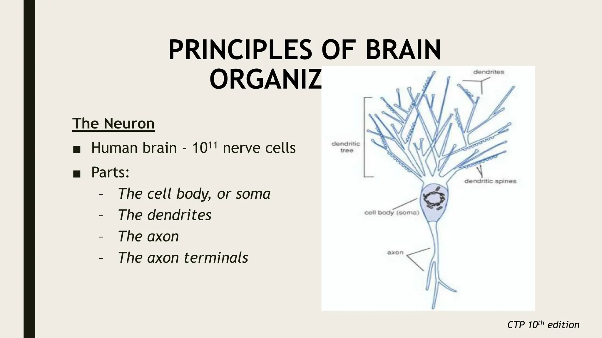
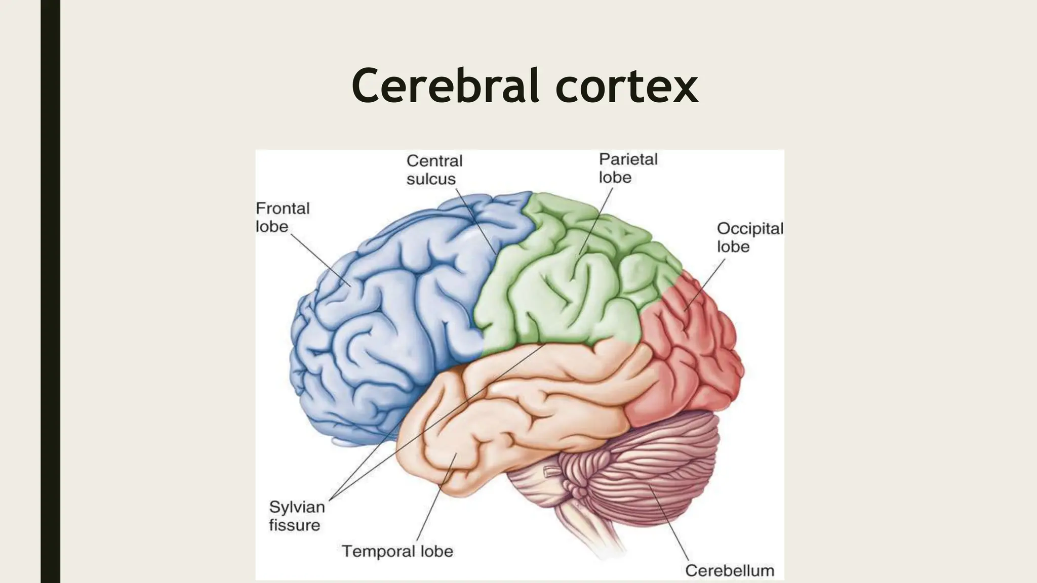
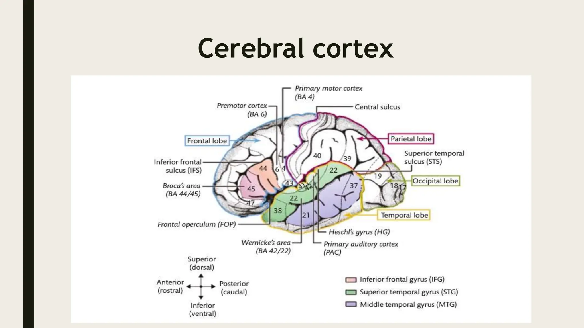
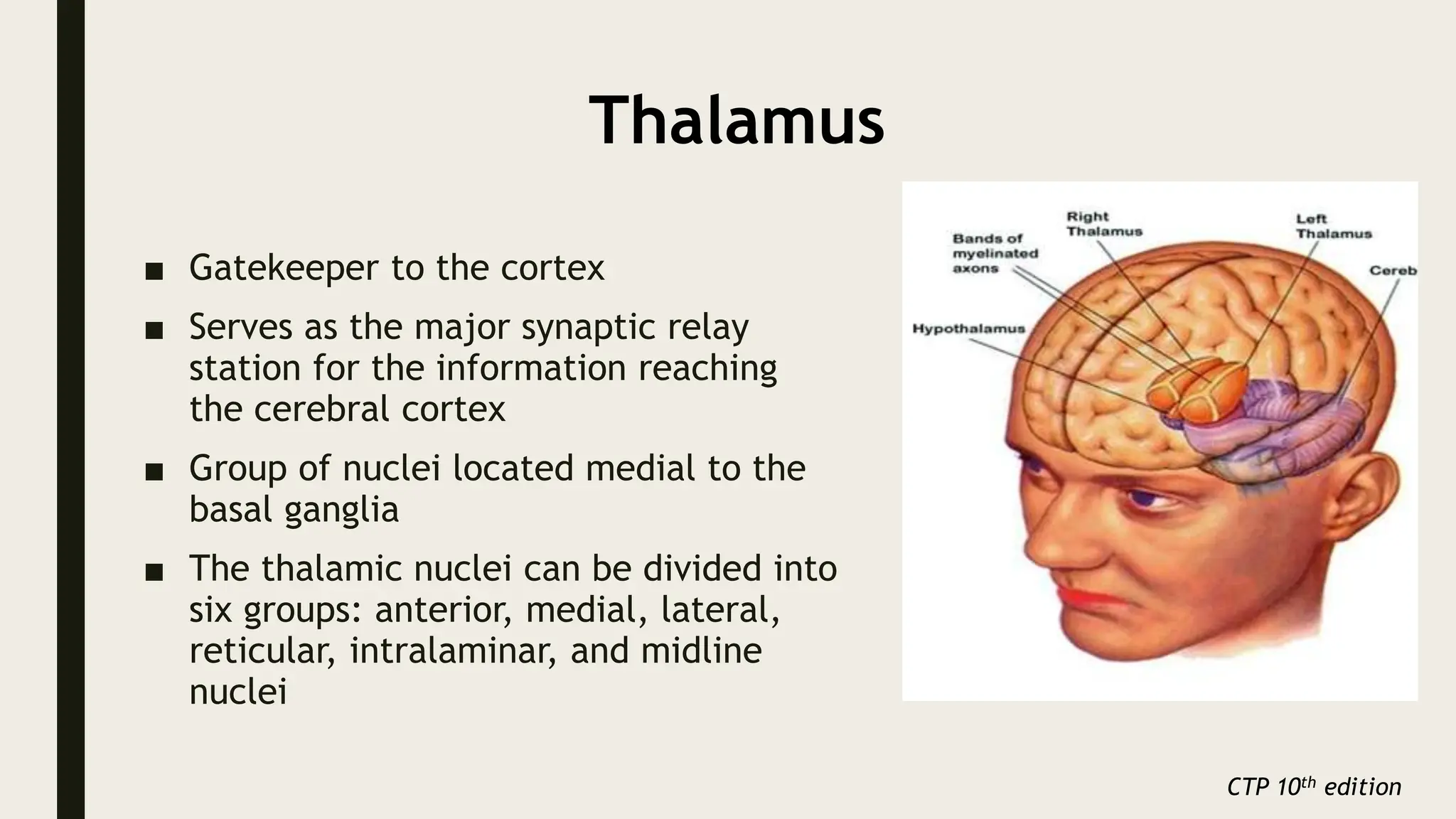
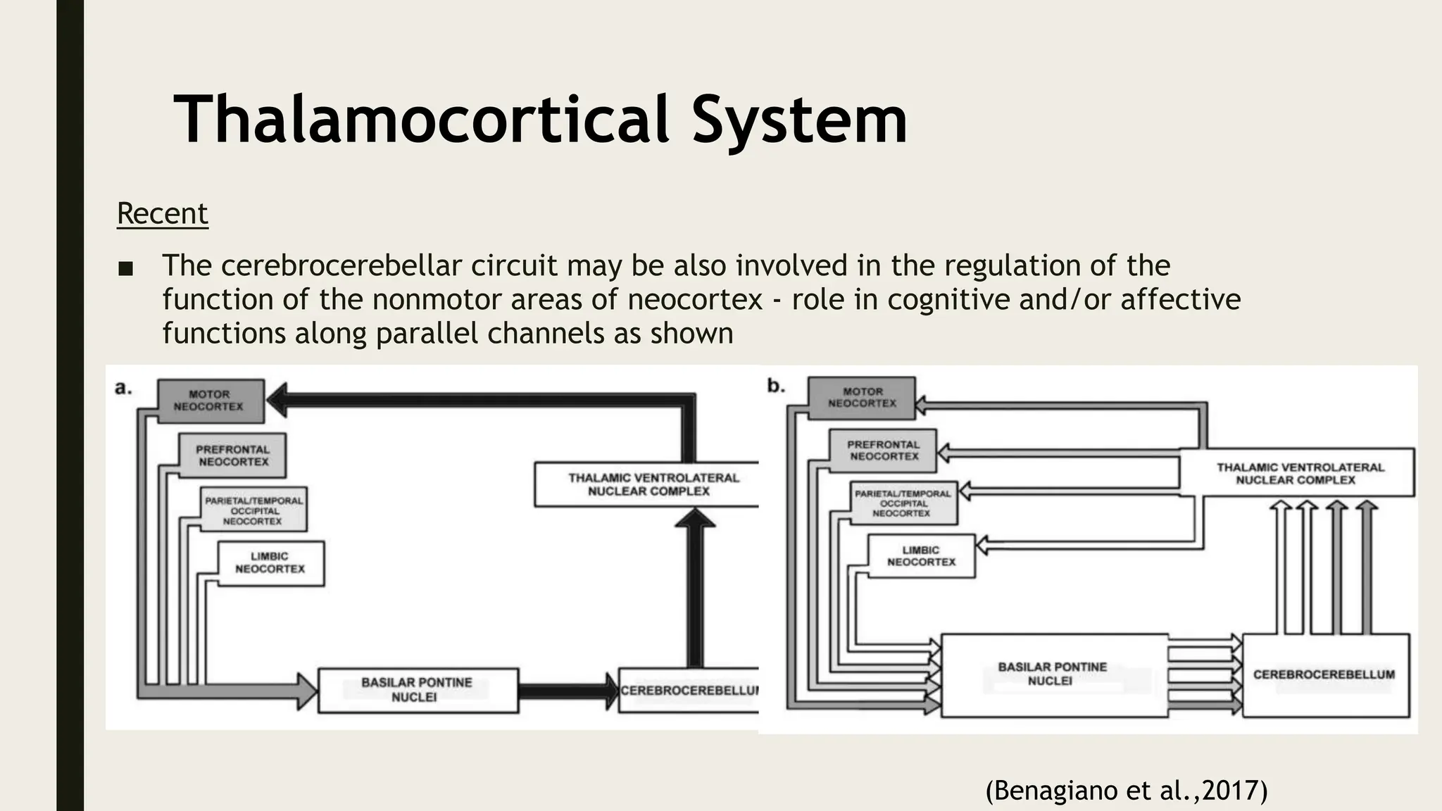
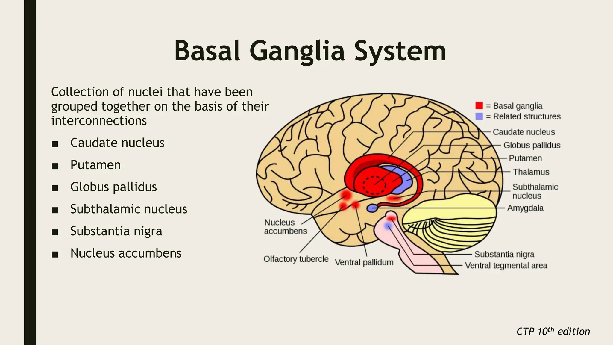
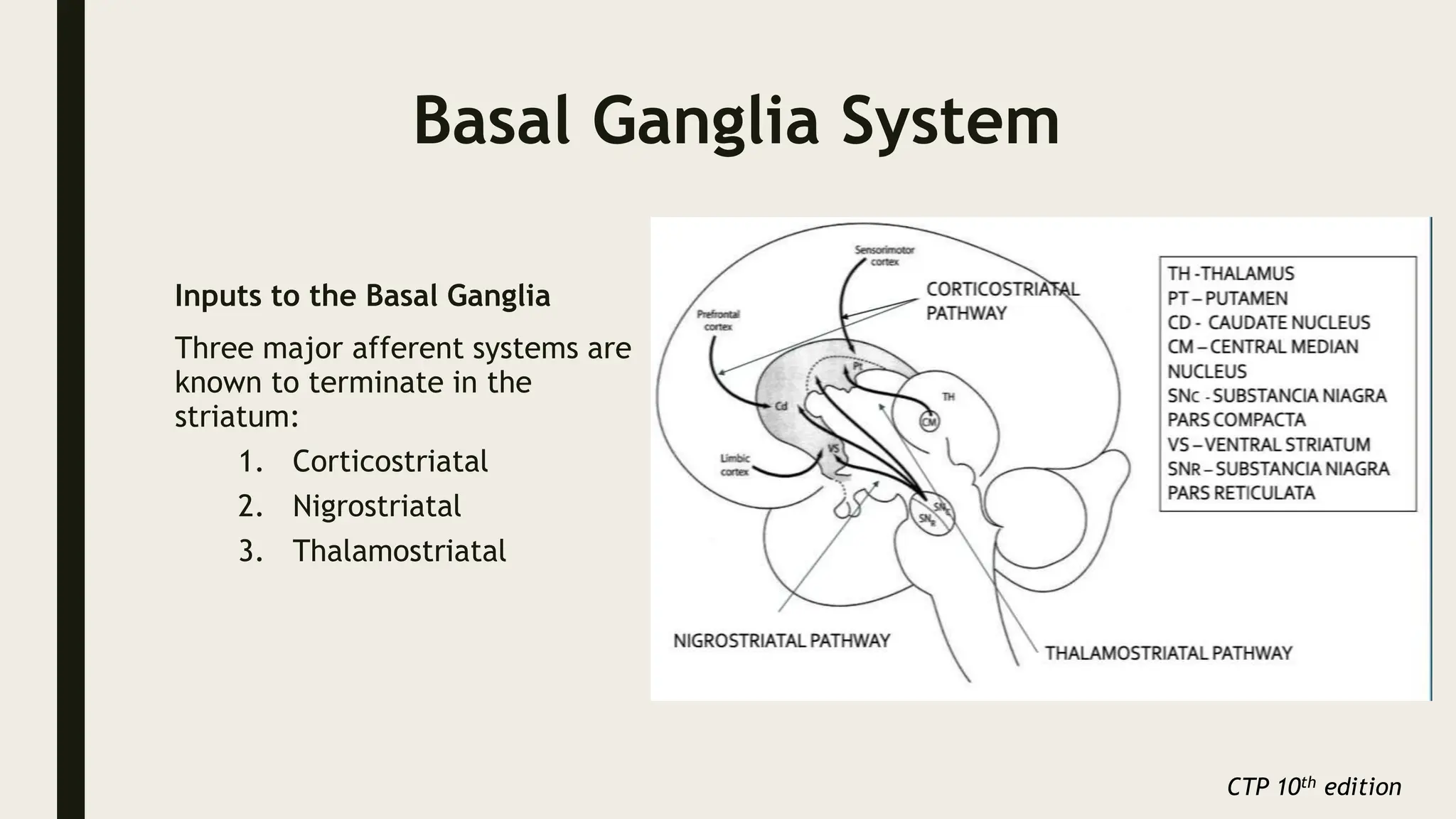
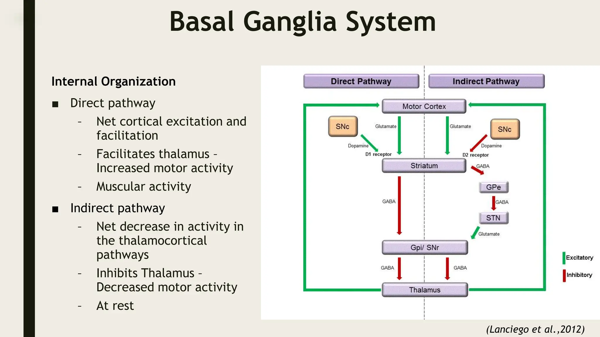
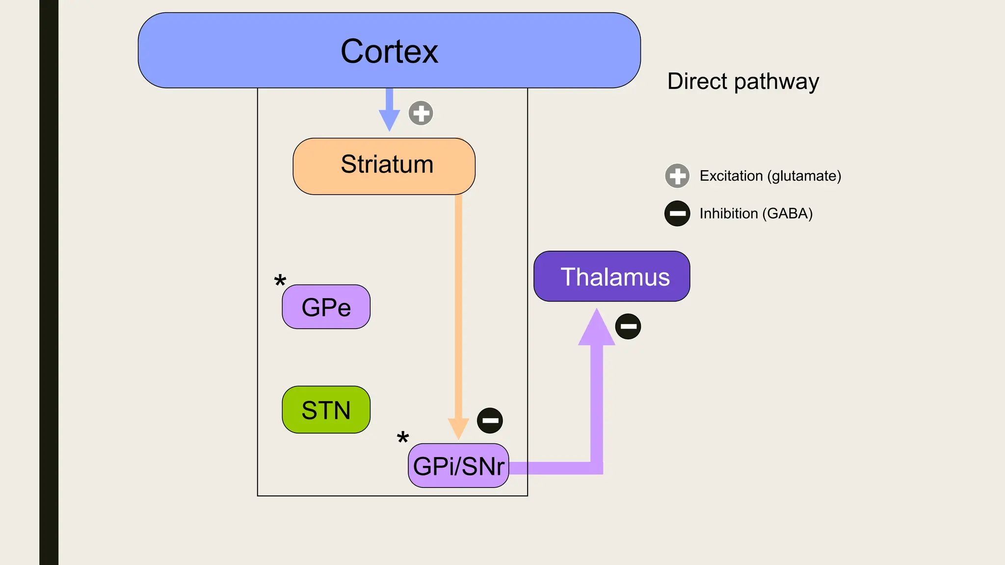
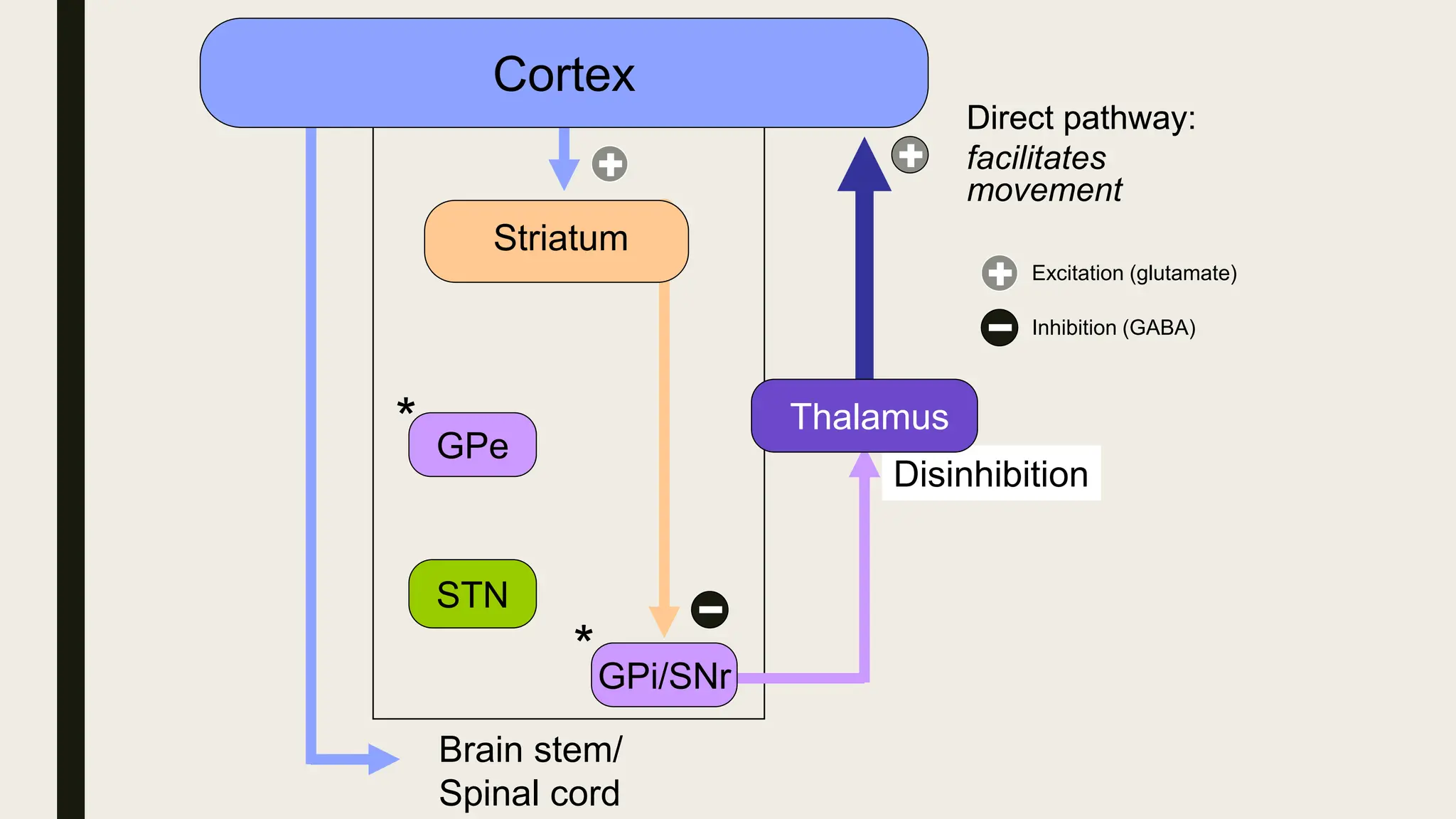
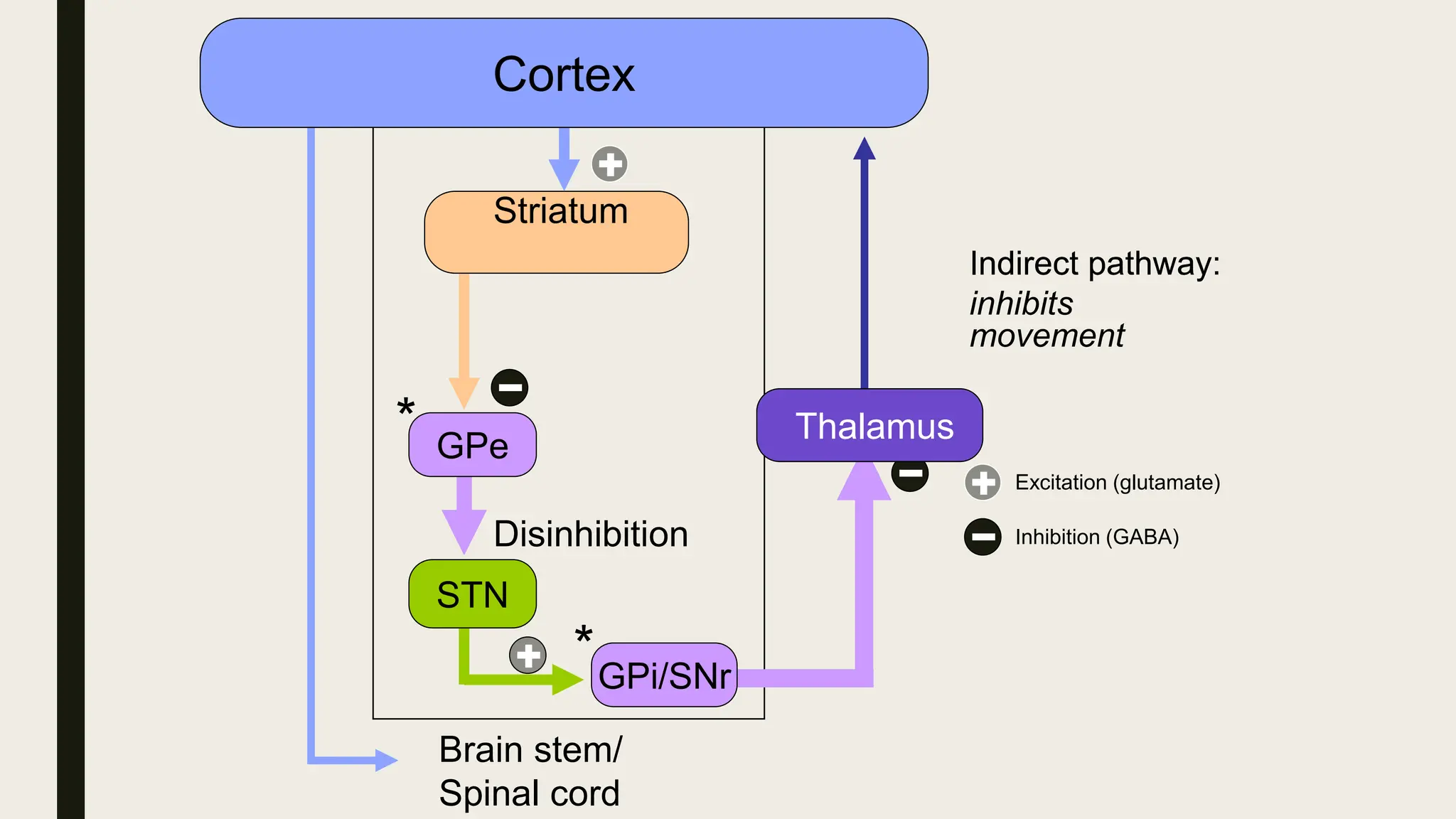
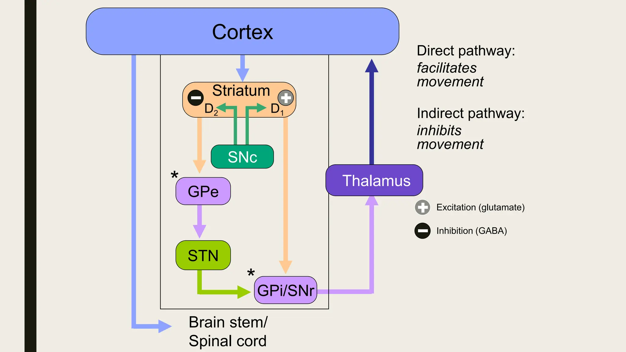
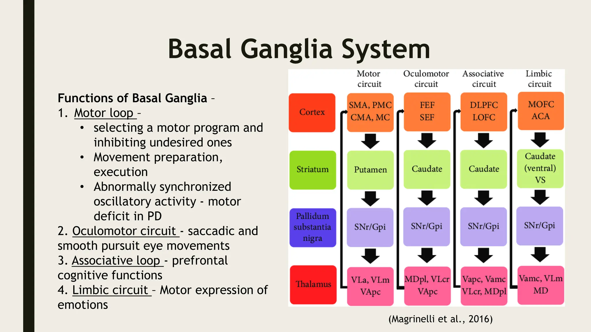
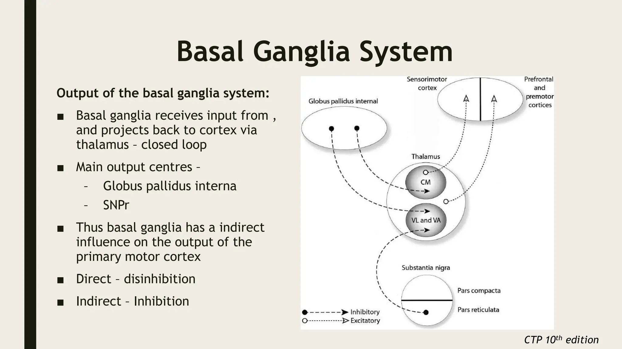
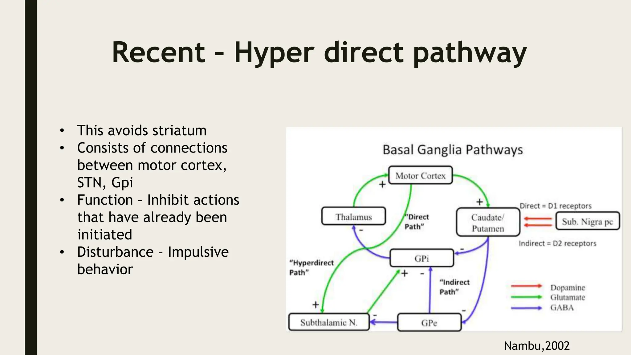
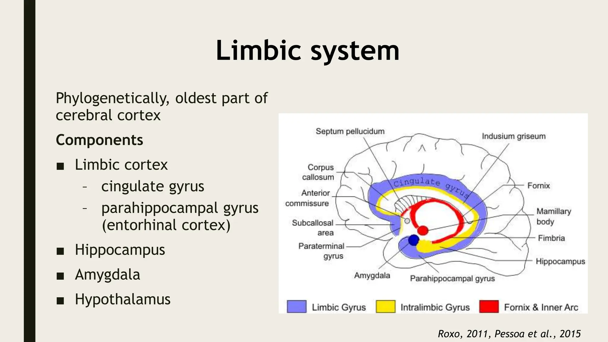
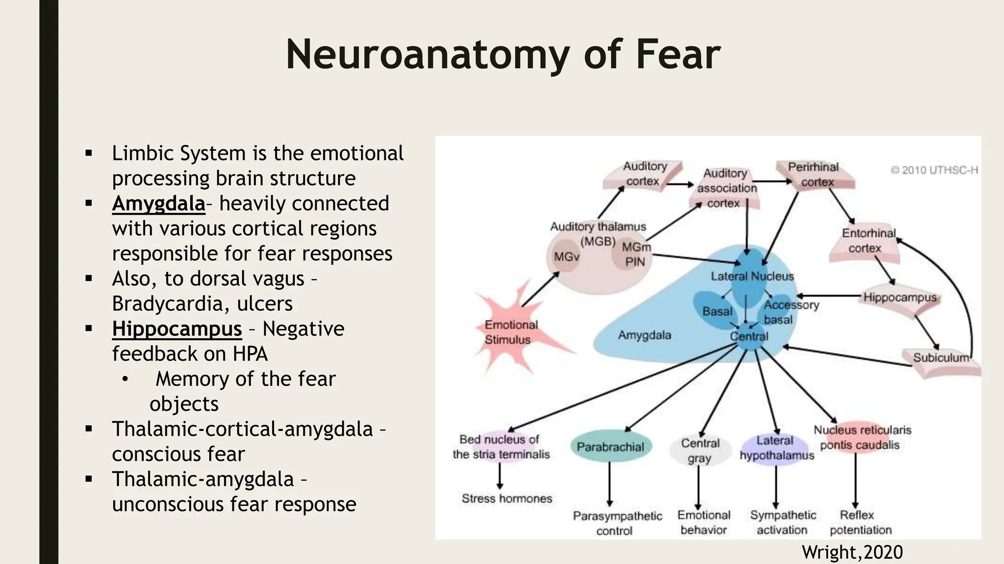
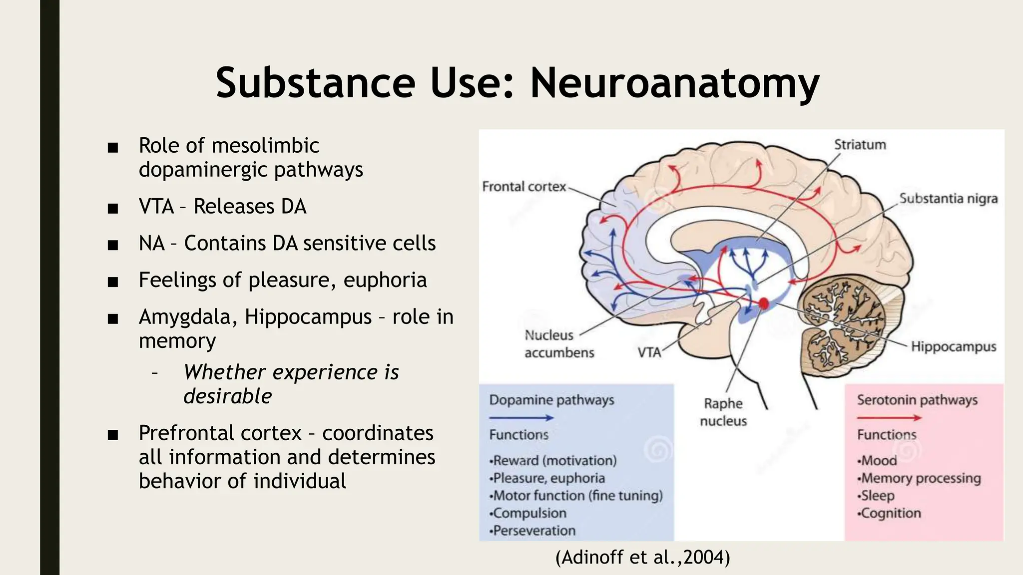
FUNCTIONAL NEUROANATOMY IN
RELATION TO
PSYCHIATRY: CURRENT
UNDERSTANDING
Chairperson: Prof. RK Chadda
Discussant: Dr. Nishtha Laspal
Presenter: Dr. Ilambaridhi
OUTLINE
■ Introduction
■ Principles of brain organization
■ Functional brain systems
– Cerebral cortex and Frontal Lobe
– Thalamus
– Basal ganglia
– Limbic System
■ Neuroanatomical correlates of major psychiatric disorders
■ Conclusion
INTRODUCTIO
N
INTRODUCTION
■ Functional Neuroanatomy is the study of interacting neurons,
glia and brain regions.
■ It combines structural knowledge with behavioral data
■ Mental functioning derives from the operation of large groups of
neurons - organized into nuclei, brain regions, and neural circuits
■ Understanding the neurobiological bases in psychiatric disorders
requires appreciation of the functional organization and
connections in the human brain.
PRINCIPLES OF BRAIN
ORGANIZATION
PRINCIPLES OF BRAIN
ORGANIZATION
The Neuron
■ Human brain - 1011 nerve cells
■ Parts:
– The cell body, or soma
– The dendrites
– The axon
– The axon terminals
CTP 10th edition
PRINCIPLES OF BRAIN
ORGANIZATION
Other Cells
■ Glial cells
– Astrocytes
– Oligodendrocytes/Schwann cells
– Microglia
CTP 10th edition
PRINCIPLES OF BRAIN
ORGANIZATION
■ Types of connection
– Reciprocal - Most brain regions
– Divergent - Locus coeruleus
– Convergent - Entorhinal region
■ Hierarchical pathways - Visual
pathway
– different types of visual information
(e.g., motion and form) – in parallel
fashion
CTP 10th edition
FUNCTIONAL BRAIN
SYSTEMS
Cerebral cortex
Cerebral cortex
■ Laminated sheet of neuron
cell bodies
■ Neocortex - more than 90%
 6 layered architecture
 70% pyramidal
 Projection neurons
 Excitatory
neurotransmitters
 30% stellate
 Inter neurons
 Inhibitory neurotransmitter
■ Allocortex - rest (old cortex)
CTP 10th edition
Cerebral cortex
■ Cortex has 6 layers marked : I
to VI
■ Homotypical cortex:
– all 6 layers present
■ Heterotypical cortex:
– Do not retain a 6 layer structure
– Primary motor cortex
■ no layer IV
– Primary sensory cortex
■ expanded layer IV
CTP 10th edition
Cerebral cortex
■ The neocortex can be divided further into discrete areas, based
on distinctive architecture and role in particular brain functions
■ Most widely used system  by Korbinian Brodmann - 47 areas
Cerebral cortex
FRONTAL LOBE
Functional regions
1. Primary motor area
2. Premotor area
3. Frontal eye fields
4. Dorsolateral prefrontal
cortex
5. Orbital and basal areas
6. Supplementary motor area
and anterior cingulated
gyrus area
Functional circuits - frontal-
subcortical circuits
1. Motor circuit – from motor
and pre-motor cortex
2. Oculomotor unit - frontal
eye fields
3. Dorsolateral prefrontal
circuit
4. Anterior cingulate circuit
5. Orbitofrontal circuit
CTP 10th edition
Areas Function Neuropsychiatric
Manifestations
Dorsolateral prefrontal cortex
(DLPFC)
1. Executive functions
2. Working Memory
3. Planning and Abstract
thinking
1. Cognitive dysfunction
2. Diminished judgement,
insight
3. Concrete and inflexible ideas
4. Decreased spontaneous
behavior
5. Stereotyped response
And Perseveration
Orbitofrontal Cortex Personality (socially
appropriate behavior,
empathy)
Disinhibition, irritability,
lability, recklessness, euphoria
Anterior cingulate Motivation Amotivation, Apathy
(Foster et al.,2001, Mansouri et al.,2017)
Thalamus
■ Gatekeeper to the cortex
■ Serves as the major synaptic relay
station for the information reaching
the cerebral cortex
■ Group of nuclei located medial to the
basal ganglia
■ The thalamic nuclei can be divided into
six groups: anterior, medial, lateral,
reticular, intralaminar, and midline
nuclei
CTP 10th edition
Thalamus
Divided by thin Y shaped sheet of myelinated
fibers the Internal Medullary Lamina into
various parts:
Part Nuclei
Anterior part Anterior nucleus
Medial part Medial dorsal nucleus, Medial ventral
nucleus
Lateral part – Dorsal tier Lateral dorsal, Lateral posterior,
Pulvinar
Lateral part - Ventral tier Ventral anterior, Ventral lateral, Ventral
posterior
Other parts Intralaminar nucleus, Reticular nucleus CTP 10th edition
Thalamus
Functional organization
■ Relay nucleus
– Project and receive from→ cerebral cortex
– Modulate thalamic input
■ Specific relay nucleus
– Process input either from a single sensory modality or from a distinct
part of the motor system
– Visual input from optic tract → Lateral geniculate
■ Association relay nuclei
– receive highly processed input from more than one source and project to
larger areas of the association cortex
– Medical dorsal nucleus → Hypothalamus & prefrontal cortex
■ Reticular nucleus
– receive input from diverse sources and project to widespread areas of
the cerebral cortex and to the thalamus
– Inhibitory neuron → regulate thalamic activity CTP 10th edition
Thalamocortical System
4 major thalamic systems exemplify the principles of brain
organization
• Thalamocortical sensory system
• Thalamocortical motor system
• Thalamocortical association system
• Cerebellothalamo-cortical system
Thalamocortical System
Thalamocortical Sensory System
■ Skin sensation (Somatosensory system)
■ Ventral posterior nucleus of the thalamus
■ Topography
– Body – VPL
– Head – VPM
■ Distorted representations – fingers highest
■ Sub modalities are parallelly processed
– E.g. Proprioceptive in medial lemniscus
Pain & temperature in spinothalamic
tract
CTP 10th edition
Thalamocortical System
Thalamocortical Motor System
■ Corticospinal tract from layer V of primary motor and premotor cortex
■ Terminates in the spinal cord to influence motor behavior
Thalamocortical Association Systems
■ Association regions receive a convergence of input from a variety of sources
■ The prefrontal cortex receives afferents from
– Parietal lobe - Contralateral prefrontal cortex
– Temporal lobe - Medial dorsal nucleus of thalamus
– Cingulate cortex
■ This patterns of connectivity is related to the ability to perform spatial delayed-
response tasks
CTP 10th edition
Thalamocortical System
Cerebello-Thalamocortical-systems
■ Chief efferent: Dentatothalamic fibres
Superior cerebellar peduncle (Dentate nucleus) →opposite thalamus→ cortex
■ Responsible for smooth and coordinated movements
■ Learning motor skills
■ Lesions –
– Decreased muscle tone
– Intention tremor
– Dysdiadochokinesia
– Rebound phenomenon
CTP 10th edition
Thalamocortical System
Recent
■ The cerebrocerebellar circuit may be also involved in the regulation of the
function of the nonmotor areas of neocortex - role in cognitive and/or affective
functions along parallel channels as shown
(Benagiano et al.,2017)
Basal Ganglia System
Collection of nuclei that have been
grouped together on the basis of their
interconnections
■ Caudate nucleus
■ Putamen
■ Globus pallidus
■ Subthalamic nucleus
■ Substantia nigra
■ Nucleus accumbens
CTP 10th edition
Basal Ganglia System
Inputs to the Basal Ganglia
Three major afferent systems are
known to terminate in the
striatum:
1. Corticostriatal
2. Nigrostriatal
3. Thalamostriatal
CTP 10th edition
Basal Ganglia System
Internal Organization
■ Direct pathway
– Net cortical excitation and
facilitation
– Facilitates thalamus –
Increased motor activity
– Muscular activity
■ Indirect pathway
– Net decrease in activity in
the thalamocortical
pathways
– Inhibits Thalamus –
Decreased motor activity
– At rest
(Lanciego et al.,2012)
Cortex
Thalamus
GPi/SNr
Striatum
*
GPe
STN
*
Direct pathway
Excitation (glutamate)
Inhibition (GABA)
Direct pathway
Brain stem/
Spinal cord
Striatum
Direct pathway:
facilitates
movement
*
GPe
STN
*
Disinhibition
Cortex
GPi/SNr
Excitation (glutamate)
Inhibition (GABA)
Thalamus
Disinhibition
Brain stem/
Spinal cord
Striatum
*
STN
*
Indirect pathway:
inhibits
movement
Cortex
Indirect pathway
GPe
GPi/SNr
Excitation (glutamate)
Inhibition (GABA)
Thalamus
Cortex
Brain stem/
Spinal cord
GPi/SNr
Striatum
Direct pathway:
facilitates
movement
*
GPe
STN
*
Indirect pathway:
inhibits
movement
D1
D2
SNc
Excitation (glutamate)
Inhibition (GABA)
Thalamus
Basal Ganglia System
Functions of Basal Ganglia –
1. Motor loop –
• selecting a motor program and
inhibiting undesired ones
• Movement preparation,
execution
• Abnormally synchronized
oscillatory activity - motor
deficit in PD
2. Oculomotor circuit - saccadic and
smooth pursuit eye movements
3. Associative loop - prefrontal
cognitive functions
4. Limbic circuit – Motor expression of
emotions
(Magrinelli et al., 2016)
Basal Ganglia System
Output of the basal ganglia system:
■ Basal ganglia receives input from ,
and projects back to cortex via
thalamus – closed loop
■ Main output centres –
– Globus pallidus interna
– SNPr
■ Thus basal ganglia has a indirect
influence on the output of the
primary motor cortex
■ Direct – disinhibition
■ Indirect – Inhibition
CTP 10th edition
Recent – Hyper direct pathway
• This avoids striatum
• Consists of connections
between motor cortex,
STN, Gpi
• Function – Inhibit actions
that have already been
initiated
• Disturbance – Impulsive
behavior
Nambu,2002
Limbic system
Phylogenetically, oldest part of
cerebral cortex
Components
■ Limbic cortex
– cingulate gyrus
– parahippocampal gyrus
(entorhinal cortex)
■ Hippocampus
■ Amygdala
■ Hypothalamus
Roxo, 2011, Pessoa et al., 2015
Limbic system- Hippocampus
 Curved elevation of
grey matter in floor of
the inferior horn of LV
 Resembles seahorse in coronal
section and is called Ammon’s
horn / cornu ammonis
Hippocampal formation
• Dentate gyrus
• Hippocampus
• Subicular complex
 On basis of
cytoarchitecture –
 CA1, CA2, CA3
Pessoa et al., 2015
Limbic system – Hippocampus
Hippocampal Afferents –
- Involves fibers from
entorhinal area (layer II,III),
dentate gyrus, subiculum
- Trisynaptic pathway–
- Perforant pathway
- Mossy fibers
- Schaffer collaterals
Hippocampal Efferents –
Via fornix
CTP 10th edition ; Rajmohan 2007
Limbic system – Amygdala
 Integrates information between
prefrontal/temporal association
cortices and hypothalamus
 Major output pathways –
• Ventral amygdalofugal pathway
• Stria terminalis
• Direct connections –
• Hippocampus
• Thalamus
• Brainstem
• Entorhinal cortex
• Controls somatic responses to internal
drives
CTP 10th ed; Rajmohan 2007
Limbic system – Functional Circuits
Integration of information between the
cerebral cortex ,hypothalamus, brainstem
Functions -
1. Emotional aspects of behavior
2. Visceral responses accompanying these
emotions
3. Recent memory - hippocampus
4. Integration of olfactory, visceral, somatic
impulses reaching brain – Amygdala
(Rajmohan, 2007)
Neuroanatomical correlates of major
psychiatric disorders
■ Schizophrenia
■ Mood Disorders
■ Anxiety disorders
■ Substance Use
■ Obsessive Compulsive Disorder
■ Autism
■ ADHD
Schizophrenia
■ Neurodevelopmental vs
Neurodegenerative theory
■ Lesions of the dopaminergic pathways
■ Involvement of prefrontal and limbic
system
■ DLPFC – Primary negative symptoms
■ ACC – Psychotic symptoms
■ Wernicke’s and Broca’s areas –
Hallucinations, Delusions, Formal
thought disorder
■ Attention and information processing
abnormalities
(CTP 10th ed; Frangou et al.,2017)
Schizophrenia
■ PFC volume: 3-12% smaller than healthy
■ Gray matter volume primarily reduced
■ Negative and cognitive Symptoms resemble DLPFC damage
■ Reduction in hippocampal volume
(Frangou et al.,2017)
■ Significant negative correlation between the severity of hallucinations and gray
matter volume in the left insula and right superior temporal gyrus
(Palaniyappan et al.,2012)
Schizophrenia
■ Lesions in the insula and abnormal insular response in emotion based tasks
(Wylie et al.,2010)
■ fMRI - Reduction in hippocampal activation during tasks assessing verbal memory
(Tamminga et al.,2010)
■ Task-activated hypofrontality (executive, vigilance, and memory tasks)
(Hill et al.,2004)
■ Increased activation in the ACC and left frontal pole regions
(Glahn et al., 2005)
■ Hypoactivation in VMPFC, left hippocampus, and hyperactivation in the bilateral
lingual gyrus compared with controls (Kuhn et al.,
2014)
■ Altered metabolic or hemodynamic activity in frontal, cingulate, parietal, and
occipital brain regions
Mood Disorders
Normal sadness is associated with an increase in blood flow and neuronal
activity in the thalamus and medial PFC ( a nonspecific change associated
with diverse emotional responses )
Depression
■ MRI - Hyperintensities in subcortical regions
■ Activation in left amygdala, hippocampal formation, and Para
hippocampal gyrus
■ PET - decreased anterior brain metabolism - more pronounced on the
left side
CTP 10th edition
Mood Disorders
■ Abnormalities of regional cerebral metabolism have been observed in unipolar and
bipolar depressions – state dependent
■ Reversal of hypo frontality after shifts from depression into hypomania
■ Depression - Greater left hemisphere reductions
■ Mania - Greater right hemisphere reductions
■ During episodes of depression, increased glucose metabolism is correlated with
intrusive ruminations
CTP 10th ed; Strakowski et al., 2005 ; Cerullo et al,
2009
Mood Disorders
■ Reduced cerebral blood flow or metabolism - in the dopamine tracts of the
mesocortical and mesolimbic systems in depression
■ Prefrontal and anterior cingulate cortex
– increased activation in bipolar patients compared to healthy subjects
– increased blood flow in the anterior cingulate during mania as compared
to euthymia
– significant variability in the direction of activation and the subregions of
the PFC activated
CTP 10th ed; Strakowski et al., 2005 ; Cerullo et al,
2009
Mood Disorders
■ Amygdala
– over-activation of the amygdala
during the appraisal of emotional
and non-emotional stimuli in
bipolar patients
■ Basal ganglia and thalamus
– increased activation in bipolar
compared to healthy subjects
CTP 10th ed; Strakowski et al., 2005 ; Cerullo et al,
2009
Neuroanatomy of Fear
 Limbic System is the emotional
processing brain structure
 Amygdala– heavily connected
with various cortical regions
responsible for fear responses
 Also, to dorsal vagus –
Bradycardia, ulcers
 Hippocampus – Negative
feedback on HPA
• Memory of the fear
objects
 Thalamic-cortical-amygdala –
conscious fear
 Thalamic-amygdala –
unconscious fear response
Wright,2020
Anxiety disorders
PTSD
■ MRI – Hippocampal atrophy
– Decreased ACC volume
■ PET – Inverse U relationship between anxiety and cortical functions
■ Functional imaging – Decrease NAA in hippocampus
– Failure of hippocampal activation during memory task in PTSD
– Provocative studies (e.g., stimulation of Noradrenergic system with Yohimbine)
• Increase in PTSD symptoms
• Decrease in prefrontal function (Fitzgerald
et al.,2019)
Phobia
■ Increase amygdala function in response to particular contexts
■ Increase visual association (Garcia
et al.,2017)
Anxiety disorders
GAD
■ Heterogenous findings
■ Increased vs. decreased amygdala volume
(De Bellis et al., 2002; Milham et al., 2005)
■ Reduced hippocampal volume as a possible indicator of anxiety‐related
memory dysregulation (Abdallah et al., 2013,
Moon et al., 2014)
■ Hypoactivation of prefrontal cortex during emotion regulation tasks
(Schienle et al.,2020)
Substance Use: Neuroanatomy
■ Role of mesolimbic
dopaminergic pathways
■ VTA – Releases DA
■ NA – Contains DA sensitive cells
■ Feelings of pleasure, euphoria
■ Amygdala, Hippocampus – role in
memory
– Whether experience is
desirable
■ Prefrontal cortex – coordinates
all information and determines
behavior of individual
(Adinoff et al.,2004)
Substance Use: Neuroanatomy
Drug induced relapse
• Activates dopamine in NA
•Cross priming – suggesting
common mechanism
Cue induced relapse
•Involves classical conditioning
•Amygdala is a critical substrate
Stress induced relapse
• Excitatory projection from PFC
to VTA is a major pathway
Carter,2011
Obsessive Compulsive & Related
Disorder
■ Imbalance between direct and indirect pathways of CSTC – Prevents from
inhibiting behaviors related to certain stimuli and switching to alternate
behaviors
■ Imaging studies – Atrophy of caudate
– Decreased volume or increased grey matter density in CSTC circuits
– Functional imaging - increased activity in OFC, cingulate, and striatum at
rest, and especially during exposure to feared stimuli
■ Similar findings seen in Tourette syndrome
– Involvement of head of caudate and putamen
– Hence, OCD and Tourette frequently comorbid
(Neuener et al.,2013, Martino et al.,2018)
■ Decreased putamen volume seen in Trichotillomania (Lamothe et al.,2020)
Autism
■ Task-Based Studies
– Hyper-activation in bilateral thalamus, caudate and right precuneus during
emotional face processing
(Aoki
et al., 2014)
■ Resting State Connectivity and Default- Mode Network
• ASD vs Control - Under connectivity (medial prefrontal cortex–posterior
cingulate cortex)
• Within ASD group - Lower connectivity in PCC–MPFC correlated with
poorer social function
(Verys et al.,2015)
Eating Disorders
Anorexia nervosa
• Hypoactivity: inferior parietal lobe; lateral PFC
• Hyperactivity: medial PFC; posterior cingulate cortex
(Kim et al., 2012; Uher et al., 2003)
• Decreased responses in the hypothalamus, amygdala, hippocampus, OFC
and insula
(Holsen et al., 2012)
Bulimia Nervosa
• Hypoactivity: temporal lobe; visual cortex, inferior parietal lobule;
postcentral gyrus
(Brooks et al., 2011; Joos et al., 2011),
(Kim et al., 2012)
ADHD
■ MRI
• Smaller Intracranial volume in individuals with ADHD compared with
controls
• Smaller volumes of Nucleus Accumbens, Amygdala, Caudate, Hippocampus,
Putamen
(Hoogman., 2017)
■ SPECT
• Hypoperfusion in frontal, prefrontal cortex, striatum and cerebellum
• Decreased blood flow in frontal, prefrontal and basal ganglia-
normalization of blood flow after methylphenidate
• Decreased metabolism in prefrontal cortex and reduced metabolic rate
■ fNIRS
• Reduced concentration of oxygenated haemoglobin (oxy-Hb) in VLPFC of
adults with ADHD, compared to a healthy control group, during a working
memory task
Neuroanatomical correlates of neuro-
psychiatric disorders
■ Parkinson’s Disease
■ Disorders of movement
■ Dementia
Parkinson’s Disease
(Maiti et al.,2017)
Decrease in DA levels in the SNpc and striatum - causes loss of control of striatal neuronal
firing, leading to withdrawal of inhibitory effects on globus pallidus as well as thalamus -
thalamus becomes over-excitable, which activates the motor cortex excessively - Motor
incoordination
Movement Disorders
Disorders Characteristics Pathology Mechanism
Parkinson’s
Disease
Resting tremors,
Bradykinesia, Rigidity
C/L Substantia Nigra Less inhibition of the
activity of striatal
neurons, fire
excessively
Hemiballismus movements are of large am
plitude and intensity, invol
ving proximal muscle group
s
C/L subthalamic
nucleus
Inhibits indirect
pathway- remove
ability to suppress
movements
Chorea Involuntary, irregular, purp
oseless, nonrhythmic, abru
pt, rapid
Caudate
nucleus/putamen
Inhibits indirect
pathway- remove
ability to suppress
movements
Memory
■ James Papez
■ Originally for emotional
expression
■ Later, McLean termed ‘limbic
system”
■ Significant role in memory
functions
■ Spatial and Episodic memory
■ Disconnection of mammillary
body from the circuit – amnesia
(Aggleton et al.,2016)
Memory
(Lee et al.,2017)
Neurodegenerative Disorders
■ PFC is involved not only in FTD, but also Alzheimer disease, MCI and normal aging
■ Recent memory impairment in AD was associated with increased cerebral
metabolism in inferior and lateral temporal regions in FDG-PET
(Staffaroni et al.,2016)
■ Studies in MCI and sporadic Alzheimer’s disease show reduced functional
connectivity involving the cerebellum
(Jacobs et al.,2018)
■ Ongoing clinical studies about the use of DBS in patients with AD targeting
mammillothalamic tract (Balak
et al.,2018)
■ bvFTD - Emotion-specific functional abnormalities in frontal and limbic regions
– Decreased activity in posterior ventral visual regions, specifically the fusiform
cortex, possibly reflecting reduced afferent input from limbic regions
(Virani et al.,2013)
RECENT ADVANCE
Human Connectome
■ Functional connectivity was originally studied in the context of simultaneous
recordings of neuronal spike trains
■ If two regions have highly correlated neuronal activity (i.e., have high functional
connectivity )  they are more likely to be relevant to a shared or common set of
processing mechanisms
■ Tool for understanding
– which brain regions may be communicating during the completion of
cognitive or effective demands
– which brain circuits support performance in different domains of cognition,
emotion, or social processing
(Barch,2017; Fox,2018)
Human Connectome
■ Major shift: Biswal and colleagues reported that spontaneous activity from
regions in the right and left motor cortices was highly correlated even while
an individual was resting.
■ Functional connectivity between brain regions, even when people are not
performing a specifically targeted task
■ Such resting-state activity of the brain may consume a major portion of the
body’s energy (~20%)
■ Changes in metabolism due to engagement in a specific task are typically less
than 5%
(Biswal et al,1995; Barch,2017)
Human Connectome
■ Resting state functional connectivity reveals organized networks of brain
regions that are consistently functionally connected, even in the absence of
task-induced perturbations in ongoing brain activity
■ A major project to study the human functional and structural connectome –
Human Connectome Project
Brain Networks
■ The frontal- parietal (FPN) and the cingulo-opercular (CON) networks -
cognitive control functions
■ The dorsal and ventral attention networks - stimulus-driven and endogenous
attention
(Barch,2017)
Human Connectome
■ The default mode network (DMN) - attention to internal emotional states and
the ability to distinguish or shift between internal and external modes of
attention
■ The salience network - strong connectivity with limbic and subcortical regions
– serves to process and coordinate reactions to salient events in the
environment
– Increased connectivity of the salience network has been associated with
anxiety and arousal.
(Barch,2017)
Human Connectome
• Connectomics offers an intermediate
phenotype, between the domains of genetics
at the lower end and human behaviour at the
upper end of the scale
• Numerous applications in clinical studies
have shown patterns of change in
connectome topology across the lifespan, as
well as the network basis of brain disorders
• Future work will illuminate the role of the
connectome in the functioning of the healthy
and diseased brain
New Oxford Textbook of Psychiatry,
3rd edition
CONCLUSION
• Functional neuroanatomy is a rapidly emerging field
• Combines structural knowledge with behavioral data
• Structure and function of CNS is highly interrelated
• Most psychiatric illnesses have neurobiological correlates
• Understanding neural circuits can provide new insights in aetiopathogenesis as
well as management in psychiatric illnesses
THANK YOU

Functional Neuroanatomy in relation to Psychiatry

  • 1.
    FUNCTIONAL NEUROANATOMY IN RELATIONTO PSYCHIATRY: CURRENT UNDERSTANDING Chairperson: Prof. RK Chadda Discussant: Dr. Nishtha Laspal Presenter: Dr. Ilambaridhi
  • 2.
    OUTLINE ■ Introduction ■ Principlesof brain organization ■ Functional brain systems – Cerebral cortex and Frontal Lobe – Thalamus – Basal ganglia – Limbic System ■ Neuroanatomical correlates of major psychiatric disorders ■ Conclusion
  • 3.
  • 4.
    INTRODUCTION ■ Functional Neuroanatomyis the study of interacting neurons, glia and brain regions. ■ It combines structural knowledge with behavioral data ■ Mental functioning derives from the operation of large groups of neurons - organized into nuclei, brain regions, and neural circuits ■ Understanding the neurobiological bases in psychiatric disorders requires appreciation of the functional organization and connections in the human brain.
  • 5.
  • 6.
    PRINCIPLES OF BRAIN ORGANIZATION TheNeuron ■ Human brain - 1011 nerve cells ■ Parts: – The cell body, or soma – The dendrites – The axon – The axon terminals CTP 10th edition
  • 7.
    PRINCIPLES OF BRAIN ORGANIZATION OtherCells ■ Glial cells – Astrocytes – Oligodendrocytes/Schwann cells – Microglia CTP 10th edition
  • 8.
    PRINCIPLES OF BRAIN ORGANIZATION ■Types of connection – Reciprocal - Most brain regions – Divergent - Locus coeruleus – Convergent - Entorhinal region ■ Hierarchical pathways - Visual pathway – different types of visual information (e.g., motion and form) – in parallel fashion CTP 10th edition
  • 9.
  • 10.
  • 11.
    Cerebral cortex ■ Laminatedsheet of neuron cell bodies ■ Neocortex - more than 90%  6 layered architecture  70% pyramidal  Projection neurons  Excitatory neurotransmitters  30% stellate  Inter neurons  Inhibitory neurotransmitter ■ Allocortex - rest (old cortex) CTP 10th edition
  • 12.
    Cerebral cortex ■ Cortexhas 6 layers marked : I to VI ■ Homotypical cortex: – all 6 layers present ■ Heterotypical cortex: – Do not retain a 6 layer structure – Primary motor cortex ■ no layer IV – Primary sensory cortex ■ expanded layer IV CTP 10th edition
  • 13.
    Cerebral cortex ■ Theneocortex can be divided further into discrete areas, based on distinctive architecture and role in particular brain functions ■ Most widely used system  by Korbinian Brodmann - 47 areas
  • 14.
  • 15.
    FRONTAL LOBE Functional regions 1.Primary motor area 2. Premotor area 3. Frontal eye fields 4. Dorsolateral prefrontal cortex 5. Orbital and basal areas 6. Supplementary motor area and anterior cingulated gyrus area Functional circuits - frontal- subcortical circuits 1. Motor circuit – from motor and pre-motor cortex 2. Oculomotor unit - frontal eye fields 3. Dorsolateral prefrontal circuit 4. Anterior cingulate circuit 5. Orbitofrontal circuit CTP 10th edition
  • 16.
    Areas Function Neuropsychiatric Manifestations Dorsolateralprefrontal cortex (DLPFC) 1. Executive functions 2. Working Memory 3. Planning and Abstract thinking 1. Cognitive dysfunction 2. Diminished judgement, insight 3. Concrete and inflexible ideas 4. Decreased spontaneous behavior 5. Stereotyped response And Perseveration Orbitofrontal Cortex Personality (socially appropriate behavior, empathy) Disinhibition, irritability, lability, recklessness, euphoria Anterior cingulate Motivation Amotivation, Apathy (Foster et al.,2001, Mansouri et al.,2017)
  • 17.
    Thalamus ■ Gatekeeper tothe cortex ■ Serves as the major synaptic relay station for the information reaching the cerebral cortex ■ Group of nuclei located medial to the basal ganglia ■ The thalamic nuclei can be divided into six groups: anterior, medial, lateral, reticular, intralaminar, and midline nuclei CTP 10th edition
  • 18.
    Thalamus Divided by thinY shaped sheet of myelinated fibers the Internal Medullary Lamina into various parts: Part Nuclei Anterior part Anterior nucleus Medial part Medial dorsal nucleus, Medial ventral nucleus Lateral part – Dorsal tier Lateral dorsal, Lateral posterior, Pulvinar Lateral part - Ventral tier Ventral anterior, Ventral lateral, Ventral posterior Other parts Intralaminar nucleus, Reticular nucleus CTP 10th edition
  • 19.
    Thalamus Functional organization ■ Relaynucleus – Project and receive from→ cerebral cortex – Modulate thalamic input ■ Specific relay nucleus – Process input either from a single sensory modality or from a distinct part of the motor system – Visual input from optic tract → Lateral geniculate ■ Association relay nuclei – receive highly processed input from more than one source and project to larger areas of the association cortex – Medical dorsal nucleus → Hypothalamus & prefrontal cortex ■ Reticular nucleus – receive input from diverse sources and project to widespread areas of the cerebral cortex and to the thalamus – Inhibitory neuron → regulate thalamic activity CTP 10th edition
  • 20.
    Thalamocortical System 4 majorthalamic systems exemplify the principles of brain organization • Thalamocortical sensory system • Thalamocortical motor system • Thalamocortical association system • Cerebellothalamo-cortical system
  • 21.
    Thalamocortical System Thalamocortical SensorySystem ■ Skin sensation (Somatosensory system) ■ Ventral posterior nucleus of the thalamus ■ Topography – Body – VPL – Head – VPM ■ Distorted representations – fingers highest ■ Sub modalities are parallelly processed – E.g. Proprioceptive in medial lemniscus Pain & temperature in spinothalamic tract CTP 10th edition
  • 22.
    Thalamocortical System Thalamocortical MotorSystem ■ Corticospinal tract from layer V of primary motor and premotor cortex ■ Terminates in the spinal cord to influence motor behavior Thalamocortical Association Systems ■ Association regions receive a convergence of input from a variety of sources ■ The prefrontal cortex receives afferents from – Parietal lobe - Contralateral prefrontal cortex – Temporal lobe - Medial dorsal nucleus of thalamus – Cingulate cortex ■ This patterns of connectivity is related to the ability to perform spatial delayed- response tasks CTP 10th edition
  • 23.
    Thalamocortical System Cerebello-Thalamocortical-systems ■ Chiefefferent: Dentatothalamic fibres Superior cerebellar peduncle (Dentate nucleus) →opposite thalamus→ cortex ■ Responsible for smooth and coordinated movements ■ Learning motor skills ■ Lesions – – Decreased muscle tone – Intention tremor – Dysdiadochokinesia – Rebound phenomenon CTP 10th edition
  • 24.
    Thalamocortical System Recent ■ Thecerebrocerebellar circuit may be also involved in the regulation of the function of the nonmotor areas of neocortex - role in cognitive and/or affective functions along parallel channels as shown (Benagiano et al.,2017)
  • 25.
    Basal Ganglia System Collectionof nuclei that have been grouped together on the basis of their interconnections ■ Caudate nucleus ■ Putamen ■ Globus pallidus ■ Subthalamic nucleus ■ Substantia nigra ■ Nucleus accumbens CTP 10th edition
  • 26.
    Basal Ganglia System Inputsto the Basal Ganglia Three major afferent systems are known to terminate in the striatum: 1. Corticostriatal 2. Nigrostriatal 3. Thalamostriatal CTP 10th edition
  • 27.
    Basal Ganglia System InternalOrganization ■ Direct pathway – Net cortical excitation and facilitation – Facilitates thalamus – Increased motor activity – Muscular activity ■ Indirect pathway – Net decrease in activity in the thalamocortical pathways – Inhibits Thalamus – Decreased motor activity – At rest (Lanciego et al.,2012)
  • 28.
  • 29.
    Direct pathway Brain stem/ Spinalcord Striatum Direct pathway: facilitates movement * GPe STN * Disinhibition Cortex GPi/SNr Excitation (glutamate) Inhibition (GABA) Thalamus
  • 30.
    Disinhibition Brain stem/ Spinal cord Striatum * STN * Indirectpathway: inhibits movement Cortex Indirect pathway GPe GPi/SNr Excitation (glutamate) Inhibition (GABA) Thalamus
  • 31.
    Cortex Brain stem/ Spinal cord GPi/SNr Striatum Directpathway: facilitates movement * GPe STN * Indirect pathway: inhibits movement D1 D2 SNc Excitation (glutamate) Inhibition (GABA) Thalamus
  • 32.
    Basal Ganglia System Functionsof Basal Ganglia – 1. Motor loop – • selecting a motor program and inhibiting undesired ones • Movement preparation, execution • Abnormally synchronized oscillatory activity - motor deficit in PD 2. Oculomotor circuit - saccadic and smooth pursuit eye movements 3. Associative loop - prefrontal cognitive functions 4. Limbic circuit – Motor expression of emotions (Magrinelli et al., 2016)
  • 33.
    Basal Ganglia System Outputof the basal ganglia system: ■ Basal ganglia receives input from , and projects back to cortex via thalamus – closed loop ■ Main output centres – – Globus pallidus interna – SNPr ■ Thus basal ganglia has a indirect influence on the output of the primary motor cortex ■ Direct – disinhibition ■ Indirect – Inhibition CTP 10th edition
  • 34.
    Recent – Hyperdirect pathway • This avoids striatum • Consists of connections between motor cortex, STN, Gpi • Function – Inhibit actions that have already been initiated • Disturbance – Impulsive behavior Nambu,2002
  • 35.
    Limbic system Phylogenetically, oldestpart of cerebral cortex Components ■ Limbic cortex – cingulate gyrus – parahippocampal gyrus (entorhinal cortex) ■ Hippocampus ■ Amygdala ■ Hypothalamus Roxo, 2011, Pessoa et al., 2015
  • 36.
    Limbic system- Hippocampus Curved elevation of grey matter in floor of the inferior horn of LV  Resembles seahorse in coronal section and is called Ammon’s horn / cornu ammonis Hippocampal formation • Dentate gyrus • Hippocampus • Subicular complex  On basis of cytoarchitecture –  CA1, CA2, CA3 Pessoa et al., 2015
  • 37.
    Limbic system –Hippocampus Hippocampal Afferents – - Involves fibers from entorhinal area (layer II,III), dentate gyrus, subiculum - Trisynaptic pathway– - Perforant pathway - Mossy fibers - Schaffer collaterals Hippocampal Efferents – Via fornix CTP 10th edition ; Rajmohan 2007
  • 38.
    Limbic system –Amygdala  Integrates information between prefrontal/temporal association cortices and hypothalamus  Major output pathways – • Ventral amygdalofugal pathway • Stria terminalis • Direct connections – • Hippocampus • Thalamus • Brainstem • Entorhinal cortex • Controls somatic responses to internal drives CTP 10th ed; Rajmohan 2007
  • 39.
    Limbic system –Functional Circuits Integration of information between the cerebral cortex ,hypothalamus, brainstem Functions - 1. Emotional aspects of behavior 2. Visceral responses accompanying these emotions 3. Recent memory - hippocampus 4. Integration of olfactory, visceral, somatic impulses reaching brain – Amygdala (Rajmohan, 2007)
  • 40.
    Neuroanatomical correlates ofmajor psychiatric disorders ■ Schizophrenia ■ Mood Disorders ■ Anxiety disorders ■ Substance Use ■ Obsessive Compulsive Disorder ■ Autism ■ ADHD
  • 41.
    Schizophrenia ■ Neurodevelopmental vs Neurodegenerativetheory ■ Lesions of the dopaminergic pathways ■ Involvement of prefrontal and limbic system ■ DLPFC – Primary negative symptoms ■ ACC – Psychotic symptoms ■ Wernicke’s and Broca’s areas – Hallucinations, Delusions, Formal thought disorder ■ Attention and information processing abnormalities (CTP 10th ed; Frangou et al.,2017)
  • 42.
    Schizophrenia ■ PFC volume:3-12% smaller than healthy ■ Gray matter volume primarily reduced ■ Negative and cognitive Symptoms resemble DLPFC damage ■ Reduction in hippocampal volume (Frangou et al.,2017) ■ Significant negative correlation between the severity of hallucinations and gray matter volume in the left insula and right superior temporal gyrus (Palaniyappan et al.,2012)
  • 43.
    Schizophrenia ■ Lesions inthe insula and abnormal insular response in emotion based tasks (Wylie et al.,2010) ■ fMRI - Reduction in hippocampal activation during tasks assessing verbal memory (Tamminga et al.,2010) ■ Task-activated hypofrontality (executive, vigilance, and memory tasks) (Hill et al.,2004) ■ Increased activation in the ACC and left frontal pole regions (Glahn et al., 2005) ■ Hypoactivation in VMPFC, left hippocampus, and hyperactivation in the bilateral lingual gyrus compared with controls (Kuhn et al., 2014) ■ Altered metabolic or hemodynamic activity in frontal, cingulate, parietal, and occipital brain regions
  • 44.
    Mood Disorders Normal sadnessis associated with an increase in blood flow and neuronal activity in the thalamus and medial PFC ( a nonspecific change associated with diverse emotional responses ) Depression ■ MRI - Hyperintensities in subcortical regions ■ Activation in left amygdala, hippocampal formation, and Para hippocampal gyrus ■ PET - decreased anterior brain metabolism - more pronounced on the left side CTP 10th edition
  • 45.
    Mood Disorders ■ Abnormalitiesof regional cerebral metabolism have been observed in unipolar and bipolar depressions – state dependent ■ Reversal of hypo frontality after shifts from depression into hypomania ■ Depression - Greater left hemisphere reductions ■ Mania - Greater right hemisphere reductions ■ During episodes of depression, increased glucose metabolism is correlated with intrusive ruminations CTP 10th ed; Strakowski et al., 2005 ; Cerullo et al, 2009
  • 46.
    Mood Disorders ■ Reducedcerebral blood flow or metabolism - in the dopamine tracts of the mesocortical and mesolimbic systems in depression ■ Prefrontal and anterior cingulate cortex – increased activation in bipolar patients compared to healthy subjects – increased blood flow in the anterior cingulate during mania as compared to euthymia – significant variability in the direction of activation and the subregions of the PFC activated CTP 10th ed; Strakowski et al., 2005 ; Cerullo et al, 2009
  • 47.
    Mood Disorders ■ Amygdala –over-activation of the amygdala during the appraisal of emotional and non-emotional stimuli in bipolar patients ■ Basal ganglia and thalamus – increased activation in bipolar compared to healthy subjects CTP 10th ed; Strakowski et al., 2005 ; Cerullo et al, 2009
  • 48.
    Neuroanatomy of Fear Limbic System is the emotional processing brain structure  Amygdala– heavily connected with various cortical regions responsible for fear responses  Also, to dorsal vagus – Bradycardia, ulcers  Hippocampus – Negative feedback on HPA • Memory of the fear objects  Thalamic-cortical-amygdala – conscious fear  Thalamic-amygdala – unconscious fear response Wright,2020
  • 49.
    Anxiety disorders PTSD ■ MRI– Hippocampal atrophy – Decreased ACC volume ■ PET – Inverse U relationship between anxiety and cortical functions ■ Functional imaging – Decrease NAA in hippocampus – Failure of hippocampal activation during memory task in PTSD – Provocative studies (e.g., stimulation of Noradrenergic system with Yohimbine) • Increase in PTSD symptoms • Decrease in prefrontal function (Fitzgerald et al.,2019) Phobia ■ Increase amygdala function in response to particular contexts ■ Increase visual association (Garcia et al.,2017)
  • 50.
    Anxiety disorders GAD ■ Heterogenousfindings ■ Increased vs. decreased amygdala volume (De Bellis et al., 2002; Milham et al., 2005) ■ Reduced hippocampal volume as a possible indicator of anxiety‐related memory dysregulation (Abdallah et al., 2013, Moon et al., 2014) ■ Hypoactivation of prefrontal cortex during emotion regulation tasks (Schienle et al.,2020)
  • 51.
    Substance Use: Neuroanatomy ■Role of mesolimbic dopaminergic pathways ■ VTA – Releases DA ■ NA – Contains DA sensitive cells ■ Feelings of pleasure, euphoria ■ Amygdala, Hippocampus – role in memory – Whether experience is desirable ■ Prefrontal cortex – coordinates all information and determines behavior of individual (Adinoff et al.,2004)
  • 52.
    Substance Use: Neuroanatomy Druginduced relapse • Activates dopamine in NA •Cross priming – suggesting common mechanism Cue induced relapse •Involves classical conditioning •Amygdala is a critical substrate Stress induced relapse • Excitatory projection from PFC to VTA is a major pathway Carter,2011
  • 53.
    Obsessive Compulsive &Related Disorder ■ Imbalance between direct and indirect pathways of CSTC – Prevents from inhibiting behaviors related to certain stimuli and switching to alternate behaviors ■ Imaging studies – Atrophy of caudate – Decreased volume or increased grey matter density in CSTC circuits – Functional imaging - increased activity in OFC, cingulate, and striatum at rest, and especially during exposure to feared stimuli ■ Similar findings seen in Tourette syndrome – Involvement of head of caudate and putamen – Hence, OCD and Tourette frequently comorbid (Neuener et al.,2013, Martino et al.,2018) ■ Decreased putamen volume seen in Trichotillomania (Lamothe et al.,2020)
  • 54.
    Autism ■ Task-Based Studies –Hyper-activation in bilateral thalamus, caudate and right precuneus during emotional face processing (Aoki et al., 2014) ■ Resting State Connectivity and Default- Mode Network • ASD vs Control - Under connectivity (medial prefrontal cortex–posterior cingulate cortex) • Within ASD group - Lower connectivity in PCC–MPFC correlated with poorer social function (Verys et al.,2015)
  • 55.
    Eating Disorders Anorexia nervosa •Hypoactivity: inferior parietal lobe; lateral PFC • Hyperactivity: medial PFC; posterior cingulate cortex (Kim et al., 2012; Uher et al., 2003) • Decreased responses in the hypothalamus, amygdala, hippocampus, OFC and insula (Holsen et al., 2012) Bulimia Nervosa • Hypoactivity: temporal lobe; visual cortex, inferior parietal lobule; postcentral gyrus (Brooks et al., 2011; Joos et al., 2011), (Kim et al., 2012)
  • 56.
    ADHD ■ MRI • SmallerIntracranial volume in individuals with ADHD compared with controls • Smaller volumes of Nucleus Accumbens, Amygdala, Caudate, Hippocampus, Putamen (Hoogman., 2017) ■ SPECT • Hypoperfusion in frontal, prefrontal cortex, striatum and cerebellum • Decreased blood flow in frontal, prefrontal and basal ganglia- normalization of blood flow after methylphenidate • Decreased metabolism in prefrontal cortex and reduced metabolic rate ■ fNIRS • Reduced concentration of oxygenated haemoglobin (oxy-Hb) in VLPFC of adults with ADHD, compared to a healthy control group, during a working memory task
  • 57.
    Neuroanatomical correlates ofneuro- psychiatric disorders ■ Parkinson’s Disease ■ Disorders of movement ■ Dementia
  • 58.
    Parkinson’s Disease (Maiti etal.,2017) Decrease in DA levels in the SNpc and striatum - causes loss of control of striatal neuronal firing, leading to withdrawal of inhibitory effects on globus pallidus as well as thalamus - thalamus becomes over-excitable, which activates the motor cortex excessively - Motor incoordination
  • 59.
    Movement Disorders Disorders CharacteristicsPathology Mechanism Parkinson’s Disease Resting tremors, Bradykinesia, Rigidity C/L Substantia Nigra Less inhibition of the activity of striatal neurons, fire excessively Hemiballismus movements are of large am plitude and intensity, invol ving proximal muscle group s C/L subthalamic nucleus Inhibits indirect pathway- remove ability to suppress movements Chorea Involuntary, irregular, purp oseless, nonrhythmic, abru pt, rapid Caudate nucleus/putamen Inhibits indirect pathway- remove ability to suppress movements
  • 60.
    Memory ■ James Papez ■Originally for emotional expression ■ Later, McLean termed ‘limbic system” ■ Significant role in memory functions ■ Spatial and Episodic memory ■ Disconnection of mammillary body from the circuit – amnesia (Aggleton et al.,2016)
  • 61.
  • 62.
    Neurodegenerative Disorders ■ PFCis involved not only in FTD, but also Alzheimer disease, MCI and normal aging ■ Recent memory impairment in AD was associated with increased cerebral metabolism in inferior and lateral temporal regions in FDG-PET (Staffaroni et al.,2016) ■ Studies in MCI and sporadic Alzheimer’s disease show reduced functional connectivity involving the cerebellum (Jacobs et al.,2018) ■ Ongoing clinical studies about the use of DBS in patients with AD targeting mammillothalamic tract (Balak et al.,2018) ■ bvFTD - Emotion-specific functional abnormalities in frontal and limbic regions – Decreased activity in posterior ventral visual regions, specifically the fusiform cortex, possibly reflecting reduced afferent input from limbic regions (Virani et al.,2013)
  • 63.
  • 64.
    Human Connectome ■ Functionalconnectivity was originally studied in the context of simultaneous recordings of neuronal spike trains ■ If two regions have highly correlated neuronal activity (i.e., have high functional connectivity )  they are more likely to be relevant to a shared or common set of processing mechanisms ■ Tool for understanding – which brain regions may be communicating during the completion of cognitive or effective demands – which brain circuits support performance in different domains of cognition, emotion, or social processing (Barch,2017; Fox,2018)
  • 65.
    Human Connectome ■ Majorshift: Biswal and colleagues reported that spontaneous activity from regions in the right and left motor cortices was highly correlated even while an individual was resting. ■ Functional connectivity between brain regions, even when people are not performing a specifically targeted task ■ Such resting-state activity of the brain may consume a major portion of the body’s energy (~20%) ■ Changes in metabolism due to engagement in a specific task are typically less than 5% (Biswal et al,1995; Barch,2017)
  • 66.
    Human Connectome ■ Restingstate functional connectivity reveals organized networks of brain regions that are consistently functionally connected, even in the absence of task-induced perturbations in ongoing brain activity ■ A major project to study the human functional and structural connectome – Human Connectome Project Brain Networks ■ The frontal- parietal (FPN) and the cingulo-opercular (CON) networks - cognitive control functions ■ The dorsal and ventral attention networks - stimulus-driven and endogenous attention (Barch,2017)
  • 67.
    Human Connectome ■ Thedefault mode network (DMN) - attention to internal emotional states and the ability to distinguish or shift between internal and external modes of attention ■ The salience network - strong connectivity with limbic and subcortical regions – serves to process and coordinate reactions to salient events in the environment – Increased connectivity of the salience network has been associated with anxiety and arousal. (Barch,2017)
  • 68.
    Human Connectome • Connectomicsoffers an intermediate phenotype, between the domains of genetics at the lower end and human behaviour at the upper end of the scale • Numerous applications in clinical studies have shown patterns of change in connectome topology across the lifespan, as well as the network basis of brain disorders • Future work will illuminate the role of the connectome in the functioning of the healthy and diseased brain New Oxford Textbook of Psychiatry, 3rd edition
  • 69.
    CONCLUSION • Functional neuroanatomyis a rapidly emerging field • Combines structural knowledge with behavioral data • Structure and function of CNS is highly interrelated • Most psychiatric illnesses have neurobiological correlates • Understanding neural circuits can provide new insights in aetiopathogenesis as well as management in psychiatric illnesses
  • 70.