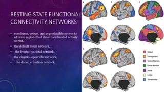
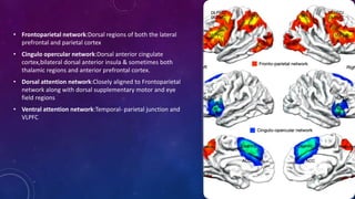
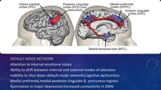
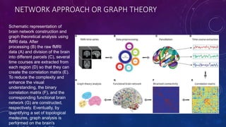
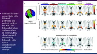
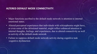
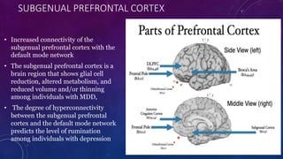
FUNCTIONAL BRAIN
CONNECTIVITY &
PSYCHOPATHOLOGY
CONNECTIVITY
• Structural connectivity-How are different areas of brain structurally connected?
• Functional connectivity-How are different areas of brain functionally connected?
• Effective connectivity-What is the influence of one brain region over another?
TASK BASED ACTIVATION STUDY
• Bilateral finger tapping task – bilateral
motor cortices simultaneous activation.
DEFENITION
Functional connectivity is defined as the “temporal correlations between spatially
remote neurophysiological events”
A main inference of functional connectivity is that, if two regions have highly
correlated neuronal activity (i.e., have high functional connectivity), then they
are more likely to engaged in a common set of processing mechanisms.
HOW TO MEASURE FUNCTIONAL CONNECTIVITY?
PECT-Glucose or oxygen metabolism
SPECT-Blood flow
BOLD Activity
BOLD
RESTING STATE FUNCTIONAL CONNECTIVITY
Result from the original Biswal et al. 1995
paper. Panel (a) shows task-related activity
in the motor cortices, while panel (b) shows
functional connectivity during a resting-
state scan, using the left motor cortex as a
seed region. Note the high degree of
overlap between the task and the resting-
state images.
Intrinsically organised networks of brain
regions that are consistently functionally
connected even in absence of task induced
pertuberations in ongoing brain activity.
RESTING STATE FUNCTIONAL
CONNECTIVITY NETWORKS
• consistent, robust, and reproducible networks
of brain regions that show coordinated activity
at rest.
• the default mode network,
• the frontal–parietal network,
• the cingulo–opercular network,
• the dorsal attention network,
• Frontoparietal network:Dorsal regions of both the lateral
prefrontal and parietal cortex
• Cingulo opercular network:Dorsal anterior cingulate
cortex,bilateral dorsal anterior insula & sometimes both
thalamic regions and anterior prefrontal cortex.
• Dorsal attention network:Closely aligned to Frontoparietal
network along with dorsal supplementary motor and eye
field regions
• Ventral attention network:Temporal- parietal junction and
VLPFC
DEFAULT MODE NETWORK
Attention to internal emotional states
Ability to shift between internal and external modes of attention
Inability to shut down default mode network:Cognitive dysfunction
Medial prefrontal,medial posterior cingulate & precuneus regions
Rumination in major depression:Increased connectivity in DMN
• Launched in
2009 as a
Blueprint Grand
Challenge, the
NIH Human
Connectome
Project (HCP),Is
a recent major
project to study
the human
structural and
functional
connectome
across multiple
scales and
modalities.The
results have
confirmed the
relationship
between
variations in
functional
connectivity &
range of
behaviors
MEASURING RESTING STATE FUNCTIONAL
CONNECTIVITY
NON INVASIVE INVASIVE
fMRI with BOLD related activity Electrocorticography
EEG
MEG
ANALYSING RESTING STATE FUNCTIONAL
CONNECTIVITY
• ROI-ROI or a Priori Network based analysis
• Seed/ROI to whole brain connectivity analysis
• Independent component analysis
ROI-ROI OR NETWORK BASED CONNECTIVITY
Specific set of
regions/specific network
already defined and
identified
Extracting time series
from these regions
Analysing connections
among those specific
regions of interest or
among the regions in
that specific network.
ROI/SEED BASED CONNECTIVITY
SEED
Connectivity
between SEED and
rest of the regions
Seed as a robust
member of network
,thus identifying
functional networks
Eg:Connectivity of amygdala or subregions of amygdala in relation to depression.
ROI/SEED BASED CONNECTIVITY
ADVANTAGES DISADVANTAGES
DIRECT TESTING OF RESEARCH QUESTION NEGLECT ADJASENT FUNCTIONAL AREAS OF BRAIN
FLEXIBLE ANATOMICAL ABNORMALITIES
STREETLAMP PROBLEM
INDEPENDENT COMPONENT ANALYSIS
• Multivariate data driven approach
• Algorithm that decomposes the BOLD data into spatially seggregarted ,statistically independent
components.
• Does not require definition of priori regions.
• Limitations:Only identifies componenets of data ,researcher should determine what each component
represents.
NETWORK APPROACH OR GRAPH THEORY
Schematic representation of
brain network construction and
graph theoretical analysis using
fMRI data. After
processing (B) the raw fMRI
data (A) and division of the brain
into different parcels (C), several
time courses are extracted from
each region (D) so that they can
create the correlation matrix (E).
To reduce the complexity and
enhance the visual
understanding, the binary
correlation matrix (F), and the
corresponding functional brain
network (G) are constructed,
respectively. Eventually, by
quantifying a set of topological
measures, graph analysis is
performed on the brain's
VARIATIONS IN GRAPH PROPERTIES
• Global efficiency:How easy it is for information to be communicated across
brain regions
• Local efficiency: How brain regions neighbours continue to communicate
following with each other following the removal of that region.
• Partcipation coefficient: Number of networks with which a particular brain
region communicates.
• Hubs:Regions with high participation coefficient.eg DLPFC
FUNCTIONAL CONNECTIVITY ABNORMALITIES IN
SCHIZOPHRENIA
ALTERED THALAMIC CONNECTIVITY
Altered Default mode connectivity
Graph theory analysis
ALTERED THALAMIC CONNECTIVITY
• Schizophrenia is considered as a disconnection syndrome .
• Andreasen et al:Disruption of cortical, subcortical &cerebellar circuits: COGNITIVE
DYSMETRIA
• Individuals with schizophrenia in this sample show reduced thalamic connectivity
with bilateral prefrontal and parietal cortices, the ACC, and bilateral striatum and
cerebellum. In contrast, they show increased connectivity bilaterally with motor,
somatosensory and visual cortices.
• reduced connectivity between the thalamus and the DLPFC predicts cognitive
impairment.
• Altered thalamic connectivity :potential biomarker for the risk of psychosis.
• Reduced thalamic
connectivity with
bilateral
prefrontal and
parietal cortices,
the ACC, and
bilateral striatum
and cerebellum.
In contrast, they
show increased
connectivity
bilaterally with
motor,
somatosensory
and visual
cortices.
ALTERED DEFAULT MODE CONNECTIVITY.
• Major functions ascribed to the default mode network is attention to internal
emotional states.
• Altered perceptual experiences that individuals with schizophrenia might have,
or even some of the delusional material, might reflect enhanced attention to
internal thoughts, feelings, and experiences, due to altered connectivity as well
as activity of the default mode network
• Failure to suppress default mode network activity during cognitive task
:cognitive dysfunction
GRAPH THEORY ANALYSES OF FUNCTIONAL BRAIN CONNECTIVITY IN
SCHIZOPHRENIA.
• Individuals with schizophrenia show reductions in both global and local efficiency, particularly
when examining specific networks
• Reduced functional connectivity in or between frontal–parietal and cingulo–opercular
networks in schizophrenia
• Reductions in both the local and global connectivity of the frontal–parietal and cingulo–
opercular networks have also been associated with impaired cognition in schizophrenia,
• Significant reductions in the connections among regions characterized as hubs, including the
insula, the superior frontal cortex, the precuneus, and the superior parietal lobule.
• Reduced gray matter volume in at least some of these hub regions, including the anterior
insula.
Summary of Schizophrenia Functional Connectivity Literature.
reduced thalamic connectivity with frontal,
cingulate, striatal, and cerebellar regions,
reduced global and local efficiency in
schizophrenia, particularly in
frontal–parietal and cingulo–
opercular networks,
reduced connectivity of hub regions
in this illness, both of which predict
cognitive function
default mode
connectivity
FUNCTIONAL CONNECTIVITY ABNORMALITIES IN MAJOR DEPRESSION
• Amygdala connectivity & Emotional regulation
• Subcortical structure that has its role in fear conditioning, threat sensitivity, and general
responding to salient events in the environment.
• Amygdala shows positive resting state functional connectivity with other regions in the medial
and anterior temporal lobes, including the hippocampus, as well with as the ventral medial
prefrontal cortex :Emotional and stress responsivity
• The amygdala also shows negative functional connectivity— anticorrelations—with bilateral
dorsal prefrontal and parietal regions, as well as the dorsal anterior cingulate , are part of the
frontal–parietal and cingulo–opercular networks, suggesting a critical role for the integrity of
these connections between amygdala and frontal–parietal and cingulo–opercular networks in
mood and effective function.
early life
stress and/or
maternal
deprivation
effective
emotion
regulation
altered
connectivity
between
amygdala and
ventral medial
prefrontal cortex
and pregenual
anterior
cingulate
emotion
dysregulation.
MDD &Thalamic
connectivity
prefrontal cortex
dorsolateral
prefrontal,
rostral
prefrontal&
ventral prefrontal
anterior cingulate
DEFAULT MODE NETWORK CONNECTIVITY AND RUMINATION.
• one of the major functions ascribed to the default mode network is to modulate attention to
internal emotional states and to govern self- referential processing
• increase in connectivity among default mode regions in major depression, especially those
forming the more anterior part of the default mode network, including the dorsomedial
prefrontal cortex and rostral anterior insula.
• work has also shown an increase in connectivity between the dorsomedial prefrontal cortex and
the rostral anterior insula, part of the salience network, among individuals with major
depression.
SUBGENUAL PREFRONTAL CORTEX
• Increased connectivity of the
subgenual prefrontal cortex with the
default mode network
• The subgenual prefrontal cortex is a
brain region that shows glial cell
reduction, altered metabolism, and
reduced volume and/or thinning
among individuals with MDD,
• The degree of hyperconnectivity
between the subgenual prefrontal
cortex and the default mode network
predicts the level of rumination
among individuals with depression

FUNCTIONAL BRAIN default mode, central executive network.pptx

  • 1.
  • 2.
    CONNECTIVITY • Structural connectivity-Howare different areas of brain structurally connected? • Functional connectivity-How are different areas of brain functionally connected? • Effective connectivity-What is the influence of one brain region over another?
  • 3.
    TASK BASED ACTIVATIONSTUDY • Bilateral finger tapping task – bilateral motor cortices simultaneous activation.
  • 4.
    DEFENITION Functional connectivity isdefined as the “temporal correlations between spatially remote neurophysiological events” A main inference of functional connectivity is that, if two regions have highly correlated neuronal activity (i.e., have high functional connectivity), then they are more likely to engaged in a common set of processing mechanisms.
  • 5.
    HOW TO MEASUREFUNCTIONAL CONNECTIVITY? PECT-Glucose or oxygen metabolism SPECT-Blood flow BOLD Activity
  • 6.
  • 7.
    RESTING STATE FUNCTIONALCONNECTIVITY Result from the original Biswal et al. 1995 paper. Panel (a) shows task-related activity in the motor cortices, while panel (b) shows functional connectivity during a resting- state scan, using the left motor cortex as a seed region. Note the high degree of overlap between the task and the resting- state images. Intrinsically organised networks of brain regions that are consistently functionally connected even in absence of task induced pertuberations in ongoing brain activity.
  • 8.
    RESTING STATE FUNCTIONAL CONNECTIVITYNETWORKS • consistent, robust, and reproducible networks of brain regions that show coordinated activity at rest. • the default mode network, • the frontal–parietal network, • the cingulo–opercular network, • the dorsal attention network,
  • 9.
    • Frontoparietal network:Dorsalregions of both the lateral prefrontal and parietal cortex • Cingulo opercular network:Dorsal anterior cingulate cortex,bilateral dorsal anterior insula & sometimes both thalamic regions and anterior prefrontal cortex. • Dorsal attention network:Closely aligned to Frontoparietal network along with dorsal supplementary motor and eye field regions • Ventral attention network:Temporal- parietal junction and VLPFC
  • 10.
    DEFAULT MODE NETWORK Attentionto internal emotional states Ability to shift between internal and external modes of attention Inability to shut down default mode network:Cognitive dysfunction Medial prefrontal,medial posterior cingulate & precuneus regions Rumination in major depression:Increased connectivity in DMN
  • 11.
    • Launched in 2009as a Blueprint Grand Challenge, the NIH Human Connectome Project (HCP),Is a recent major project to study the human structural and functional connectome across multiple scales and modalities.The results have confirmed the relationship between variations in functional connectivity & range of behaviors
  • 12.
    MEASURING RESTING STATEFUNCTIONAL CONNECTIVITY NON INVASIVE INVASIVE fMRI with BOLD related activity Electrocorticography EEG MEG
  • 13.
    ANALYSING RESTING STATEFUNCTIONAL CONNECTIVITY • ROI-ROI or a Priori Network based analysis • Seed/ROI to whole brain connectivity analysis • Independent component analysis
  • 14.
    ROI-ROI OR NETWORKBASED CONNECTIVITY Specific set of regions/specific network already defined and identified Extracting time series from these regions Analysing connections among those specific regions of interest or among the regions in that specific network.
  • 15.
    ROI/SEED BASED CONNECTIVITY SEED Connectivity betweenSEED and rest of the regions Seed as a robust member of network ,thus identifying functional networks Eg:Connectivity of amygdala or subregions of amygdala in relation to depression.
  • 16.
    ROI/SEED BASED CONNECTIVITY ADVANTAGESDISADVANTAGES DIRECT TESTING OF RESEARCH QUESTION NEGLECT ADJASENT FUNCTIONAL AREAS OF BRAIN FLEXIBLE ANATOMICAL ABNORMALITIES STREETLAMP PROBLEM
  • 17.
    INDEPENDENT COMPONENT ANALYSIS •Multivariate data driven approach • Algorithm that decomposes the BOLD data into spatially seggregarted ,statistically independent components. • Does not require definition of priori regions. • Limitations:Only identifies componenets of data ,researcher should determine what each component represents.
  • 18.
    NETWORK APPROACH ORGRAPH THEORY Schematic representation of brain network construction and graph theoretical analysis using fMRI data. After processing (B) the raw fMRI data (A) and division of the brain into different parcels (C), several time courses are extracted from each region (D) so that they can create the correlation matrix (E). To reduce the complexity and enhance the visual understanding, the binary correlation matrix (F), and the corresponding functional brain network (G) are constructed, respectively. Eventually, by quantifying a set of topological measures, graph analysis is performed on the brain's
  • 19.
    VARIATIONS IN GRAPHPROPERTIES • Global efficiency:How easy it is for information to be communicated across brain regions • Local efficiency: How brain regions neighbours continue to communicate following with each other following the removal of that region. • Partcipation coefficient: Number of networks with which a particular brain region communicates. • Hubs:Regions with high participation coefficient.eg DLPFC
  • 20.
    FUNCTIONAL CONNECTIVITY ABNORMALITIESIN SCHIZOPHRENIA ALTERED THALAMIC CONNECTIVITY Altered Default mode connectivity Graph theory analysis
  • 21.
    ALTERED THALAMIC CONNECTIVITY •Schizophrenia is considered as a disconnection syndrome . • Andreasen et al:Disruption of cortical, subcortical &cerebellar circuits: COGNITIVE DYSMETRIA • Individuals with schizophrenia in this sample show reduced thalamic connectivity with bilateral prefrontal and parietal cortices, the ACC, and bilateral striatum and cerebellum. In contrast, they show increased connectivity bilaterally with motor, somatosensory and visual cortices. • reduced connectivity between the thalamus and the DLPFC predicts cognitive impairment. • Altered thalamic connectivity :potential biomarker for the risk of psychosis.
  • 22.
    • Reduced thalamic connectivitywith bilateral prefrontal and parietal cortices, the ACC, and bilateral striatum and cerebellum. In contrast, they show increased connectivity bilaterally with motor, somatosensory and visual cortices.
  • 23.
    ALTERED DEFAULT MODECONNECTIVITY. • Major functions ascribed to the default mode network is attention to internal emotional states. • Altered perceptual experiences that individuals with schizophrenia might have, or even some of the delusional material, might reflect enhanced attention to internal thoughts, feelings, and experiences, due to altered connectivity as well as activity of the default mode network • Failure to suppress default mode network activity during cognitive task :cognitive dysfunction
  • 24.
    GRAPH THEORY ANALYSESOF FUNCTIONAL BRAIN CONNECTIVITY IN SCHIZOPHRENIA. • Individuals with schizophrenia show reductions in both global and local efficiency, particularly when examining specific networks • Reduced functional connectivity in or between frontal–parietal and cingulo–opercular networks in schizophrenia • Reductions in both the local and global connectivity of the frontal–parietal and cingulo– opercular networks have also been associated with impaired cognition in schizophrenia, • Significant reductions in the connections among regions characterized as hubs, including the insula, the superior frontal cortex, the precuneus, and the superior parietal lobule. • Reduced gray matter volume in at least some of these hub regions, including the anterior insula.
  • 25.
    Summary of SchizophreniaFunctional Connectivity Literature. reduced thalamic connectivity with frontal, cingulate, striatal, and cerebellar regions, reduced global and local efficiency in schizophrenia, particularly in frontal–parietal and cingulo– opercular networks, reduced connectivity of hub regions in this illness, both of which predict cognitive function default mode connectivity
  • 26.
    FUNCTIONAL CONNECTIVITY ABNORMALITIESIN MAJOR DEPRESSION • Amygdala connectivity & Emotional regulation • Subcortical structure that has its role in fear conditioning, threat sensitivity, and general responding to salient events in the environment. • Amygdala shows positive resting state functional connectivity with other regions in the medial and anterior temporal lobes, including the hippocampus, as well with as the ventral medial prefrontal cortex :Emotional and stress responsivity • The amygdala also shows negative functional connectivity— anticorrelations—with bilateral dorsal prefrontal and parietal regions, as well as the dorsal anterior cingulate , are part of the frontal–parietal and cingulo–opercular networks, suggesting a critical role for the integrity of these connections between amygdala and frontal–parietal and cingulo–opercular networks in mood and effective function.
  • 27.
    early life stress and/or maternal deprivation effective emotion regulation altered connectivity between amygdalaand ventral medial prefrontal cortex and pregenual anterior cingulate emotion dysregulation.
  • 28.
  • 29.
    DEFAULT MODE NETWORKCONNECTIVITY AND RUMINATION. • one of the major functions ascribed to the default mode network is to modulate attention to internal emotional states and to govern self- referential processing • increase in connectivity among default mode regions in major depression, especially those forming the more anterior part of the default mode network, including the dorsomedial prefrontal cortex and rostral anterior insula. • work has also shown an increase in connectivity between the dorsomedial prefrontal cortex and the rostral anterior insula, part of the salience network, among individuals with major depression.
  • 30.
    SUBGENUAL PREFRONTAL CORTEX •Increased connectivity of the subgenual prefrontal cortex with the default mode network • The subgenual prefrontal cortex is a brain region that shows glial cell reduction, altered metabolism, and reduced volume and/or thinning among individuals with MDD, • The degree of hyperconnectivity between the subgenual prefrontal cortex and the default mode network predicts the level of rumination among individuals with depression