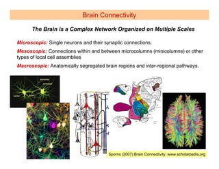
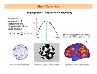
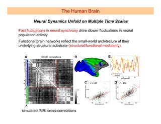
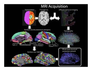
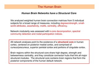
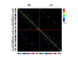
Kavli Institute 2008


 Brain Networks for Efficient Computation
                         Olaf Sporns
       Department of Psychological and Brain Sciences
           Indiana University, Bloomington, IN 47405
    http://www.indiana.edu/~cortex , osporns@indiana.edu


Outline
Brain Connectivity
       Network Science Approaches
Brain Dynamics
       Structure, Function, Information, Complexity
The Human Brain
     Building a Map of the Human Brain
Outline


Brain Connectivity

   Brain Dynamics

   The Human Brain
Brain Connectivity

      The Brain is a Complex Network Organized on Multiple Scales

Microscopic: Single neurons and their synaptic connections.
Mesoscopic: Connections within and between microcolumns (minicolumns) or other
types of local cell assemblies
Macroscopic: Anatomically segregated brain regions and inter-regional pathways.


                                        Sporns (2007) Brain Connectivity. www.scholarpedia.org
Brain Connectivity

    Structure and Function of the Brain are Intricately Linked

Anatomical (Structural) Connectivity: Pattern of structural connections
between neurons, neuronal populations, or brain regions.
Functional Connectivity: Pattern of statistical dependencies (e.g. temporal
correlations) between distinct (often remote) neuronal elements.
Effective Connectivity: Network of causal effects, combination of functional
connectivity and structural model.
Brain Connectivity
                       Brain Networks Form a Small World

In highly evolved brains, structural brain
connectivity forms a small-world (high clustering,
short path length, low wiring cost, modules,
hubs)                               Sporns and Zwi (2004)

Highly clustered connection patterns at the large-
scale reflect functional relations between sets of
brain regions. These functional relations may be
a result of clustered connectivity.


                                   Hilgetag et al., 2000


                                        Kaiser and Hilgetag, 2006

                                            Short path lengths indicate that all cortical areas
                                            can be linked in very few processing steps.
Outline


Brain Connectivity

Brain Dynamics

 The Human Brain
Brain Dynamics

The Brain is Organized to Efficiently Extract and Coordinate Information

  Two major organizational principles of cortex:

      Segregation (anatomical/functional)                clustering

      Integration (anatomical/functional)                path length

  These principles are complementary and interdependent.


  Two major challenges for information processing in the brain:

      Rapid extraction of information (elimination of redundant dimensions,
      efficient coding, maximum information transfer)

      Coordination of distributed resources to create coherent states

  Both challenges must be solved simultaneously, within a common neural
  architecture.
Brain Dynamics

                            Segregation + Integration = Complexity

complexity:
coexistence of
                                    complexity
segregation and
integration (local and
global structure)
C ( X ) = H ( X ) − ∑i H ( xi X − xi ).
                                                                                   Movie courtesy of Vincent, Raichle,
                                                                                   Snyder et al (Washington University)


   small-world structural network         spontaneous activity in a neural model    spontaneous activity in a human brain
Outline


 Brain Connectivity

  Brain Dynamics

The Human Brain
The Human Brain

                 The Brain is Always Active – Even “at Rest”

Slow fluctuations in fMRI signal at rest may reflect neuronal baseline activity.
Patterns of resting state BOLD signal change are consistent across subjects.
Spontaneous fluctuations reveal the existence of two distributed and anti-
correlated resting state networks.
                                                                 Damoiseaux et al., PNAS (2006)


                                         Fox et al.,
                                         PNAS (2005)


fMRI resting state functional networks of wavelet coefficients show small-world
attributes. Small-world networks (in wavelet space) may be fractal across
multiple frequency ranges.
                                 Achard et al., J Neurosci. (2006), Bassett et al., PNAS (2006)
The Human Brain

Connectivity + Dynamics = Endogenous Brain Activity

                     Connection matrix of macaque cortex
                                       +
                 Dynamic equations describing the physiology of a
                               neural population


                                                 =
                                   Spontaneous (endogenous)
                                        neural dynamics
                                    (chaoticity, metastability)


                                      Honey, Breakspear, Kötter,
                                      Sporns (2007) PNAS
The Human Brain

         Neural Dynamics Unfold on Multiple Time Scales

Fast fluctuations in neural synchrony drive slower fluctuations in neural
population activity.
Functional brain networks reflect the small-world architecture of their
underlying structural substrate (structural/functional modularity).


  simulated fMRI cross-correlations
The Human Brain

Functional Brain Networks form a Variable Repertoire


                                 static pattern (anatomy)


                                variable pattern (functional
                                relations)
The Human Brain

  The Connectome is Necessary for Understanding Brain Function


The human connectome represents a comprehensive structural description of the
network of elements and connections forming the human brain.


Proposed initial focus: thalamocortical system
Possible scales of the human connectome:
    Microscale (neurons, synapses)
    Macroscale (parcellated brain regions, voxels)
    Mesoscale (columns, minicolumns)

Most feasible approach: macroscale (first draft), followed by “filling-in” at the
mesoscale.


 Sporns, O., Tononi, G., and Kötter, R. (2005) The human connectome: A structural description of the
 human brain. PLoS Comp. Biol.
The Human Brain

Fiber Pathways of the Cerebral Cortex can be Mapped with MRI

Diffusion Spectrum Imaging (DSI) and Computational Tractography


     Hagmann, Cammoun, Gigandet, Meuli, Honey, Wedeen, Sporns (2008) PLoS Biology
A        B


C


             RH
    LH
The Human Brain

           Human Brain Networks have a Structural Core

We analyzed weighted human brain connection matrices from 5 individual
subjects for a broad range of measures, including degrees/strength, small-
world attributes, assortativity, motifs, centrality, efficiency.

Network modularity was assessed with k-core decomposition, spectral
community detection and nodal participation indices.


All network analyses point to the existence of a structural core in human
cortex, centered on posterior medial cortex, and comprised of
cuneus/precuneus, superior parietal cortex and portions of cingulate cortex.

Brain regions within the structural core share high degree, strength and
betweenness centrality, and they constitute connector hubs that link all major
structural modules. The structural core contains brain regions that form the
posterior components of the human default network.
The Human Brain
A


    scan 1               scan 2
             subject A            subject B   subject C   subject D   subject E

B
                    subject A-E
                                                C
The Human Brain
     Human Brain Networks Have Numerous Hubs

connector hub distribution      centrality distribution
The Human Brain
Human Brain Networks Show Individual Variations
The Human Brain

    Structural and Functional Connections are Highly Correlated

A


B                                            C
    all subjects, PCUN + PC                      all subjects, all areas


                              r2 = 0.53                                    r2 = 0.62
                                             C
RH   LH
RH   LH
The Human Brain

     Computational Models Capture Large-Scale Human Brain Activity

                               Structural connections of the human brain shape
                               functional activations and dynamic states.

                                                                                         r = 0.85
                                                                  rPC        r = 0.76               r = 0.87


                               Honey et al. (PNAS, in revision)


                rsFC                  rsFC
SC              (empirical)           (nonlinear model)


                                                                                        empirical        nonlinear model
                                                                        SC
                                                                                                    rsFC
Summary

The Brain is a Complex Network Organized on Multiple Scales
   Structure-function relationship, plasticity, turnover, redundancy


Brain Networks Form a Small World
   Allows the brain to efficiently process information, promotes complexity


The Brain is Always Active – Even “at Rest”
   Endogenous processes vs. exogenous perturbations, multiple time scales


Human Brain Networks have a Structural Core and Hubs
   Core located in medial parietal cortex – a region central to self and consciousness
   Hubs may serve as integrators of cortico-cortical signal traffic
   Individual variations – clinical disturbances


Computational Models Capture Large-Scale Human Brain Activity
   Possibility of a global brain simulator
   Models as tools for exploring mechanistic substrates of human cognition


                   Funded by the JS McDonnell Foundation
Summary

The Brain is a Complex Network Organized on Multiple Scales
   Cells to systems
   Scalable architecture – common principles?


Structure and Function of the Brain are Intricately Linked
   Structure shapes function shapes structure …
   Reorganization and plasticity


Brain Networks Form a Small World
   High clustering, short path length
   Reflects volume and processing constraints

The Brain is Organized to Efficiently Extract and Coordinate Information
   A dual challenge addressed in a common architecture
   Small-world attributes map onto information processing requirements


Segregation + Integration = Complexity
   Complexity is a mixture of randomness and regularity
   Complexity emerges from structural small-world networks
Summary


The Brain is Always Active – Even “at Rest”
   Endogenous processes vs. exogenous perturbations


Connectivity + Dynamics = Endogenous Brain Activity
   Coupled dynamic models
   Metastability, itinerancy


Neural Dynamics Unfold on Multiple Time Scales
   Milliseconds to seconds
   Fractal (self-similar) functional connectivity
   Long-term averages more stable than short-term averages


Functional Brain Networks form a Variable Repertoire
   Cognitive microstates?
   Robustness versus flexibility
Summary
Fiber Pathways of the Cerebral Cortex can be Mapped with MRI
   Noninvasive methodology
   Rapid technological development
   Increasingly refined maps

Human Brain Networks have a Structural Core and Hubs
   Core located in medial parietal cortex – a region central to self and consciousness
   Hubs may serve as integrators of cortico-cortical signal traffic

Human Brain Networks Show Individual Variations
   Relation to cognitive/behavioral variation unknown
   Network disturbances can help to diagnose brain disease

Structural and Functional Connections are Highly Correlated
   Topological principles shared between anatomical and functional
   networks
   Endogenous brain activity – an expression of structural linkages

Computational Models Capture Large-Scale Human Brain Activity
   Possibility of a global brain simulator
   Models as tools for exploring mechanistic substrates of human cognition


                  Funded by the JS McDonnell Foundation
The Human Brain

1) High consistency of DSI tractography between hemispheres.
2) High consistency of DSI tractography in repeat scans.
                                  r2 = 0.78
                     scan 1                     scan 2


        RH

 r2 = 0.94


         LH


3) Connection patterns are robust to degradation (simulation scanning and
   tractography noise).
4) Comparison between macaque DSI tractography and connection patterns
   derived by anatomical tract tracing shows significant overlap.
5) Comparison between structural and functional connections in human
   brain shows significant correlation.
Macaque Brain Imaging

DSI acquisition from a single fixed m. fascicularis cortical hemisphere
Macaque Brain Imaging

A                                     Comparison of DSI
                                      tractography data with
                                      classical tract tracing
                                      neuroanatomical data


                                       B                                                  B
DSI                   Cocomac
fiber                 data
density               (symmetrized)


                                                 known present


                                                                 unknown


                                                                           known absent

Sporns kavli2008

  • 1.
    Kavli Institute 2008 Brain Networks for Efficient Computation Olaf Sporns Department of Psychological and Brain Sciences Indiana University, Bloomington, IN 47405 http://www.indiana.edu/~cortex , osporns@indiana.edu Outline Brain Connectivity Network Science Approaches Brain Dynamics Structure, Function, Information, Complexity The Human Brain Building a Map of the Human Brain
  • 2.
    Outline Brain Connectivity Brain Dynamics The Human Brain
  • 3.
    Brain Connectivity The Brain is a Complex Network Organized on Multiple Scales Microscopic: Single neurons and their synaptic connections. Mesoscopic: Connections within and between microcolumns (minicolumns) or other types of local cell assemblies Macroscopic: Anatomically segregated brain regions and inter-regional pathways. Sporns (2007) Brain Connectivity. www.scholarpedia.org
  • 4.
    Brain Connectivity Structure and Function of the Brain are Intricately Linked Anatomical (Structural) Connectivity: Pattern of structural connections between neurons, neuronal populations, or brain regions. Functional Connectivity: Pattern of statistical dependencies (e.g. temporal correlations) between distinct (often remote) neuronal elements. Effective Connectivity: Network of causal effects, combination of functional connectivity and structural model.
  • 5.
    Brain Connectivity Brain Networks Form a Small World In highly evolved brains, structural brain connectivity forms a small-world (high clustering, short path length, low wiring cost, modules, hubs) Sporns and Zwi (2004) Highly clustered connection patterns at the large- scale reflect functional relations between sets of brain regions. These functional relations may be a result of clustered connectivity. Hilgetag et al., 2000 Kaiser and Hilgetag, 2006 Short path lengths indicate that all cortical areas can be linked in very few processing steps.
  • 6.
  • 7.
    Brain Dynamics The Brainis Organized to Efficiently Extract and Coordinate Information Two major organizational principles of cortex: Segregation (anatomical/functional) clustering Integration (anatomical/functional) path length These principles are complementary and interdependent. Two major challenges for information processing in the brain: Rapid extraction of information (elimination of redundant dimensions, efficient coding, maximum information transfer) Coordination of distributed resources to create coherent states Both challenges must be solved simultaneously, within a common neural architecture.
  • 8.
    Brain Dynamics Segregation + Integration = Complexity complexity: coexistence of complexity segregation and integration (local and global structure) C ( X ) = H ( X ) − ∑i H ( xi X − xi ). Movie courtesy of Vincent, Raichle, Snyder et al (Washington University) small-world structural network spontaneous activity in a neural model spontaneous activity in a human brain
  • 9.
    Outline Brain Connectivity Brain Dynamics The Human Brain
  • 10.
    The Human Brain The Brain is Always Active – Even “at Rest” Slow fluctuations in fMRI signal at rest may reflect neuronal baseline activity. Patterns of resting state BOLD signal change are consistent across subjects. Spontaneous fluctuations reveal the existence of two distributed and anti- correlated resting state networks. Damoiseaux et al., PNAS (2006) Fox et al., PNAS (2005) fMRI resting state functional networks of wavelet coefficients show small-world attributes. Small-world networks (in wavelet space) may be fractal across multiple frequency ranges. Achard et al., J Neurosci. (2006), Bassett et al., PNAS (2006)
  • 11.
    The Human Brain Connectivity+ Dynamics = Endogenous Brain Activity Connection matrix of macaque cortex + Dynamic equations describing the physiology of a neural population = Spontaneous (endogenous) neural dynamics (chaoticity, metastability) Honey, Breakspear, Kötter, Sporns (2007) PNAS
  • 12.
    The Human Brain Neural Dynamics Unfold on Multiple Time Scales Fast fluctuations in neural synchrony drive slower fluctuations in neural population activity. Functional brain networks reflect the small-world architecture of their underlying structural substrate (structural/functional modularity). simulated fMRI cross-correlations
  • 13.
    The Human Brain FunctionalBrain Networks form a Variable Repertoire static pattern (anatomy) variable pattern (functional relations)
  • 14.
    The Human Brain The Connectome is Necessary for Understanding Brain Function The human connectome represents a comprehensive structural description of the network of elements and connections forming the human brain. Proposed initial focus: thalamocortical system Possible scales of the human connectome: Microscale (neurons, synapses) Macroscale (parcellated brain regions, voxels) Mesoscale (columns, minicolumns) Most feasible approach: macroscale (first draft), followed by “filling-in” at the mesoscale. Sporns, O., Tononi, G., and Kötter, R. (2005) The human connectome: A structural description of the human brain. PLoS Comp. Biol.
  • 15.
    The Human Brain FiberPathways of the Cerebral Cortex can be Mapped with MRI Diffusion Spectrum Imaging (DSI) and Computational Tractography Hagmann, Cammoun, Gigandet, Meuli, Honey, Wedeen, Sporns (2008) PLoS Biology
  • 17.
    A B C RH LH
  • 18.
    The Human Brain Human Brain Networks have a Structural Core We analyzed weighted human brain connection matrices from 5 individual subjects for a broad range of measures, including degrees/strength, small- world attributes, assortativity, motifs, centrality, efficiency. Network modularity was assessed with k-core decomposition, spectral community detection and nodal participation indices. All network analyses point to the existence of a structural core in human cortex, centered on posterior medial cortex, and comprised of cuneus/precuneus, superior parietal cortex and portions of cingulate cortex. Brain regions within the structural core share high degree, strength and betweenness centrality, and they constitute connector hubs that link all major structural modules. The structural core contains brain regions that form the posterior components of the human default network.
  • 19.
    The Human Brain A scan 1 scan 2 subject A subject B subject C subject D subject E B subject A-E C
  • 20.
    The Human Brain Human Brain Networks Have Numerous Hubs connector hub distribution centrality distribution
  • 21.
    The Human Brain HumanBrain Networks Show Individual Variations
  • 22.
    The Human Brain Structural and Functional Connections are Highly Correlated A B C all subjects, PCUN + PC all subjects, all areas r2 = 0.53 r2 = 0.62 C
  • 23.
    RH LH
  • 24.
    RH LH
  • 25.
    The Human Brain Computational Models Capture Large-Scale Human Brain Activity Structural connections of the human brain shape functional activations and dynamic states. r = 0.85 rPC r = 0.76 r = 0.87 Honey et al. (PNAS, in revision) rsFC rsFC SC (empirical) (nonlinear model) empirical nonlinear model SC rsFC
  • 26.
    Summary The Brain isa Complex Network Organized on Multiple Scales Structure-function relationship, plasticity, turnover, redundancy Brain Networks Form a Small World Allows the brain to efficiently process information, promotes complexity The Brain is Always Active – Even “at Rest” Endogenous processes vs. exogenous perturbations, multiple time scales Human Brain Networks have a Structural Core and Hubs Core located in medial parietal cortex – a region central to self and consciousness Hubs may serve as integrators of cortico-cortical signal traffic Individual variations – clinical disturbances Computational Models Capture Large-Scale Human Brain Activity Possibility of a global brain simulator Models as tools for exploring mechanistic substrates of human cognition Funded by the JS McDonnell Foundation
  • 27.
    Summary The Brain isa Complex Network Organized on Multiple Scales Cells to systems Scalable architecture – common principles? Structure and Function of the Brain are Intricately Linked Structure shapes function shapes structure … Reorganization and plasticity Brain Networks Form a Small World High clustering, short path length Reflects volume and processing constraints The Brain is Organized to Efficiently Extract and Coordinate Information A dual challenge addressed in a common architecture Small-world attributes map onto information processing requirements Segregation + Integration = Complexity Complexity is a mixture of randomness and regularity Complexity emerges from structural small-world networks
  • 28.
    Summary The Brain isAlways Active – Even “at Rest” Endogenous processes vs. exogenous perturbations Connectivity + Dynamics = Endogenous Brain Activity Coupled dynamic models Metastability, itinerancy Neural Dynamics Unfold on Multiple Time Scales Milliseconds to seconds Fractal (self-similar) functional connectivity Long-term averages more stable than short-term averages Functional Brain Networks form a Variable Repertoire Cognitive microstates? Robustness versus flexibility
  • 29.
    Summary Fiber Pathways ofthe Cerebral Cortex can be Mapped with MRI Noninvasive methodology Rapid technological development Increasingly refined maps Human Brain Networks have a Structural Core and Hubs Core located in medial parietal cortex – a region central to self and consciousness Hubs may serve as integrators of cortico-cortical signal traffic Human Brain Networks Show Individual Variations Relation to cognitive/behavioral variation unknown Network disturbances can help to diagnose brain disease Structural and Functional Connections are Highly Correlated Topological principles shared between anatomical and functional networks Endogenous brain activity – an expression of structural linkages Computational Models Capture Large-Scale Human Brain Activity Possibility of a global brain simulator Models as tools for exploring mechanistic substrates of human cognition Funded by the JS McDonnell Foundation
  • 30.
    The Human Brain 1)High consistency of DSI tractography between hemispheres. 2) High consistency of DSI tractography in repeat scans. r2 = 0.78 scan 1 scan 2 RH r2 = 0.94 LH 3) Connection patterns are robust to degradation (simulation scanning and tractography noise). 4) Comparison between macaque DSI tractography and connection patterns derived by anatomical tract tracing shows significant overlap. 5) Comparison between structural and functional connections in human brain shows significant correlation.
  • 31.
    Macaque Brain Imaging DSIacquisition from a single fixed m. fascicularis cortical hemisphere
  • 32.
    Macaque Brain Imaging A Comparison of DSI tractography data with classical tract tracing neuroanatomical data B B DSI Cocomac fiber data density (symmetrized) known present unknown known absent