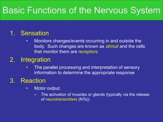
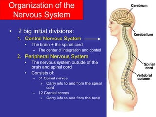
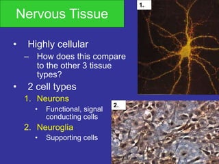
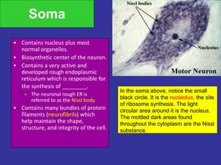
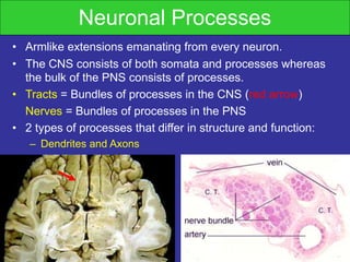
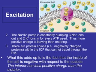
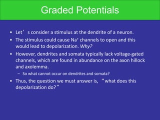
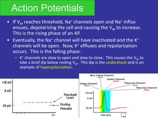
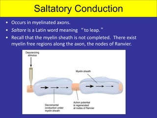
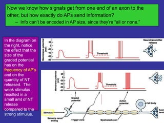
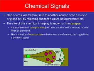
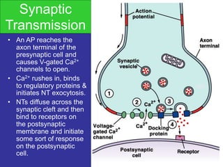
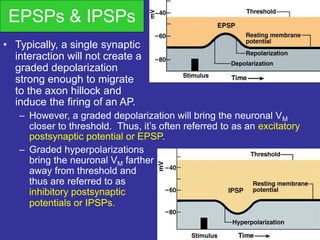
SlideShare a Scribd company logo
1 of 53
The Nervous System
• A network of billions of nerve cells linked
together in a highly organized fashion to
form the rapid control center of the body.
• Functions include:
– Integrating center for homeostasis,
movement, and almost all other body
functions.
– The mysterious source of those traits that we
think of as setting humans apart from animals
Basic Functions of the Nervous System
1. Sensation
• Monitors changes/events occurring in and outside the
body. Such changes are known as stimuli and the cells
that monitor them are receptors.
2. Integration
• The parallel processing and interpretation of sensory
information to determine the appropriate response
3. Reaction
• Motor output.
– The activation of muscles or glands (typically via the release
of neurotransmitters (NTs))
Nervous vs. Endocrine System
• Similarities:
– They both monitor stimuli and react so as to
maintain homeostasis.
• Differences:
– The NS is a rapid, fast-acting system whose
effects do not always persevere.
– The ES acts slower (via blood-borne chemical
signals called H _ _ _ _ _ _ _) and its actions
are usually much longer lasting.
Organization of the
Nervous System
• 2 big initial divisions:
1. Central Nervous System
• The brain + the spinal cord
– The center of integration and control
2. Peripheral Nervous System
• The nervous system outside of the
brain and spinal cord
• Consists of:
– 31 Spinal nerves
» Carry info to and from the spinal
cord
– 12 Cranial nerves
» Carry info to and from the brain
Peripheral Nervous System
• Responsible for communication btwn the CNS
and the rest of the body.
• Can be divided into:
– Sensory Division
• Afferent division
– Conducts impulses from receptors to the CNS
– Informs the CNS of the state of the body interior and exterior
– Sensory nerve fibers can be somatic (from skin, skeletal
muscles or joints) or visceral (from organs w/i the ventral body
cavity)
– Motor Division
• Efferent division
– Conducts impulses from CNS to effectors (muscles/glands)
– Motor nerve fibers
Motor Efferent Division
• Can be divided further:
– Somatic nervous system
• VOLUNTARY (generally)
• Somatic nerve fibers that conduct impulses from
the CNS to skeletal muscles
– Autonomic nervous system
• INVOLUNTARY (generally)
• Conducts impulses from the CNS to smooth
muscle, cardiac muscle, and glands.
Autonomic Nervous System
• Can be divided into:
– Sympathetic Nervous
System
• “Fight or Flight”
– Parasympathetic
Nervous System
• “Rest and Digest”
These 2 systems are antagonistic.
Typically, we balance these 2 to keep ourselves in a
state of dynamic balance.
We’ll go further into the difference btwn these 2
later!
Nervous Tissue
• Highly cellular
– How does this compare
to the other 3 tissue
types?
• 2 cell types
1. Neurons
• Functional, signal
conducting cells
2. Neuroglia
• Supporting cells
1.
2.
Neuroglia
• Outnumber neurons by about
10 to 1 (the guy on the right had an
inordinate amount of them).
• 6 types of supporting cells
– 4 are found in the CNS:
1. Astrocytes
• Star-shaped, abundant, and
versatile
• Guide the migration of developing
neurons
• Act as K+ and NT buffers
• Involved in the formation of the
blood brain barrier
• Function in nutrient transfer
Neuroglia
2. Microglia
• Specialized immune cells that act
as the macrophages of the CNS
• Why is it important for the CNS to
have its own army of immune
cells?
3. Ependymal Cells
• Low columnar epithelial-esque
cells that line the ventricles of the
brain and the central canal of the
spinal cord
• Some are ciliated which
facilitates the movement of
cerebrospinal fluid
Neuroglia
4. Oligodendrocytes
• Produce the
myelin
sheath
which
provides the
electrical
insulation for
certain
neurons in
the CNS
• 2 types of glia in the
PNS
1. Satellite cells
• Surround clusters of
neuronal cell bodies in the
PNS
• Unknown function
2. Schwann cells
• Form myelin sheaths
around the larger nerve
fibers in the PNS.
• Vital to neuronal
regeneration
Neuroglia
Neurons• The functional and structural unit
of the nervous system
• Specialized to conduct information from one part of the
body to another
• There are many, many different types of neurons but most
have certain structural and functional characteristics in
common:
- Cell body (soma)
- One or more
specialized, slender
processes
(axons/dendrites)
- An input region
(dendrites/soma)
- A conducting
component (axon)
- A secretory (output)
region (axon terminal)
Soma
• Contains nucleus plus most
normal organelles.
• Biosynthetic center of the neuron.
• Contains a very active and
developed rough endoplasmic
reticulum which is responsible for
the synthesis of ________.
– The neuronal rough ER is
referred to as the Nissl body.
• Contains many bundles of protein
filaments (neurofibrils) which
help maintain the shape,
structure, and integrity of the cell.
In the soma above, notice the small
black circle. It is the nucleolus, the site
of ribosome synthesis. The light
circular area around it is the nucleus.
The mottled dark areas found
throughout the cytoplasm are the Nissl
substance.
Somata
• Contain multiple
mitochondria. Why?
• Acts as a receptive service for interaction
with other neurons.
• Most somata are found in the bony
environs of the CNS. Why?
• Clusters of somata in the CNS are known
as nuclei. Clusters of somata in the PNS
are known as ganglia.
Neuronal Processes
• Armlike extensions emanating from every neuron.
• The CNS consists of both somata and processes whereas
the bulk of the PNS consists of processes.
• Tracts = Bundles of processes in the CNS (red arrow)
Nerves = Bundles of processes in the PNS
• 2 types of processes that differ in structure and function:
– Dendrites and Axons
• Dendrites are thin, branched processes whose main
function is to receive incoming signals.
• They effectively increase the surface area of a neuron to
increase its ability to communicate with other neurons.
• Small, mushroom-shaped dendritic spines further increase
the SA
• Convey info towards the soma thru the use of graded
potentials – which are somewhat similar to action potentials.
Notice the multiple
processes extending
from the neuron on the
right. Also notice the
multiple dark circular
dots in the slide. They’re
not neurons, so they
must be…
• Most neurons have a single
axon – a long (up to 1m)
process designed to convey
info away from the cell body.
• Originates from a special
region of the cell body called
the axon hillock.
• Transmit APs from the soma
toward the end of the axon
where they cause NT release.
• Often branch sparsely, forming
collaterals.
• Each collateral may split into
telodendria which end in a
synaptic knob, which contains
synaptic vesicles –
membranous bags of NTs.
Axons
• Axolemma = axon plasma
membrane.
• Surrounded by a myelin sheath, a
wrapping of lipid which:
– Protects the axon and electrically isolates it
– Increases the rate of AP transmission
• The myelin sheath is made by ________ in the CNS and by
_________ in the PNS.
• This wrapping is never complete. Interspersed along the axon are
gaps where there is no myelin – these are nodes of Ranvier.
• In the PNS, the exterior of the Schwann cell surrounding an axon
is the neurilemma
Myelination in the CNS
Myelination in the PNS
• A bundle of processes in the PNS is a nerve.
• Within a nerve, each axon is surrounded by an
endoneurium (too small to see on the photomicrograph) –
a layer of loose CT.
• Groups of fibers
are bound
together into
bundles
(fascicles) by a
perineurium (red
arrow).
• All the fascicles
of a nerve are
enclosed by a
epineurium
(black arrow).
Communication
• Begins with the stimulation of a neuron.
– One neuron may be stimulated by another, by a receptor cell, or
even by some physical event such as pressure.
• Once stimulated, a neuron will communicate information
about the causative event.
– Such neurons are sensory neurons and they provide info about
both the internal and external environments.
– Sensory neurons (a.k.a. afferent neurons) will send info to
neurons in the brain and spinal cord. There, association
neurons (a.k.a. interneurons) will integrate the information and
then perhaps send commands to motor neurons (efferent
neurons) which synapse with muscles or glands.
Communication
• Thus, neurons need to be able to
conduct information in 2 ways:
1. From one end of a neuron to the other end.
2. Across the minute space separating one
neuron from another. (What is this called?)
• The 1st is accomplished electrically via APs.
• The 2nd is accomplished chemically via
neurotransmitters.
• All cells have a voltage difference across their
plasma membrane. This is the result of several
things:
1. The ECF is very high in Na+ while the ICF is very high in K+.
The PM is impermeable to Na+ but slightly permeable to K+. As
a result, K+ is constantly leaking out of the cell. In other words,
positive charge is constantly leaking out of the cell.
Excitation
2. The Na+/K+ pump is constantly pumping 3 Na+ ions
out and 2 K+ ions in for every ATP used. Thus more
positive charge is leaving than entering.
3. There are protein anions (i.e., negatively charged
proteins) within the ICF that cannot travel through the
PM.
• What this adds up to is the fact that the inside of
the cell is negative with respect to the outside.
The interior has less positive charge than the
exterior.
Excitation
• This charge separation is known as a membrane
potential (abbreviated Vm).
• Cells that exhibit a Vm are said to be polarized.
• Vm can be changed by influx or efflux of
charge.
Resting Potential
Neurons are highly polarized (w/ a VM of about
–70mV) due to:
» Differential membrane permeability to K+ and Na+
» The electrogenic nature of the Na+/K+ pump
» The presence of intracellular impermeable anions
• Changes in VM allow for the generation of
action potentials and thus informative
intercellular communication.
Graded Potentials
• Let’s consider a stimulus at the dendrite of a neuron.
• The stimulus could cause Na+ channels to open and this
would lead to depolarization. Why?
• However, dendrites and somata typically lack voltage-gated
channels, which are found in abundance on the axon hillock
and axolemma.
– So what cannot occur on dendrites and somata?
• Thus, the question we must answer is, “what does this
depolarization do?”
Graded Potentials
• The positive charge carried by the Na+ spreads as a wave of
depolarization through the cytoplasm (much like the ripples
created by a stone tossed into a pond).
• As the Na+ drifts, some of it will leak back out of the
membrane.
– What this means is that the degree of depolarization caused by the
graded potential decreases with distance from the origin.
Graded Potentials
• Their initial amplitude may be of almost any size
– it simply depends on how much Na+ originally
entered the cell.
• If the initial amplitude of the GP is sufficient, it
will spread all the way to the axon hillock where
V-gated channels reside.
• If the arriving potential change is suprathreshold,
an AP will be initiated in the axon hillock and it
will travel down the axon to the synaptic knob
where it will cause NT exocytosis. If the
potential change is subthreshold, then no AP will
ensue and nothing will happen.
Action Potentials
• If VM reaches threshold, Na+ channels open and Na+ influx
ensues, depolarizing the cell and causing the VM to increase.
This is the rising phase of an AP.
• Eventually, the Na+ channel will have inactivated and the K+
channels will be open. Now, K+ effluxes and repolarization
occurs. This is the falling phase.
– K+ channels are slow to open and slow to close. This causes the VM to
take a brief dip below resting VM. This dip is the undershoot and is an
example of hyperpolarization.
Na+ Channels
• They have 2 gates.
– At rest, one is closed (the
activation gate) and the
other is open (the
inactivation gate).
– Suprathreshold
depolarization affects
both of them.
1
2
3
4 5
Absolute Refractory Period
• During the time interval between the opening of
the Na+ channel activation gate and the opening
of the inactivation gate, a Na+ channel CANNOT
be stimulated.
– This is the ABSOLUTE REFRACTORY PERIOD.
– A Na+ channel cannot be involved in another AP until
the inactivation gate has been reset.
– This being said, can you determine why an AP is said
to be unidirectional.
• What are the advantages of such a scenario?
Relative Refractory Period
• Could an AP be generated during the undershoot?
• Yes! But it would take an initial stimulus that is much,
much stronger than usual.
– WHY?
• This situation is known as the relative refractory period.
Imagine, if you will, a toilet.
When you pull the handle, water floods the bowl. This event takes a
couple of seconds and you cannot stop it in the middle. Once the
bowl empties, the flush is complete. Now the upper tank is empty. If
you try pulling the handle at this point, nothing happens (absolute
refractory). Wait for the upper tank to begin refilling. You can now
flush again, but the intensity of the flushes increases as the upper
tank refills (relative refractory)
TIME
VM
In this figure, what do the red
and blue box represent?
Some Action Potential Questions
• What does it mean when we say an AP is
“all or none?”
– Can you ever have ½ an AP?
• How does the concept of threshold relate
to the “all or none” notion?
• Will one AP ever be bigger than another?
– Why or why not?
Action Potential Conduction
• If an AP is generated at the axon hillock, it will
travel all the way down to the synaptic knob.
• The manner in which it travels depends on
whether the neuron is myelinated or
unmyelinated.
• Unmyelinated neurons undergo the continuous
conduction of an AP whereas myelinated
neurons undergo saltatory conduction of an AP.
Continuous Conduction
• Occurs in unmyelinated axons.
• In this situation, the wave of de- and repolarization
simply travels from one patch of membrane to the next
adjacent
patch.
• APs moved
in this fashion
along the
sarcolemma
of a muscle
fiber as well.
• Analogous to
dominoes
falling.
Saltatory Conduction
• Occurs in myelinated axons.
• Saltare is a Latin word meaning “to leap.”
• Recall that the myelin sheath is not completed. There exist
myelin free regions along the axon, the nodes of Ranvier.
Rates of AP Conduction
1. Which do you think has a faster rate of AP
conduction – myelinated or unmyelinated axons?
2. Which do you think would conduct an AP faster –
an axon with a large diameter or an axon with a
small diameter?
The answer to #1 is a myelinated axon. If you can’t see why, then answer this
question: could you move 100ft faster if you walked heel to toe or if you
bounded in a way that there were 3ft in between your feet with each step?
The answer to #2 is an axon with a large diameter. If you can’t see why, then
answer this question: could you move faster if you walked through a hallway
that was 6ft wide or if you walked through a hallway that was 1ft wide?
Types of Nerve Fibers
1. Group A
– Axons of the somatic sensory neurons and motor neurons
serving the skin, skeletal muscles, and joints.
– Large diameters and thick myelin sheaths.
• How does this influence their AP conduction?
2. Group B
– Type B are lightly myelinated and of intermediate diameter.
3. Group C
– Type C are unmyelinated and have the smallest diameter.
– Autonomic nervous system fibers serving the visceral organs,
visceral sensory fibers, and small somatic sensory fibers are
Type B and Type C fibers.
Now we know how signals get from one end of an axon to the
other, but how exactly do APs send information?
– Info can’t be encoded in AP size, since they’re “all or none.”
In the diagram on
the right, notice
the effect that the
size of the
graded potential
has on the
frequency of AP’s
and on the
quantity of NT
released. The
weak stimulus
resulted in a
small amt of NT
release
compared to the
strong stimulus.
Chemical Signals
• One neuron will transmit info to another neuron or to a muscle
or gland cell by releasing chemicals called neurotransmitters.
• The site of this chemical interplay is known as the synapse.
– An axon terminal (synaptic knob) will abut another cell, a neuron, muscle
fiber, or gland cell.
– This is the site of transduction – the conversion of an electrical signal into
a chemical signal.
Synaptic
Transmission
• An AP reaches the
axon terminal of the
presynaptic cell and
causes V-gated Ca2+
channels to open.
• Ca2+ rushes in, binds
to regulatory proteins &
initiates NT exocytosis.
• NTs diffuse across the
synaptic cleft and then
bind to receptors on
the postsynaptic
membrane and initiate
some sort of response
on the postsynaptic
cell.
Effects of the Neurotransmitter
• Different neurons can contain different NTs.
• Different postsynaptic cells may contain different
receptors.
– Thus, the effects of an NT can vary.
• Some NTs cause cation channels to open, which
results in a graded depolarization.
• Some NTs cause anion channels to open, which
results in a graded hyperpolarization.
EPSPs & IPSPs
• Typically, a single synaptic
interaction will not create a
graded depolarization
strong enough to migrate
to the axon hillock and
induce the firing of an AP.
– However, a graded depolarization will bring the neuronal VM
closer to threshold. Thus, it’s often referred to as an excitatory
postsynaptic potential or EPSP.
– Graded hyperpolarizations
bring the neuronal VM farther
away from threshold and
thus are referred to as
inhibitory postsynaptic
potentials or IPSPs.
Summation
• One EPSP is usually
not strong enough
to cause an AP.
• However, EPSPs may
be summed.
• Temporal summation
– The same presynaptic
neuron stimulates the
postsynaptic neuron
multiple times in a brief period. The depolarization
resulting from the combination of all the EPSPs may be
able to cause an AP.
• Spatial summation
• Multiple neurons all stimulate a postsynaptic neuron resulting
in a combination of EPSPs which may yield an AP
• Communication btwn
neurons is not typically a
one-to-one event.
– Sometimes a single neuron
branches and its collaterals
synapse on multiple target
neurons. This is known as
divergence.
– A single postsynaptic neuron
may have synapses with as
many as 10,000 postsynaptic
neurons. This is
convergence.
– Can you think of an
advantage to having
convergent and divergent
circuits?
• Neurons may also form reverberating
circuits.
• A chain of neurons where many give off collaterals
that go back and synapse on previous neurons.
– What might be a benefit of this arrangement?
Neurotransmitter Removal
• Why did we want
to remove ACh
from the neuro-
muscular junction?
• How was ACh
removed from
the NMJ?
• NTs are removed
from the synaptic
cleft via:
– Enzymatic
degradation
– Diffusion
– Reuptake

More Related Content

What's hot (20)

Nerve muscle physiology 1
Nerve muscle physiology  1Nerve muscle physiology  1
Nerve muscle physiology 1
 
Lecture 5 (membrane potential and action potential)
Lecture 5 (membrane potential and action potential)Lecture 5 (membrane potential and action potential)
Lecture 5 (membrane potential and action potential)
 
SYNAPSE
SYNAPSESYNAPSE
SYNAPSE
 
Muscle physiology
Muscle physiologyMuscle physiology
Muscle physiology
 
Nerve & Action potential
 Nerve &  Action potential Nerve &  Action potential
Nerve & Action potential
 
Sensory receptors
Sensory receptorsSensory receptors
Sensory receptors
 
Action potential
Action potentialAction potential
Action potential
 
Synapse
SynapseSynapse
Synapse
 
Resting Membrane potential
Resting Membrane potentialResting Membrane potential
Resting Membrane potential
 
what is nervous tissue
what is nervous tissuewhat is nervous tissue
what is nervous tissue
 
Neuroglia
NeurogliaNeuroglia
Neuroglia
 
Muscle contraction
 Muscle contraction Muscle contraction
Muscle contraction
 
Smooth Muscles
Smooth MusclesSmooth Muscles
Smooth Muscles
 
Nerve fibers
Nerve fibersNerve fibers
Nerve fibers
 
Mechanism of muscle contraction
Mechanism of muscle contractionMechanism of muscle contraction
Mechanism of muscle contraction
 
Cardiac muscle physiology
Cardiac muscle physiologyCardiac muscle physiology
Cardiac muscle physiology
 
Properties of cardiac muscle
Properties of cardiac muscleProperties of cardiac muscle
Properties of cardiac muscle
 
Blood Supply of Long Bone by Aakash Pandit
Blood Supply of Long Bone by Aakash PanditBlood Supply of Long Bone by Aakash Pandit
Blood Supply of Long Bone by Aakash Pandit
 
#The heart as a pump and function of the heart valves
#The heart as a pump and function of the heart valves#The heart as a pump and function of the heart valves
#The heart as a pump and function of the heart valves
 
Structure of the Cardiac muscles
Structure of the Cardiac muscles Structure of the Cardiac muscles
Structure of the Cardiac muscles
 

Similar to Nerve physiology

Nerve1
Nerve1Nerve1
Nerve1HQAED
 
The Nervous System
The Nervous SystemThe Nervous System
The Nervous SystemVijay A Raj
 
UNIT VII. THE NERVOUS SYSTEM ANATOMY.pptx
UNIT VII. THE NERVOUS SYSTEM ANATOMY.pptxUNIT VII. THE NERVOUS SYSTEM ANATOMY.pptx
UNIT VII. THE NERVOUS SYSTEM ANATOMY.pptxJRRolfNeuqelet
 
Nervous system ppt
Nervous system pptNervous system ppt
Nervous system pptArsha P Babu
 
Nervous system unit iii stds
Nervous system unit iii stdsNervous system unit iii stds
Nervous system unit iii stdsshafaatullahkhatt
 
Peripheral Nervous System, Audumbar Mali
Peripheral Nervous System, Audumbar MaliPeripheral Nervous System, Audumbar Mali
Peripheral Nervous System, Audumbar MaliAudumbar Mali
 
Nervous system
Nervous systemNervous system
Nervous systemShohrat266
 
INTRODUCTION TO NERVOUS SYSTEM.pptx
INTRODUCTION TO NERVOUS SYSTEM.pptxINTRODUCTION TO NERVOUS SYSTEM.pptx
INTRODUCTION TO NERVOUS SYSTEM.pptxKinfeGebrealEsu
 
NERVOUS SYSTEM - DIVISION OF NS.pdf
NERVOUS SYSTEM - DIVISION OF NS.pdfNERVOUS SYSTEM - DIVISION OF NS.pdf
NERVOUS SYSTEM - DIVISION OF NS.pdfpriyagarcia1
 
Nervous system ( anatomy and physiology)
Nervous system ( anatomy and physiology)Nervous system ( anatomy and physiology)
Nervous system ( anatomy and physiology)Ravish Yadav
 
central nervous system
central nervous systemcentral nervous system
central nervous systemlaraib jameel
 
central nervous system
central nervous systemcentral nervous system
central nervous systemVenkeyReddy
 
Organization and nerve cells
Organization and nerve cellsOrganization and nerve cells
Organization and nerve cellsSado Anatomist
 
9. The Nervous System ppt presentation.pdf
9. The Nervous System ppt presentation.pdf9. The Nervous System ppt presentation.pdf
9. The Nervous System ppt presentation.pdfTakondwaMitomoni
 

Similar to Nerve physiology (20)

The nervous system
The nervous systemThe nervous system
The nervous system
 
Nerve1
Nerve1Nerve1
Nerve1
 
The Nervous System
The Nervous SystemThe Nervous System
The Nervous System
 
UNIT VII. THE NERVOUS SYSTEM ANATOMY.pptx
UNIT VII. THE NERVOUS SYSTEM ANATOMY.pptxUNIT VII. THE NERVOUS SYSTEM ANATOMY.pptx
UNIT VII. THE NERVOUS SYSTEM ANATOMY.pptx
 
Nervous system ppt
Nervous system pptNervous system ppt
Nervous system ppt
 
Nervous system unit iii stds
Nervous system unit iii stdsNervous system unit iii stds
Nervous system unit iii stds
 
Nervous
NervousNervous
Nervous
 
Peripheral Nervous System, Audumbar Mali
Peripheral Nervous System, Audumbar MaliPeripheral Nervous System, Audumbar Mali
Peripheral Nervous System, Audumbar Mali
 
Nervous system
Nervous systemNervous system
Nervous system
 
INTRODUCTION TO NERVOUS SYSTEM.pptx
INTRODUCTION TO NERVOUS SYSTEM.pptxINTRODUCTION TO NERVOUS SYSTEM.pptx
INTRODUCTION TO NERVOUS SYSTEM.pptx
 
NERVOUS SYSTEM - DIVISION OF NS.pdf
NERVOUS SYSTEM - DIVISION OF NS.pdfNERVOUS SYSTEM - DIVISION OF NS.pdf
NERVOUS SYSTEM - DIVISION OF NS.pdf
 
Nervous system
Nervous systemNervous system
Nervous system
 
Nervous system ( anatomy and physiology)
Nervous system ( anatomy and physiology)Nervous system ( anatomy and physiology)
Nervous system ( anatomy and physiology)
 
central nervous system
central nervous systemcentral nervous system
central nervous system
 
central nervous system
central nervous systemcentral nervous system
central nervous system
 
Organization and nerve cells
Organization and nerve cellsOrganization and nerve cells
Organization and nerve cells
 
Nervous system
Nervous systemNervous system
Nervous system
 
9. The Nervous System ppt presentation.pdf
9. The Nervous System ppt presentation.pdf9. The Nervous System ppt presentation.pdf
9. The Nervous System ppt presentation.pdf
 
Human Nervous system
Human Nervous systemHuman Nervous system
Human Nervous system
 
Ana physio 1
Ana physio 1Ana physio 1
Ana physio 1
 

Recently uploaded

Speech, hearing, noise, intelligibility.pptx
Speech, hearing, noise, intelligibility.pptxSpeech, hearing, noise, intelligibility.pptx
Speech, hearing, noise, intelligibility.pptxpriyankatabhane
 
Temporomandibular joint Muscles of Mastication
Temporomandibular joint Muscles of MasticationTemporomandibular joint Muscles of Mastication
Temporomandibular joint Muscles of Masticationvidulajaib
 
Analytical Profile of Coleus Forskohlii | Forskolin .pptx
Analytical Profile of Coleus Forskohlii | Forskolin .pptxAnalytical Profile of Coleus Forskohlii | Forskolin .pptx
Analytical Profile of Coleus Forskohlii | Forskolin .pptxSwapnil Therkar
 
TOPIC 8 Temperature and Heat.pdf physics
TOPIC 8 Temperature and Heat.pdf physicsTOPIC 8 Temperature and Heat.pdf physics
TOPIC 8 Temperature and Heat.pdf physicsssuserddc89b
 
Call Us ≽ 9953322196 ≼ Call Girls In Mukherjee Nagar(Delhi) |
Call Us ≽ 9953322196 ≼ Call Girls In Mukherjee Nagar(Delhi) |Call Us ≽ 9953322196 ≼ Call Girls In Mukherjee Nagar(Delhi) |
Call Us ≽ 9953322196 ≼ Call Girls In Mukherjee Nagar(Delhi) |aasikanpl
 
Bentham & Hooker's Classification. along with the merits and demerits of the ...
Bentham & Hooker's Classification. along with the merits and demerits of the ...Bentham & Hooker's Classification. along with the merits and demerits of the ...
Bentham & Hooker's Classification. along with the merits and demerits of the ...Nistarini College, Purulia (W.B) India
 
Artificial Intelligence In Microbiology by Dr. Prince C P
Artificial Intelligence In Microbiology by Dr. Prince C PArtificial Intelligence In Microbiology by Dr. Prince C P
Artificial Intelligence In Microbiology by Dr. Prince C PPRINCE C P
 
Welcome to GFDL for Take Your Child To Work Day
Welcome to GFDL for Take Your Child To Work DayWelcome to GFDL for Take Your Child To Work Day
Welcome to GFDL for Take Your Child To Work DayZachary Labe
 
Microphone- characteristics,carbon microphone, dynamic microphone.pptx
Microphone- characteristics,carbon microphone, dynamic microphone.pptxMicrophone- characteristics,carbon microphone, dynamic microphone.pptx
Microphone- characteristics,carbon microphone, dynamic microphone.pptxpriyankatabhane
 
BIOETHICS IN RECOMBINANT DNA TECHNOLOGY.
BIOETHICS IN RECOMBINANT DNA TECHNOLOGY.BIOETHICS IN RECOMBINANT DNA TECHNOLOGY.
BIOETHICS IN RECOMBINANT DNA TECHNOLOGY.PraveenaKalaiselvan1
 
Call Girls in Hauz Khas Delhi 💯Call Us 🔝9953322196🔝 💯Escort.
Call Girls in Hauz Khas Delhi 💯Call Us 🔝9953322196🔝 💯Escort.Call Girls in Hauz Khas Delhi 💯Call Us 🔝9953322196🔝 💯Escort.
Call Girls in Hauz Khas Delhi 💯Call Us 🔝9953322196🔝 💯Escort.aasikanpl
 
Forest laws, Indian forest laws, why they are important
Forest laws, Indian forest laws, why they are importantForest laws, Indian forest laws, why they are important
Forest laws, Indian forest laws, why they are importantadityabhardwaj282
 
Twin's paradox experiment is a meassurement of the extra dimensions.pptx
Twin's paradox experiment is a meassurement of the extra dimensions.pptxTwin's paradox experiment is a meassurement of the extra dimensions.pptx
Twin's paradox experiment is a meassurement of the extra dimensions.pptxEran Akiva Sinbar
 
Grafana in space: Monitoring Japan's SLIM moon lander in real time
Grafana in space: Monitoring Japan's SLIM moon lander  in real timeGrafana in space: Monitoring Japan's SLIM moon lander  in real time
Grafana in space: Monitoring Japan's SLIM moon lander in real timeSatoshi NAKAHIRA
 
SOLUBLE PATTERN RECOGNITION RECEPTORS.pptx
SOLUBLE PATTERN RECOGNITION RECEPTORS.pptxSOLUBLE PATTERN RECOGNITION RECEPTORS.pptx
SOLUBLE PATTERN RECOGNITION RECEPTORS.pptxkessiyaTpeter
 
Call Girls In Nihal Vihar Delhi ❤️8860477959 Looking Escorts In 24/7 Delhi NCR
Call Girls In Nihal Vihar Delhi ❤️8860477959 Looking Escorts In 24/7 Delhi NCRCall Girls In Nihal Vihar Delhi ❤️8860477959 Looking Escorts In 24/7 Delhi NCR
Call Girls In Nihal Vihar Delhi ❤️8860477959 Looking Escorts In 24/7 Delhi NCRlizamodels9
 
Behavioral Disorder: Schizophrenia & it's Case Study.pdf
Behavioral Disorder: Schizophrenia & it's Case Study.pdfBehavioral Disorder: Schizophrenia & it's Case Study.pdf
Behavioral Disorder: Schizophrenia & it's Case Study.pdfSELF-EXPLANATORY
 
Spermiogenesis or Spermateleosis or metamorphosis of spermatid
Spermiogenesis or Spermateleosis or metamorphosis of spermatidSpermiogenesis or Spermateleosis or metamorphosis of spermatid
Spermiogenesis or Spermateleosis or metamorphosis of spermatidSarthak Sekhar Mondal
 

Recently uploaded (20)

Speech, hearing, noise, intelligibility.pptx
Speech, hearing, noise, intelligibility.pptxSpeech, hearing, noise, intelligibility.pptx
Speech, hearing, noise, intelligibility.pptx
 
Temporomandibular joint Muscles of Mastication
Temporomandibular joint Muscles of MasticationTemporomandibular joint Muscles of Mastication
Temporomandibular joint Muscles of Mastication
 
Analytical Profile of Coleus Forskohlii | Forskolin .pptx
Analytical Profile of Coleus Forskohlii | Forskolin .pptxAnalytical Profile of Coleus Forskohlii | Forskolin .pptx
Analytical Profile of Coleus Forskohlii | Forskolin .pptx
 
TOPIC 8 Temperature and Heat.pdf physics
TOPIC 8 Temperature and Heat.pdf physicsTOPIC 8 Temperature and Heat.pdf physics
TOPIC 8 Temperature and Heat.pdf physics
 
Call Us ≽ 9953322196 ≼ Call Girls In Mukherjee Nagar(Delhi) |
Call Us ≽ 9953322196 ≼ Call Girls In Mukherjee Nagar(Delhi) |Call Us ≽ 9953322196 ≼ Call Girls In Mukherjee Nagar(Delhi) |
Call Us ≽ 9953322196 ≼ Call Girls In Mukherjee Nagar(Delhi) |
 
Bentham & Hooker's Classification. along with the merits and demerits of the ...
Bentham & Hooker's Classification. along with the merits and demerits of the ...Bentham & Hooker's Classification. along with the merits and demerits of the ...
Bentham & Hooker's Classification. along with the merits and demerits of the ...
 
Artificial Intelligence In Microbiology by Dr. Prince C P
Artificial Intelligence In Microbiology by Dr. Prince C PArtificial Intelligence In Microbiology by Dr. Prince C P
Artificial Intelligence In Microbiology by Dr. Prince C P
 
Welcome to GFDL for Take Your Child To Work Day
Welcome to GFDL for Take Your Child To Work DayWelcome to GFDL for Take Your Child To Work Day
Welcome to GFDL for Take Your Child To Work Day
 
Microphone- characteristics,carbon microphone, dynamic microphone.pptx
Microphone- characteristics,carbon microphone, dynamic microphone.pptxMicrophone- characteristics,carbon microphone, dynamic microphone.pptx
Microphone- characteristics,carbon microphone, dynamic microphone.pptx
 
BIOETHICS IN RECOMBINANT DNA TECHNOLOGY.
BIOETHICS IN RECOMBINANT DNA TECHNOLOGY.BIOETHICS IN RECOMBINANT DNA TECHNOLOGY.
BIOETHICS IN RECOMBINANT DNA TECHNOLOGY.
 
Call Girls in Hauz Khas Delhi 💯Call Us 🔝9953322196🔝 💯Escort.
Call Girls in Hauz Khas Delhi 💯Call Us 🔝9953322196🔝 💯Escort.Call Girls in Hauz Khas Delhi 💯Call Us 🔝9953322196🔝 💯Escort.
Call Girls in Hauz Khas Delhi 💯Call Us 🔝9953322196🔝 💯Escort.
 
Engler and Prantl system of classification in plant taxonomy
Engler and Prantl system of classification in plant taxonomyEngler and Prantl system of classification in plant taxonomy
Engler and Prantl system of classification in plant taxonomy
 
Volatile Oils Pharmacognosy And Phytochemistry -I
Volatile Oils Pharmacognosy And Phytochemistry -IVolatile Oils Pharmacognosy And Phytochemistry -I
Volatile Oils Pharmacognosy And Phytochemistry -I
 
Forest laws, Indian forest laws, why they are important
Forest laws, Indian forest laws, why they are importantForest laws, Indian forest laws, why they are important
Forest laws, Indian forest laws, why they are important
 
Twin's paradox experiment is a meassurement of the extra dimensions.pptx
Twin's paradox experiment is a meassurement of the extra dimensions.pptxTwin's paradox experiment is a meassurement of the extra dimensions.pptx
Twin's paradox experiment is a meassurement of the extra dimensions.pptx
 
Grafana in space: Monitoring Japan's SLIM moon lander in real time
Grafana in space: Monitoring Japan's SLIM moon lander  in real timeGrafana in space: Monitoring Japan's SLIM moon lander  in real time
Grafana in space: Monitoring Japan's SLIM moon lander in real time
 
SOLUBLE PATTERN RECOGNITION RECEPTORS.pptx
SOLUBLE PATTERN RECOGNITION RECEPTORS.pptxSOLUBLE PATTERN RECOGNITION RECEPTORS.pptx
SOLUBLE PATTERN RECOGNITION RECEPTORS.pptx
 
Call Girls In Nihal Vihar Delhi ❤️8860477959 Looking Escorts In 24/7 Delhi NCR
Call Girls In Nihal Vihar Delhi ❤️8860477959 Looking Escorts In 24/7 Delhi NCRCall Girls In Nihal Vihar Delhi ❤️8860477959 Looking Escorts In 24/7 Delhi NCR
Call Girls In Nihal Vihar Delhi ❤️8860477959 Looking Escorts In 24/7 Delhi NCR
 
Behavioral Disorder: Schizophrenia & it's Case Study.pdf
Behavioral Disorder: Schizophrenia & it's Case Study.pdfBehavioral Disorder: Schizophrenia & it's Case Study.pdf
Behavioral Disorder: Schizophrenia & it's Case Study.pdf
 
Spermiogenesis or Spermateleosis or metamorphosis of spermatid
Spermiogenesis or Spermateleosis or metamorphosis of spermatidSpermiogenesis or Spermateleosis or metamorphosis of spermatid
Spermiogenesis or Spermateleosis or metamorphosis of spermatid
 

Nerve physiology

  • 1. The Nervous System • A network of billions of nerve cells linked together in a highly organized fashion to form the rapid control center of the body. • Functions include: – Integrating center for homeostasis, movement, and almost all other body functions. – The mysterious source of those traits that we think of as setting humans apart from animals
  • 2. Basic Functions of the Nervous System 1. Sensation • Monitors changes/events occurring in and outside the body. Such changes are known as stimuli and the cells that monitor them are receptors. 2. Integration • The parallel processing and interpretation of sensory information to determine the appropriate response 3. Reaction • Motor output. – The activation of muscles or glands (typically via the release of neurotransmitters (NTs))
  • 3. Nervous vs. Endocrine System • Similarities: – They both monitor stimuli and react so as to maintain homeostasis. • Differences: – The NS is a rapid, fast-acting system whose effects do not always persevere. – The ES acts slower (via blood-borne chemical signals called H _ _ _ _ _ _ _) and its actions are usually much longer lasting.
  • 4. Organization of the Nervous System • 2 big initial divisions: 1. Central Nervous System • The brain + the spinal cord – The center of integration and control 2. Peripheral Nervous System • The nervous system outside of the brain and spinal cord • Consists of: – 31 Spinal nerves » Carry info to and from the spinal cord – 12 Cranial nerves » Carry info to and from the brain
  • 5. Peripheral Nervous System • Responsible for communication btwn the CNS and the rest of the body. • Can be divided into: – Sensory Division • Afferent division – Conducts impulses from receptors to the CNS – Informs the CNS of the state of the body interior and exterior – Sensory nerve fibers can be somatic (from skin, skeletal muscles or joints) or visceral (from organs w/i the ventral body cavity) – Motor Division • Efferent division – Conducts impulses from CNS to effectors (muscles/glands) – Motor nerve fibers
  • 6. Motor Efferent Division • Can be divided further: – Somatic nervous system • VOLUNTARY (generally) • Somatic nerve fibers that conduct impulses from the CNS to skeletal muscles – Autonomic nervous system • INVOLUNTARY (generally) • Conducts impulses from the CNS to smooth muscle, cardiac muscle, and glands.
  • 7. Autonomic Nervous System • Can be divided into: – Sympathetic Nervous System • “Fight or Flight” – Parasympathetic Nervous System • “Rest and Digest” These 2 systems are antagonistic. Typically, we balance these 2 to keep ourselves in a state of dynamic balance. We’ll go further into the difference btwn these 2 later!
  • 8. Nervous Tissue • Highly cellular – How does this compare to the other 3 tissue types? • 2 cell types 1. Neurons • Functional, signal conducting cells 2. Neuroglia • Supporting cells 1. 2.
  • 9. Neuroglia • Outnumber neurons by about 10 to 1 (the guy on the right had an inordinate amount of them). • 6 types of supporting cells – 4 are found in the CNS: 1. Astrocytes • Star-shaped, abundant, and versatile • Guide the migration of developing neurons • Act as K+ and NT buffers • Involved in the formation of the blood brain barrier • Function in nutrient transfer
  • 10. Neuroglia 2. Microglia • Specialized immune cells that act as the macrophages of the CNS • Why is it important for the CNS to have its own army of immune cells? 3. Ependymal Cells • Low columnar epithelial-esque cells that line the ventricles of the brain and the central canal of the spinal cord • Some are ciliated which facilitates the movement of cerebrospinal fluid
  • 11. Neuroglia 4. Oligodendrocytes • Produce the myelin sheath which provides the electrical insulation for certain neurons in the CNS
  • 12. • 2 types of glia in the PNS 1. Satellite cells • Surround clusters of neuronal cell bodies in the PNS • Unknown function 2. Schwann cells • Form myelin sheaths around the larger nerve fibers in the PNS. • Vital to neuronal regeneration Neuroglia
  • 13. Neurons• The functional and structural unit of the nervous system • Specialized to conduct information from one part of the body to another • There are many, many different types of neurons but most have certain structural and functional characteristics in common: - Cell body (soma) - One or more specialized, slender processes (axons/dendrites) - An input region (dendrites/soma) - A conducting component (axon) - A secretory (output) region (axon terminal)
  • 14. Soma • Contains nucleus plus most normal organelles. • Biosynthetic center of the neuron. • Contains a very active and developed rough endoplasmic reticulum which is responsible for the synthesis of ________. – The neuronal rough ER is referred to as the Nissl body. • Contains many bundles of protein filaments (neurofibrils) which help maintain the shape, structure, and integrity of the cell. In the soma above, notice the small black circle. It is the nucleolus, the site of ribosome synthesis. The light circular area around it is the nucleus. The mottled dark areas found throughout the cytoplasm are the Nissl substance.
  • 15. Somata • Contain multiple mitochondria. Why? • Acts as a receptive service for interaction with other neurons. • Most somata are found in the bony environs of the CNS. Why? • Clusters of somata in the CNS are known as nuclei. Clusters of somata in the PNS are known as ganglia.
  • 16. Neuronal Processes • Armlike extensions emanating from every neuron. • The CNS consists of both somata and processes whereas the bulk of the PNS consists of processes. • Tracts = Bundles of processes in the CNS (red arrow) Nerves = Bundles of processes in the PNS • 2 types of processes that differ in structure and function: – Dendrites and Axons
  • 17. • Dendrites are thin, branched processes whose main function is to receive incoming signals. • They effectively increase the surface area of a neuron to increase its ability to communicate with other neurons. • Small, mushroom-shaped dendritic spines further increase the SA • Convey info towards the soma thru the use of graded potentials – which are somewhat similar to action potentials. Notice the multiple processes extending from the neuron on the right. Also notice the multiple dark circular dots in the slide. They’re not neurons, so they must be…
  • 18. • Most neurons have a single axon – a long (up to 1m) process designed to convey info away from the cell body. • Originates from a special region of the cell body called the axon hillock. • Transmit APs from the soma toward the end of the axon where they cause NT release. • Often branch sparsely, forming collaterals. • Each collateral may split into telodendria which end in a synaptic knob, which contains synaptic vesicles – membranous bags of NTs.
  • 19. Axons • Axolemma = axon plasma membrane. • Surrounded by a myelin sheath, a wrapping of lipid which: – Protects the axon and electrically isolates it – Increases the rate of AP transmission • The myelin sheath is made by ________ in the CNS and by _________ in the PNS. • This wrapping is never complete. Interspersed along the axon are gaps where there is no myelin – these are nodes of Ranvier. • In the PNS, the exterior of the Schwann cell surrounding an axon is the neurilemma
  • 20. Myelination in the CNS Myelination in the PNS
  • 21. • A bundle of processes in the PNS is a nerve. • Within a nerve, each axon is surrounded by an endoneurium (too small to see on the photomicrograph) – a layer of loose CT. • Groups of fibers are bound together into bundles (fascicles) by a perineurium (red arrow). • All the fascicles of a nerve are enclosed by a epineurium (black arrow).
  • 22. Communication • Begins with the stimulation of a neuron. – One neuron may be stimulated by another, by a receptor cell, or even by some physical event such as pressure. • Once stimulated, a neuron will communicate information about the causative event. – Such neurons are sensory neurons and they provide info about both the internal and external environments. – Sensory neurons (a.k.a. afferent neurons) will send info to neurons in the brain and spinal cord. There, association neurons (a.k.a. interneurons) will integrate the information and then perhaps send commands to motor neurons (efferent neurons) which synapse with muscles or glands.
  • 23. Communication • Thus, neurons need to be able to conduct information in 2 ways: 1. From one end of a neuron to the other end. 2. Across the minute space separating one neuron from another. (What is this called?) • The 1st is accomplished electrically via APs. • The 2nd is accomplished chemically via neurotransmitters.
  • 24. • All cells have a voltage difference across their plasma membrane. This is the result of several things: 1. The ECF is very high in Na+ while the ICF is very high in K+. The PM is impermeable to Na+ but slightly permeable to K+. As a result, K+ is constantly leaking out of the cell. In other words, positive charge is constantly leaking out of the cell.
  • 25. Excitation 2. The Na+/K+ pump is constantly pumping 3 Na+ ions out and 2 K+ ions in for every ATP used. Thus more positive charge is leaving than entering. 3. There are protein anions (i.e., negatively charged proteins) within the ICF that cannot travel through the PM. • What this adds up to is the fact that the inside of the cell is negative with respect to the outside. The interior has less positive charge than the exterior.
  • 26. Excitation • This charge separation is known as a membrane potential (abbreviated Vm). • Cells that exhibit a Vm are said to be polarized. • Vm can be changed by influx or efflux of charge.
  • 27. Resting Potential Neurons are highly polarized (w/ a VM of about –70mV) due to: » Differential membrane permeability to K+ and Na+ » The electrogenic nature of the Na+/K+ pump » The presence of intracellular impermeable anions • Changes in VM allow for the generation of action potentials and thus informative intercellular communication.
  • 28. Graded Potentials • Let’s consider a stimulus at the dendrite of a neuron. • The stimulus could cause Na+ channels to open and this would lead to depolarization. Why? • However, dendrites and somata typically lack voltage-gated channels, which are found in abundance on the axon hillock and axolemma. – So what cannot occur on dendrites and somata? • Thus, the question we must answer is, “what does this depolarization do?”
  • 29. Graded Potentials • The positive charge carried by the Na+ spreads as a wave of depolarization through the cytoplasm (much like the ripples created by a stone tossed into a pond). • As the Na+ drifts, some of it will leak back out of the membrane. – What this means is that the degree of depolarization caused by the graded potential decreases with distance from the origin.
  • 30. Graded Potentials • Their initial amplitude may be of almost any size – it simply depends on how much Na+ originally entered the cell. • If the initial amplitude of the GP is sufficient, it will spread all the way to the axon hillock where V-gated channels reside. • If the arriving potential change is suprathreshold, an AP will be initiated in the axon hillock and it will travel down the axon to the synaptic knob where it will cause NT exocytosis. If the potential change is subthreshold, then no AP will ensue and nothing will happen.
  • 31. Action Potentials • If VM reaches threshold, Na+ channels open and Na+ influx ensues, depolarizing the cell and causing the VM to increase. This is the rising phase of an AP. • Eventually, the Na+ channel will have inactivated and the K+ channels will be open. Now, K+ effluxes and repolarization occurs. This is the falling phase. – K+ channels are slow to open and slow to close. This causes the VM to take a brief dip below resting VM. This dip is the undershoot and is an example of hyperpolarization.
  • 32.
  • 33. Na+ Channels • They have 2 gates. – At rest, one is closed (the activation gate) and the other is open (the inactivation gate). – Suprathreshold depolarization affects both of them. 1 2
  • 34. 3 4 5
  • 35. Absolute Refractory Period • During the time interval between the opening of the Na+ channel activation gate and the opening of the inactivation gate, a Na+ channel CANNOT be stimulated. – This is the ABSOLUTE REFRACTORY PERIOD. – A Na+ channel cannot be involved in another AP until the inactivation gate has been reset. – This being said, can you determine why an AP is said to be unidirectional. • What are the advantages of such a scenario?
  • 36. Relative Refractory Period • Could an AP be generated during the undershoot? • Yes! But it would take an initial stimulus that is much, much stronger than usual. – WHY? • This situation is known as the relative refractory period. Imagine, if you will, a toilet. When you pull the handle, water floods the bowl. This event takes a couple of seconds and you cannot stop it in the middle. Once the bowl empties, the flush is complete. Now the upper tank is empty. If you try pulling the handle at this point, nothing happens (absolute refractory). Wait for the upper tank to begin refilling. You can now flush again, but the intensity of the flushes increases as the upper tank refills (relative refractory)
  • 37. TIME VM In this figure, what do the red and blue box represent?
  • 38. Some Action Potential Questions • What does it mean when we say an AP is “all or none?” – Can you ever have ½ an AP? • How does the concept of threshold relate to the “all or none” notion? • Will one AP ever be bigger than another? – Why or why not?
  • 39. Action Potential Conduction • If an AP is generated at the axon hillock, it will travel all the way down to the synaptic knob. • The manner in which it travels depends on whether the neuron is myelinated or unmyelinated. • Unmyelinated neurons undergo the continuous conduction of an AP whereas myelinated neurons undergo saltatory conduction of an AP.
  • 40. Continuous Conduction • Occurs in unmyelinated axons. • In this situation, the wave of de- and repolarization simply travels from one patch of membrane to the next adjacent patch. • APs moved in this fashion along the sarcolemma of a muscle fiber as well. • Analogous to dominoes falling.
  • 41. Saltatory Conduction • Occurs in myelinated axons. • Saltare is a Latin word meaning “to leap.” • Recall that the myelin sheath is not completed. There exist myelin free regions along the axon, the nodes of Ranvier.
  • 42.
  • 43. Rates of AP Conduction 1. Which do you think has a faster rate of AP conduction – myelinated or unmyelinated axons? 2. Which do you think would conduct an AP faster – an axon with a large diameter or an axon with a small diameter? The answer to #1 is a myelinated axon. If you can’t see why, then answer this question: could you move 100ft faster if you walked heel to toe or if you bounded in a way that there were 3ft in between your feet with each step? The answer to #2 is an axon with a large diameter. If you can’t see why, then answer this question: could you move faster if you walked through a hallway that was 6ft wide or if you walked through a hallway that was 1ft wide?
  • 44. Types of Nerve Fibers 1. Group A – Axons of the somatic sensory neurons and motor neurons serving the skin, skeletal muscles, and joints. – Large diameters and thick myelin sheaths. • How does this influence their AP conduction? 2. Group B – Type B are lightly myelinated and of intermediate diameter. 3. Group C – Type C are unmyelinated and have the smallest diameter. – Autonomic nervous system fibers serving the visceral organs, visceral sensory fibers, and small somatic sensory fibers are Type B and Type C fibers.
  • 45. Now we know how signals get from one end of an axon to the other, but how exactly do APs send information? – Info can’t be encoded in AP size, since they’re “all or none.” In the diagram on the right, notice the effect that the size of the graded potential has on the frequency of AP’s and on the quantity of NT released. The weak stimulus resulted in a small amt of NT release compared to the strong stimulus.
  • 46. Chemical Signals • One neuron will transmit info to another neuron or to a muscle or gland cell by releasing chemicals called neurotransmitters. • The site of this chemical interplay is known as the synapse. – An axon terminal (synaptic knob) will abut another cell, a neuron, muscle fiber, or gland cell. – This is the site of transduction – the conversion of an electrical signal into a chemical signal.
  • 47. Synaptic Transmission • An AP reaches the axon terminal of the presynaptic cell and causes V-gated Ca2+ channels to open. • Ca2+ rushes in, binds to regulatory proteins & initiates NT exocytosis. • NTs diffuse across the synaptic cleft and then bind to receptors on the postsynaptic membrane and initiate some sort of response on the postsynaptic cell.
  • 48. Effects of the Neurotransmitter • Different neurons can contain different NTs. • Different postsynaptic cells may contain different receptors. – Thus, the effects of an NT can vary. • Some NTs cause cation channels to open, which results in a graded depolarization. • Some NTs cause anion channels to open, which results in a graded hyperpolarization.
  • 49. EPSPs & IPSPs • Typically, a single synaptic interaction will not create a graded depolarization strong enough to migrate to the axon hillock and induce the firing of an AP. – However, a graded depolarization will bring the neuronal VM closer to threshold. Thus, it’s often referred to as an excitatory postsynaptic potential or EPSP. – Graded hyperpolarizations bring the neuronal VM farther away from threshold and thus are referred to as inhibitory postsynaptic potentials or IPSPs.
  • 50. Summation • One EPSP is usually not strong enough to cause an AP. • However, EPSPs may be summed. • Temporal summation – The same presynaptic neuron stimulates the postsynaptic neuron multiple times in a brief period. The depolarization resulting from the combination of all the EPSPs may be able to cause an AP. • Spatial summation • Multiple neurons all stimulate a postsynaptic neuron resulting in a combination of EPSPs which may yield an AP
  • 51. • Communication btwn neurons is not typically a one-to-one event. – Sometimes a single neuron branches and its collaterals synapse on multiple target neurons. This is known as divergence. – A single postsynaptic neuron may have synapses with as many as 10,000 postsynaptic neurons. This is convergence. – Can you think of an advantage to having convergent and divergent circuits?
  • 52. • Neurons may also form reverberating circuits. • A chain of neurons where many give off collaterals that go back and synapse on previous neurons. – What might be a benefit of this arrangement?
  • 53. Neurotransmitter Removal • Why did we want to remove ACh from the neuro- muscular junction? • How was ACh removed from the NMJ? • NTs are removed from the synaptic cleft via: – Enzymatic degradation – Diffusion – Reuptake