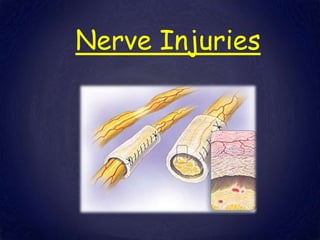
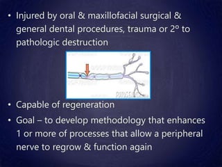
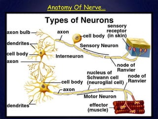
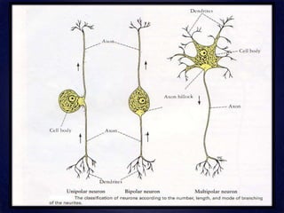
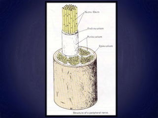
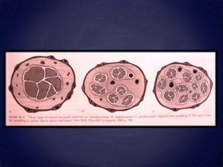
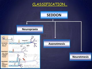
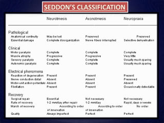
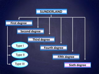
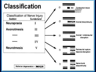
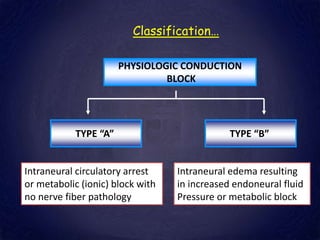
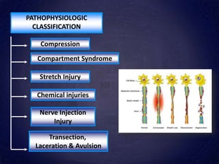
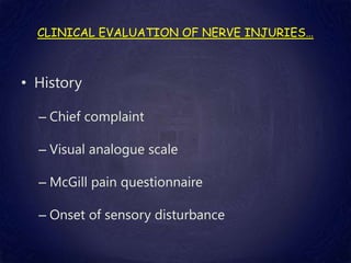
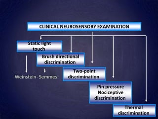
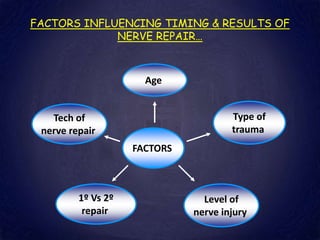
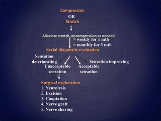
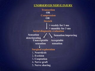
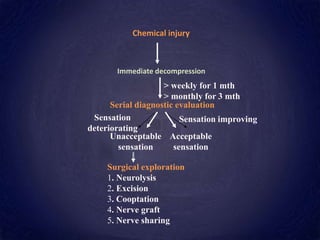
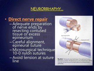
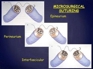
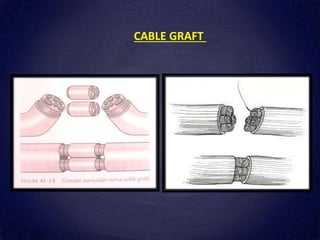
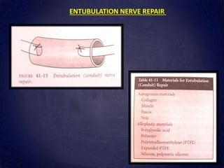
Nerve Injuries
CONTENTS
• Introduction
• Anatomy of the nerve
• Etiology of nerve injuries
• Classification of nerve injuries
• Clinical evaluation of nerve injuries
• Factors influencing the timing and results of
nerve repair
• Management of nerve injuries
INTRODUCTION…
PNS
Cranial nerves Spinal nerves
Somatic nerves Visceral nerves
Sensory Motor
• Injured by oral & maxillofacial surgical &
general dental procedures, trauma or 2º to
pathologic destruction
• Capable of regeneration
• Goal – to develop methodology that enhances
1 or more of processes that allow a peripheral
nerve to regrow & function again
HISTORY…
• Hippocrates- 460- 370 BC-1st written description of PNS
• Wlliam of Saliceto- 1210- 1277- 1st record of nerve suture
• Van Leeuwenhoek- 1632- 1723- microscopic structure of
nerve
• Haller- 1850- understanding of nerve injuries- IX & XII
nerve injuries in frog- degenerative changes distal to the
level of injury & orderly progression of regenerating
axons down the nerve fiber
History…
• Seddon- 1943- pioneered nerve grafting
techniques
• Contemporary milestones are being reached
with the research of Sunderland, Miller, Terzis
and Dykes regarding the management of
peripheral nerve injuries and the technical
consideration of their repair
ANATOMY OF NERVE…
Anatomy Of Nerve…
• AXON
– A-alpha fibers- largest myelinated fibers, range in dia
from 7-16 m, conduction velocity – 70-120 m/sec.
– A-beta fibers- next largest myelinated axons, Diameter 
6-8 m,Conduction velocity  30-70 m/sec. Sensibility of
touch is attributed to these axons
– A- delta fibers- smallest of myelinated fibers, Diameter 
2-4m, Conduction velocity  0.5-2m/sec. Transmit
stimuli encoded for slow pain, temperature & efferent
sympathetic fibers
Terminology…
• Allodynia
• Analgesia
• Anesthesia
• Anesthesia dolorosa
• Causalgia
• Paresthesia
• Dysesthesia
• Hyperesthesia
• Hypoesthesia
• Hyperalgesia
• Hypoalgesia
Etiology …
• Third molar removal
• Injections
• Infections
• Foreign bodies
• Endodontic therapy
• Maxillofacial trauma
• Orthognathic surgery
• Temporomandibular joint arthroscopy
• Miscellaneous causes
EFFECT OF NERVE INJURY…
• Depends on the type of nerve  pure
motor, pure sensory or mixed
• Motor effect
• Sensory effects
• Pseudomotor effects
• Vasomotor effect
• Loss of reflexes
Nerve Injury…
• NERVE INJURY- CHANGES DISTAL TO INJURY, SITE OF
INJURY, PROXIMAL TO INJURY & WITHIN THE CNS
• NEURON
– Hypertrophic changes- 3rd or 4th day following injury to
the axon & peak b/w 10th & 20th day
– Total RNA content ↑ as the cell ↑ in size RNA migrates to
outer edges of the cell & break up in to smaller particles
– Anabolic proteosynthetic state is maintained as long as
there are regenerative efforts – up to many years
– Chromatolytic hypertrophic changes are more pronounced in more
proximal injuries. sometimes the neuron cannot meet these metabolic
demands and cell death occurs.
– After regeneration is complete and conduction maturation has occurred,
the neuron return to normal size and electrical activity
• PROXIMAL NERVE TRUNK
– 1 hr after laceration of a nerve there is marked swelling proximally as far
as 1cm
– 7th day there is vigorous sprouting of axons
– It is not until the 28th day that axons cross the point of injury and at 42nd
day before a sizable no. of axons occupy the distal segment .
• SITE OF INJURY
–Within hrs of injury there is proliferation of
macrophages, perineurial fibroblasts,
schwann cells and epineurial fibroblasts
–7th day the schwann cell is clearly the most
active cell and assumes a phagocytic
function of debridement
• DISTAL NERVE TRUNK
–Wallerian degeneration in preparation for
the arrival of sprouting axons
–7th post injury day the majority of neural
elements break down, facilitated by
digestive enzymes present in the axons
–Majority of cellular debris has been
phagocytosed by schwann cells by 21st day
–At 42 days debridement is complete and
parts of the fascicular anatomy persist
CLASSIFICATION…
SEDDON
Neuropraxia
Axonotmesis
Neurotmesis
SEDDON’S CLASSIFICATION
SUNDERLAND
First degree
Second degree
Third degree
Fourth degree
Fifth degree
Sixth degree
Type I
Type II
Type III
Classification…
PHYSIOLOGIC CONDUCTION
BLOCK
TYPE “A” TYPE “B”
Intraneural circulatory arrest
or metabolic (ionic) block with
no nerve fiber pathology
Intraneural edema resulting
in increased endoneural fluid
Pressure or metabolic block
CLASSIFICATION…
SYMPTOMATIC
CLASSIFICATION
ANESTHESIA
DYESETHESIA
PARESTHESIA
CLASSIFICATION…
ANATOMIC
CLASSIFICATION
INTRAOSSEOUS
NERVE INJURY
SOFT TISSUE
NERVE INJURY
CLASSIFICATION…
HISTOPATHOLOGIC
CLASSIFICATION
NEUROMA FIBROSIS
Amputation Eccentric
Central
• Amputation or stump neuroma- Is a knobby, disorganized
mass of axons and collagen associated with proximal nerve
stump and completely separated from distal nerve stump-
5th degree injury
• Central neuroma or neuroma in continuity- fusiform
expansion or fibrotic narrowing of nerve with variable
degrees of fascicular disruption & disorganization- No
breach in epineurium- 4th degree & 5th degree injuries
• Lateral entrapment neuroma- Neurofibrous union between
the epineurium & lingual periosteum of mandible usually in
3rd molar region
• Lateral exophytic neuroma- Breach in epineurium
Classification…
Classification of damage by location of
the reactive fibrosis
Designation Location Prognosis
A* Epifascicular epineurium Generally good
B* Interfascicular epineurium Depends on original
damage
C* Endoneurium Poor
N In a Sunderland class IV injury, the
epineural connective tissue that
maintains continuity can be
infiltrated by a neuroma (IVN)
Poor
S Continuity in class IV injury
maintained only by scar tissue
Poor
PATHOPHYSIOLOGIC
CLASSIFICATION
Compression
Compartment Syndrome
Transection,
Laceration & Avulsion
Stretch Injury
Chemical injuries
Nerve Injection
Injury
CLINICAL EVALUATION OF NERVE INJURIES…
• History
– Chief complaint
– Visual analogue scale
– McGill pain questionnaire
– Onset of sensory disturbance
CLINICAL NEUROSENSORY EXAMINATION
Static light
touch
Brush directional
discrimination
Two-point
discrimination
Pin pressure
Nociceptive
discrimination
Thermal
discrimination
Weinstein- Semmes
FACTORS INFLUENCING TIMING & RESULTS OF
NERVE REPAIR…
FACTORS
1º Vs 2º
repair
Tech of
nerve repair
Age
Type of
trauma
Level of
nerve injury
PROGNOSIS…
• Spontaneous recovery depends on- condition of nerve, severity
of nerve injury & site of injury
– Intraosseous nerve Vs extraosseous nerve
• Conditions limiting regeneration- chronic compression or
stretch, divided nerves in which nerve ends are not
approximated, excessive scarring of either nerve/ tissue bed-
– Infected or contaminated wounds
– Poorly vascularized wounds
– Wound closure or neurorhaphy under tension
MANAGEMENT
• Observed Vs unobserved
• Timing of nerve repair- primary, delayed primary or
secondary
• Primary nerve repair- within hours of the injury,
• Delayed primary repair 14-21 days following injury &
• Secondary repair > 3 weeks following surgery
• Microsurgical operations for repair
–External decompession
–Transposition
–Internal neurolysis
–Neuroma excision
–Neurorrhaphy
–Reconstruction with autogenous
nerve, vein or muscle grafts or
alloplastic nerve guides
• Ablative nerve procedures using
thermal, chemical, capping – nerve
repair is not possible or practical.
• Non surgical treatments
• OBSERVED NERVE INJURY
Clean / sharp laceration
Immediate repair
Fibrin sealant Suture Collagen tube
Sensation improving
Sensation
deteriorating
> weekly for 1 mth
> monthly for 3 mth
Unacceptable
sensation
Acceptable
sensation
Stretch
Serial diagnostic evaluation
Surgical exploration
1. Neurolysis
2. Excision
3. Cooptation
4. Nerve graft
5. Nerve sharing
Compression
OR
Alleviate stretch ,decompression as needed
Sensation improving
Sensation
deteriorating
> weekly for 1 mo
> monthly for 3 mo
Unacceptable
sensation
Acceptable
sensation
UNOBSERVED NERVE INJURY
Compression
Serial diagnostic evaluation
Surgical exploration
1. Neurolysis
2. Excision
3. Cooptation
4. Nerve graft
5. Nerve sharing
Stretch
Transection
OR
OR
Sensation improving
Sensation
deteriorating
> weekly for 1 mth
> monthly for 3 mth
Unacceptable
sensation
Acceptable
sensation
Serial diagnostic evaluation
Surgical exploration
1. Neurolysis
2. Excision
3. Cooptation
4. Nerve graft
5. Nerve sharing
Chemical injury
Immediate decompression
SURGICAL M/M OF LINGUAL NERVE INJURIES
• Causes
• Technique
SURGICAL M/M OF INFERIOR ALVEOLAR NERVE INJURIES…
• 3 approaches
– Through the socket
– Sagittal split
– Extraoral
SURGICAL M/M OF INFRAORBITAL NERVE
INJURIES…
• Etiology and Incidence
• Incisions
• Soft tissue dissection
• Removal of bone
• Visualization
• Management
• Direct nerve repair
– Adequate preparation
of nerve ends by
resecting contused
tissue or excess
epineurium
– Careful alignment,
epineural suture
– Microsurgical technique
10-0 nylon sutures
– Avoid tension at suture
line
NEURORRHAPHY…
MICROSURGICAL
SUTURING
Interfascicular
Epineurium
Perineurium
NERVE HARVESTING PROCEDURES :
First reported by Hausamen et al in 1974
DONOR NERVE SELECTION FACTORS
1) Size of donor nerve Vs reciprocal nerve
2) Length of donor nerve available for grafting
3) Proximity to recipient area
4) Patients or surgeons preference
Nerve Diameter
(mm)
Number of
fascicles
Number
of Axons
Pattern
Inferior alveolar 2.4 18-21 16200 Polyfascicular without
grouping
Lingual 3.2 15-18 14200 Polyfascicular without
grouping
Facial 2.5 1-5 Oligofascicular
Sural 2.1 11-12 7600 Oligofascicular
Great auricular 1.5 8-9 9500 Polyfascicular with
grouping
SURAL NERVE
GREATER AURICULAR NERVE
CABLE GRAFT
ENTUBULATION NERVE REPAIR
• NON SURGICAL TREATMENT
• Indications
– Metabolic neuropathy
– Centrally mediated pain
– Atypical pain that does not conform to anatomic distribution of
peripheral nerves
– Chronic injuries – atrophy in distal nerve
– Pain unrelieved by LA block of suspected peripheral nerves
– Poor physical status
• Types
– Behavioral – counseling, meditation, yoga, psychotherapy
– Physiologic – physical therapy, acupuncture
– Pharmacologic – analgesics, anticonvulsants, antidepressants
NEURALGIAS
CONTENTS…
• Introduction
• Trigeminal Neuralgia
• Paratrigeminal neuralgia
• Sphenopalatine neuralgia
• Glossopharyngeal Neuralgia
TRIGEMINAL NEURALGIA…
• Definition
• John Locke-1677- 1st full description with its
T/t
• Nicholaus Andre- 1756- ‘Tic Doloureux’ means
painful jerking
• John Fothergill- 1773-
‘Fothergill’s disease’
Definition
• Sudden, usually unilateral, severe, brief, stabbing, recurrent pains in the
distribution of one or more branches of the fifth cranial nerve
(INTERNATIONALASSOCIATION FOR THE STUDY OF PAIN)
• Painful unilateral affliction of the face, characterized by brief electric shock like
pain limited to the distribution of one or more divisions of the trigeminal nerve.
Pain is commonly evoked by trivial stimuli including washing, shaving, smoking,
talking, and brushing the teeth, but may also occur spontaneously. The pain is
abrupt in onset and termination and may remit for varying periods
(INTERNATIONAL HEADACHE SOCIETY)
• ETIOLOGY
– Idiopathic
– Vascular factors- transient ischemia & auto- immune
hypersensitivity responses- demyelination of nerve
– Mechanical factors- pressure of
aneurysms of intrapetrous
portion of internal carotid
artery- erode the floor of
intracranial fossa to exert a
pulsatile irritation on the ventral
side of the trigeminal ganglion
– Anomaly of superior cerebellar a- lies in contact with
sensory root of trigeminal nerve- demyelination. Surgical
elevation of the artery or decompression of the sensory
root has been highly successful
• OTHER ETIOLOGIC FACTORS
– Multiple sclerosis- Olfson (1966)
– Petrous ridge (Basilar) Compression- Lee (1937)
– Post-traumatic neuralgia
– Intracranial tumours
– Intracranial vascular
abnormalities
– Viral etiology
• General Characteristics
– Incidence
– Age
– Sex prediliction
– Affliction for side
• Clinical Characteristics
– No abnormal neurologic deficit
– Sudden, u/l, intermittent paroxysmal, sharp,
shooting, lancinating, shock like pain- ‘trigger
points’
– Pain is- confined to one division- V2 or V3- rarely
crosses midline, Short duration
– Pt grimaces with pain, clutches his hands over
affected side of face, stopping all activities &
holds or rubs his face, reddening & watering of
eyes
– Refractory period
– Oral hygiene- poor
–Paroxysms occur in cycles, each cycle
lasting for weeks or months & with time-
cycle appears closer
–With each attack- pain seems to become
more intense and unbearable
• Attacks do not occur during sleep
• Poor quality of life
• Pts may undergo indiscriminate extractions
• Diagnosis
– Well-taken history
– Classic clinical pattern
– Symptoms in <35 yrs - possible intracranial space
occupying lesion or intracranial av anomalies
– MRI/ CT scan
– Response to T/t with carbamazepine
– Diagnostic injections of a LA into trigger zone
Special features Aggravating
factors
Relieving
factors
TRIGEMINAL
NEURALGIA
Unilateral isolated
attacks
Hot/cold fluids in
mouth,
blowing nose,Chewing,
carbamazepine
Phenytoin
surgical
MIGRANOUS
FACIAL
PAIN
Sporadic attacks,
often nocturnal or
early morning
Alcohol Ergotamine,
Sumatriptan
ATYPICAL
FACIAL PAIN
Bilateral,
Continuous
Interferes with sleep
None Antidepressants
,
tranquilizers
COSTEN’S
SYNDROME
Pain over TMJ when
eating
Chewing,Yawning
Talking
Bite correction
POST
HERPETIC
NEURALGIA
Tender areas between
white scars of herpes
eruption
Touching affected area
Cold wind
Phenytoin,
Anti
depressants
Differential Diagnosis:
• PROTOCOL FOR DIAGNOSTIC NERVE BLOCKS
– Armamentarium
– Begin injections at surface site of pain & then move
proximally
– Inject 0.5 cc of normal saline
– If pain persists- inject 0.5 ml of 2 % plain lignocaine at
surface site- wait for 5 min- If pain is relieved- direct therapy
at small nociceptor fibres
– If pain persists — inject little deeper & wait for 5
min- If pain is relieved- musculo skeletal origin
of pain
– If pain is not relieved- inject at more proximal
portion of nerve- If pain is relieved- direct
therapy at site
– Thus selective inferior alveolar, lingual, buccal,
infra orbital, posterior superior alveolar blocks
can be given to know involvement of branch of
trigeminal Nerve.
TREATMENT
Surgical
Medical
– Blom (1962)-anticonvulsants Carbamazepine-
highly specific
– Carbamazapine 100 mg 3times/ day is introduced
& titrated over 1- 5 wks until either remission is
achieved or side effects or toxicity are unacceptable
– Side effects-Visual blurring, dizziness, skin rashes,
ataxia & in rare cases hepatic dysfunction,
leukopenia, thrombocytopenia- aplastic anaemia
– Carbamazepine- ineffective- phenytoin or baclofen, sodium
valproate
– When carbamazepine is Contra Indicated Clonazepam 1.5
mg/day
• Side effects- Drowsiness, fatigue, lethargy.
– Tab. Phenytoin: Dose- 100 mg 3 times/ day
• Side effects- slurred speech, abnormal movements, swelling of
lymph glands, gingival hypertrophy, hirsutism, folate def
– Tab. Oxcarbazepine- 1200 mg/day
• Side effects- Hyponatremia, double vision
– Valproic acid- 600 mg/day.
• Side effects- irritability, tremors, confusion, hepatotoxicity, wt
gain
–Mephenesin Carbamate (Tolceram)- 5 to 15
ml/5 times a day
• Side effects—fatigue, vomiting
–Other less toxic agents
• Baclofen (Lioresal)
• Gabapantin (Neurontin)
• Lamotrigine
• Felbamate
• Topiramate
• Vigabtrin
Surgical M/m
Intracranial
procedures
Peripheral
procedures
Peripheral
Injections
Microvascular
decompression
Peripheral
Neurectomy
Trigeminal root
section
Gasserian Ganglion
Procedures
Cryotherapy
LA
Glycerol
Thermo
coag
Balloon
Alcohol
• Peripheral Injections
– Long-acting anesthetic agents
– Alcohol
• Peripheral Neurectomy (Nerve Avulsion)
– lnfraorbital neurectomy
• Conventional intraoral approach
• Braun’s transantral approach- 1977
– Complications, inadvertent section of vessels in
pterygopalatine fossa, inadvertent sectioning
branches of sphenopalatine ganglion or vidian
nerve
– Inferior alveolar neurectomy
• Extraoral approach
• Intraoral approach
– Lingual neurectomy
• CRYOTHERAPY OR CRYONEUROLYSIS
– Direct application of cryotherapy probe- temp < - 60°C-
Wallerian degeneration without destroying nerve sheath
– Frozen with a cryoprobe (Nitrous oxide probe) for a
period of 1-2 minutes followed by 3 minutes thaw,
repeated 3 times
• PERIPHERAL RADIOFREQUENCY NEUROLYSIS
(THERMOCOAGULATION)
– Gregg and Small- 1986- radiofrequency electrode- use of
radiofrequency (RF) lesioning of trigeminal nerve has
been used sparingly, but with success. RF neurolysis has
been shown to induce pain remission in 80 % of cases
with a 20 % recurrence rate
– PROCEDURE
• Topical anaesthesia
• Patient is grounded in an electronic circuit & the 22 gauge
lesion probe is positioned adjacent to nerve
• Tissue temp is measured with probe tip through probe
thermocouple
• Lesioning carried out at 65- 75°C for 1- 2 min
– Advantages- Low morbidity in high risk- elderly
patients.
– Disadvantages- Needs specific electronic
armamentarium & reasonable pt cooperation, In case
of inaccessibility of some pain triggering nerve trunks,
the technique will fail to achieve pain relief
• GASSERIAN GANGLION PROCEDURES
– Around 1900- 1st open surgery- gasserian ganglion for
TN- hazardous procedures
– 1910- Harris, Tapatas & Hartel separately introduced
percutaneous approaches to the ganglion via foramen
ovale - absolute alcohol or phenolglycerol mixture…
– 1931- Kirschner introduced percutaneous electro
coagulation of gasserian ganglion
– 3 percutaneous gasserian ganglion procedures
• Glycerol injection
• Thermocoagulation
• Balloon compression
• Technique
– Anaesthesia + Injection methahexitone- iv- 1.5 to 2
mg/kg body wt in increments
– Pt- lie on a table with neck well-extended-foramen
ovale is best visualized with the X-ray tube placed
for a submentovertex position
– 3 points of Hartel are marked
– on side of face
• 1ST
• 2ND
• 3RD
– During this phase- needle/electrode lies below orbit,
medial to ramus of mandible & lat to maxilla.
Engagement of the needle/electrode in foramen ovale
is best confirmed by biplanar radiology or image
intensifier
– Final position of needle/electrode is then pushed for
another half a centimeter
• Glycerol injection- 16 gauge spinal needle
• CONTROLLED RADIOFREQUENCY
THERMOCOAGULATION
– 1st by Kirschner- 1931 & later modified by Sweet-1970
– Advantages
• Avoidance of denervation of cornea
• Comparative lower rate of recurrence
• Procedure can be repeated in case of recurrence
• Zero mortality
• Well-tolerated by elderly & medically compromised
pt
• Can be performed on outpatient basis
• Preserves motor function of trigeminal nerve
• Simple, accurate, ↓ time-consuming & ↓ expensive
• Comfortable
– Indications
• Toxicity of drugs
• Failure of response to other modalities
• Dependence on drug for lifetime
• Recurrence cases
– PROCEDURE
• Probe is placed correctly in foramen ovale & advanced
into trigeminal ganglion, CSF should emerge on
removal of the stylet. Electrode is inserted then just
beyond the tip of the probe and low amplitude current
is applied using a lesion generator.
• Pt at this stage should be awake & cooperative- asked
whether on face the stimulation is felt. Stimulation is
initiated at 50 cycles/ sec slowly raising voltage until
full areas of pain covered
• A radiofrequency current- alternating current of
high frequency is passed through electrode- Heat-
ionic friction- coagulation of tissues. A facial blush
usually appears at this point & helps to localize
region of nerve root undergoing thermal destruction
• End of procedure- pt is asked to perform those
maneuvers that trigger TN
• Once pt & operator is satisfied with desired RF lesion
production, electrode is removed
NEURO-OPTHALMIC COMPLICATIONS
• BALLOON COMPRESSION
– Under general anaesthesia
– Mechanical technique to destroy root fibres partially
by advancing 4FG Fogarty catheter 1 to 2 cm within
Meckel’s cave & inflating balloon at ventral aspect of
ganglion root
– 12 gauge spinal needle is passed 1st into foramen
ovale & balloon catheter is passed through it
– Once it is in position balloon is inflated with X-ray
contrast medium upto 0.75 ml- 1min
• 1967-1976 - Jannetta
• Open craniotomy approach- gain access to trigeminal root
• Root is examined under microscope. A compressing br of superior
cerebellar a. will be seen medial to nerve
• Artery is carefully separated from nerve & interpositioned by
using sponge or Teflon wool
• Pt usually retains good facial sensation
• 75 - 80 % pts are pain free for years
• Mortality rate- 2 %- infrequent hearing loss, vertigo, CN VII
weakness
• Contraindicated in elderly patients& medically compromised pts
INTRACRANIAL
PROCEDURES
Trigeminal
root section
Microvascular
decompression
Trigeminal
root section
Extradural sensory
Root section
Frazier’s approach (1901)
Intradural root section
Wilkins (1966)
Trigeminal tractotomy
(Medullary Tractotomy)
PARATRIGEMINAL NEURALGIA
• Char by severe headache or pain in the area of
the trigeminal distribution with signs of ocular
sympathetic paralysis
• Sympathetic symptoms & homolateral pain in
head or eye occur without vasomotor or trophic
disturbances
• Signs and symptoms appear suddenly
• Most common in males- middle age
• It presents some of the signs of Homer’s
syndrome, but can be differentiated from it by
the presence of pain & little/ no change in
sweating activity on affected side of face
• Cause of the disease is unknown, but in a case
reported by Lucchesi & Topazian, dramatic
improvement occurred after elimination of
dental infection
SPHENOPALATINE NEURALGIA
• Pain syndrome originally described by Sluder as a symptom
complex referable to the nasal ganglion
• Subsequently Vial described a similar syndrome, but believed
that it involved vidian nerve & concluded that condition
reported by Sluder should be termed ‘vidian neuralgia’
• In recent years, the term ‘periodic migrainous neuralgia’
• an idiopathic syndrome consisting of recurrent brief attacks of
sudden severe, unilateral periorbital pain
• Etiology
– Not understood entirely
– Attributed to hypothalamic hormonal influences
– Pain is thought to be generated at the level of
pericarotid/cavernous sinus complex
• Clinical Features
– U/l paroxysms of intense pain in region of eyes,
maxilla, ear & mastoid, base of nose, & beneath
zygoma
– Pain- rapid onset, persist for 15 minutes- several
hours, & then disappear as rapidly as they began
– No ‘trigger zone’
–‘Alarm clock’ headache
–Apart from pain sensation- sneezing,
swelling of nasal mucosa & nasal discharge,
epiphora
–Paresthetic sensations of skin over lower
half of face also are reported
–Precipitated by either emotional stress or
injudicious intake of alcohol
–Men > women (5 : 1)
–Before the age of 40 years
• TREATMENT
– Alcohol injection
– Resection of the ganglion
– Ergotamine often produce immediate & complete
relief of symptoms- not totally effective: +
methylsergide antiserotonin agent- synergistic action
– Invasive nerve blocks and ablative neurosurgical
procedures - refractory cases
GLOSSOPHARYNGEAL NEURALGIA
• Pain similar to TN
• Not common
• Pain may be as severe & extruciating
• Clinical features
– Predilection in middle-aged or older persons
– Sharp, shooting pain in ear, pharynx, nasopharynx,
tonsil or the posterior part of tongue
– U/l & paroxysmal, rapidly subsiding type of pain
– trigger zone- post oropharynx or tonsillar fossa
– ↑ swallowing talking, yawning or coughing
• Etiology- unknown- ischemia is suggested (without
conclusive evidence)
• Treatment- resection of extracranial portion of nerve or
intracranial section. injection of alcohol into the
glossopharyngeal nerve
has not been as widely
accepted
RECENT ADVANCES IN MANAGEMENT OF
TRIGEMINAL NEURALGIA:
REFERENCES
• ORAL AND MAXILLOFACOAL TRAUMA – RAYMOND J
FONSECA & ROBERT V WALKER
• MAXILLOFACIAL SURGERY – P. WARDBOOTH
• MANAGEMENT OF BELL’S PALSY – JOMS, 1993.
• TEXT BOOK OF ORAL PATHOLOGY – SHAFERS.
• BELLS OROFACIAL PAINS—FIFTH EDITION. BY JEFFRY
P. OKESON
• TEMPEROMANDIBULAR DISODERS AND ORO-FACIAL
PAIN- RICHARD A. PERTES & SHELDON G. GROSS
Nerve injury AR.pptx

Nerve injury AR.pptx

  • 2.
  • 3.
    CONTENTS • Introduction • Anatomyof the nerve • Etiology of nerve injuries • Classification of nerve injuries • Clinical evaluation of nerve injuries • Factors influencing the timing and results of nerve repair • Management of nerve injuries
  • 4.
    INTRODUCTION… PNS Cranial nerves Spinalnerves Somatic nerves Visceral nerves Sensory Motor
  • 5.
    • Injured byoral & maxillofacial surgical & general dental procedures, trauma or 2º to pathologic destruction • Capable of regeneration • Goal – to develop methodology that enhances 1 or more of processes that allow a peripheral nerve to regrow & function again
  • 6.
    HISTORY… • Hippocrates- 460-370 BC-1st written description of PNS • Wlliam of Saliceto- 1210- 1277- 1st record of nerve suture • Van Leeuwenhoek- 1632- 1723- microscopic structure of nerve • Haller- 1850- understanding of nerve injuries- IX & XII nerve injuries in frog- degenerative changes distal to the level of injury & orderly progression of regenerating axons down the nerve fiber
  • 7.
    History… • Seddon- 1943-pioneered nerve grafting techniques • Contemporary milestones are being reached with the research of Sunderland, Miller, Terzis and Dykes regarding the management of peripheral nerve injuries and the technical consideration of their repair
  • 8.
  • 9.
  • 13.
    • AXON – A-alphafibers- largest myelinated fibers, range in dia from 7-16 m, conduction velocity – 70-120 m/sec. – A-beta fibers- next largest myelinated axons, Diameter  6-8 m,Conduction velocity  30-70 m/sec. Sensibility of touch is attributed to these axons – A- delta fibers- smallest of myelinated fibers, Diameter  2-4m, Conduction velocity  0.5-2m/sec. Transmit stimuli encoded for slow pain, temperature & efferent sympathetic fibers
  • 14.
    Terminology… • Allodynia • Analgesia •Anesthesia • Anesthesia dolorosa • Causalgia • Paresthesia • Dysesthesia • Hyperesthesia • Hypoesthesia • Hyperalgesia • Hypoalgesia
  • 15.
    Etiology … • Thirdmolar removal • Injections • Infections • Foreign bodies • Endodontic therapy • Maxillofacial trauma • Orthognathic surgery • Temporomandibular joint arthroscopy • Miscellaneous causes
  • 16.
    EFFECT OF NERVEINJURY… • Depends on the type of nerve  pure motor, pure sensory or mixed • Motor effect • Sensory effects • Pseudomotor effects • Vasomotor effect • Loss of reflexes
  • 17.
    Nerve Injury… • NERVEINJURY- CHANGES DISTAL TO INJURY, SITE OF INJURY, PROXIMAL TO INJURY & WITHIN THE CNS • NEURON – Hypertrophic changes- 3rd or 4th day following injury to the axon & peak b/w 10th & 20th day – Total RNA content ↑ as the cell ↑ in size RNA migrates to outer edges of the cell & break up in to smaller particles – Anabolic proteosynthetic state is maintained as long as there are regenerative efforts – up to many years
  • 18.
    – Chromatolytic hypertrophicchanges are more pronounced in more proximal injuries. sometimes the neuron cannot meet these metabolic demands and cell death occurs. – After regeneration is complete and conduction maturation has occurred, the neuron return to normal size and electrical activity • PROXIMAL NERVE TRUNK – 1 hr after laceration of a nerve there is marked swelling proximally as far as 1cm – 7th day there is vigorous sprouting of axons – It is not until the 28th day that axons cross the point of injury and at 42nd day before a sizable no. of axons occupy the distal segment .
  • 19.
    • SITE OFINJURY –Within hrs of injury there is proliferation of macrophages, perineurial fibroblasts, schwann cells and epineurial fibroblasts –7th day the schwann cell is clearly the most active cell and assumes a phagocytic function of debridement
  • 20.
    • DISTAL NERVETRUNK –Wallerian degeneration in preparation for the arrival of sprouting axons –7th post injury day the majority of neural elements break down, facilitated by digestive enzymes present in the axons –Majority of cellular debris has been phagocytosed by schwann cells by 21st day –At 42 days debridement is complete and parts of the fascicular anatomy persist
  • 21.
  • 22.
  • 23.
    SUNDERLAND First degree Second degree Thirddegree Fourth degree Fifth degree Sixth degree Type I Type II Type III
  • 25.
    Classification… PHYSIOLOGIC CONDUCTION BLOCK TYPE “A”TYPE “B” Intraneural circulatory arrest or metabolic (ionic) block with no nerve fiber pathology Intraneural edema resulting in increased endoneural fluid Pressure or metabolic block
  • 26.
  • 27.
  • 28.
  • 29.
    • Amputation orstump neuroma- Is a knobby, disorganized mass of axons and collagen associated with proximal nerve stump and completely separated from distal nerve stump- 5th degree injury • Central neuroma or neuroma in continuity- fusiform expansion or fibrotic narrowing of nerve with variable degrees of fascicular disruption & disorganization- No breach in epineurium- 4th degree & 5th degree injuries • Lateral entrapment neuroma- Neurofibrous union between the epineurium & lingual periosteum of mandible usually in 3rd molar region • Lateral exophytic neuroma- Breach in epineurium
  • 30.
    Classification… Classification of damageby location of the reactive fibrosis Designation Location Prognosis A* Epifascicular epineurium Generally good B* Interfascicular epineurium Depends on original damage C* Endoneurium Poor N In a Sunderland class IV injury, the epineural connective tissue that maintains continuity can be infiltrated by a neuroma (IVN) Poor S Continuity in class IV injury maintained only by scar tissue Poor
  • 31.
    PATHOPHYSIOLOGIC CLASSIFICATION Compression Compartment Syndrome Transection, Laceration &Avulsion Stretch Injury Chemical injuries Nerve Injection Injury
  • 32.
    CLINICAL EVALUATION OFNERVE INJURIES… • History – Chief complaint – Visual analogue scale – McGill pain questionnaire – Onset of sensory disturbance
  • 33.
    CLINICAL NEUROSENSORY EXAMINATION Staticlight touch Brush directional discrimination Two-point discrimination Pin pressure Nociceptive discrimination Thermal discrimination Weinstein- Semmes
  • 34.
    FACTORS INFLUENCING TIMING& RESULTS OF NERVE REPAIR… FACTORS 1º Vs 2º repair Tech of nerve repair Age Type of trauma Level of nerve injury
  • 35.
    PROGNOSIS… • Spontaneous recoverydepends on- condition of nerve, severity of nerve injury & site of injury – Intraosseous nerve Vs extraosseous nerve • Conditions limiting regeneration- chronic compression or stretch, divided nerves in which nerve ends are not approximated, excessive scarring of either nerve/ tissue bed- – Infected or contaminated wounds – Poorly vascularized wounds – Wound closure or neurorhaphy under tension
  • 36.
    MANAGEMENT • Observed Vsunobserved • Timing of nerve repair- primary, delayed primary or secondary • Primary nerve repair- within hours of the injury, • Delayed primary repair 14-21 days following injury & • Secondary repair > 3 weeks following surgery
  • 37.
    • Microsurgical operationsfor repair –External decompession –Transposition –Internal neurolysis –Neuroma excision –Neurorrhaphy –Reconstruction with autogenous nerve, vein or muscle grafts or alloplastic nerve guides • Ablative nerve procedures using thermal, chemical, capping – nerve repair is not possible or practical. • Non surgical treatments
  • 38.
    • OBSERVED NERVEINJURY Clean / sharp laceration Immediate repair Fibrin sealant Suture Collagen tube
  • 39.
    Sensation improving Sensation deteriorating > weeklyfor 1 mth > monthly for 3 mth Unacceptable sensation Acceptable sensation Stretch Serial diagnostic evaluation Surgical exploration 1. Neurolysis 2. Excision 3. Cooptation 4. Nerve graft 5. Nerve sharing Compression OR Alleviate stretch ,decompression as needed
  • 40.
    Sensation improving Sensation deteriorating > weeklyfor 1 mo > monthly for 3 mo Unacceptable sensation Acceptable sensation UNOBSERVED NERVE INJURY Compression Serial diagnostic evaluation Surgical exploration 1. Neurolysis 2. Excision 3. Cooptation 4. Nerve graft 5. Nerve sharing Stretch Transection OR OR
  • 41.
    Sensation improving Sensation deteriorating > weeklyfor 1 mth > monthly for 3 mth Unacceptable sensation Acceptable sensation Serial diagnostic evaluation Surgical exploration 1. Neurolysis 2. Excision 3. Cooptation 4. Nerve graft 5. Nerve sharing Chemical injury Immediate decompression
  • 42.
    SURGICAL M/M OFLINGUAL NERVE INJURIES • Causes • Technique
  • 44.
    SURGICAL M/M OFINFERIOR ALVEOLAR NERVE INJURIES… • 3 approaches – Through the socket – Sagittal split – Extraoral
  • 45.
    SURGICAL M/M OFINFRAORBITAL NERVE INJURIES… • Etiology and Incidence • Incisions • Soft tissue dissection • Removal of bone • Visualization • Management
  • 46.
    • Direct nerverepair – Adequate preparation of nerve ends by resecting contused tissue or excess epineurium – Careful alignment, epineural suture – Microsurgical technique 10-0 nylon sutures – Avoid tension at suture line NEURORRHAPHY…
  • 47.
  • 48.
    NERVE HARVESTING PROCEDURES: First reported by Hausamen et al in 1974 DONOR NERVE SELECTION FACTORS 1) Size of donor nerve Vs reciprocal nerve 2) Length of donor nerve available for grafting 3) Proximity to recipient area 4) Patients or surgeons preference Nerve Diameter (mm) Number of fascicles Number of Axons Pattern Inferior alveolar 2.4 18-21 16200 Polyfascicular without grouping Lingual 3.2 15-18 14200 Polyfascicular without grouping Facial 2.5 1-5 Oligofascicular Sural 2.1 11-12 7600 Oligofascicular Great auricular 1.5 8-9 9500 Polyfascicular with grouping
  • 49.
  • 50.
  • 51.
  • 52.
  • 53.
    • NON SURGICALTREATMENT • Indications – Metabolic neuropathy – Centrally mediated pain – Atypical pain that does not conform to anatomic distribution of peripheral nerves – Chronic injuries – atrophy in distal nerve – Pain unrelieved by LA block of suspected peripheral nerves – Poor physical status • Types – Behavioral – counseling, meditation, yoga, psychotherapy – Physiologic – physical therapy, acupuncture – Pharmacologic – analgesics, anticonvulsants, antidepressants
  • 55.
  • 56.
    CONTENTS… • Introduction • TrigeminalNeuralgia • Paratrigeminal neuralgia • Sphenopalatine neuralgia • Glossopharyngeal Neuralgia
  • 57.
    TRIGEMINAL NEURALGIA… • Definition •John Locke-1677- 1st full description with its T/t • Nicholaus Andre- 1756- ‘Tic Doloureux’ means painful jerking • John Fothergill- 1773- ‘Fothergill’s disease’
  • 58.
    Definition • Sudden, usuallyunilateral, severe, brief, stabbing, recurrent pains in the distribution of one or more branches of the fifth cranial nerve (INTERNATIONALASSOCIATION FOR THE STUDY OF PAIN) • Painful unilateral affliction of the face, characterized by brief electric shock like pain limited to the distribution of one or more divisions of the trigeminal nerve. Pain is commonly evoked by trivial stimuli including washing, shaving, smoking, talking, and brushing the teeth, but may also occur spontaneously. The pain is abrupt in onset and termination and may remit for varying periods (INTERNATIONAL HEADACHE SOCIETY)
  • 59.
    • ETIOLOGY – Idiopathic –Vascular factors- transient ischemia & auto- immune hypersensitivity responses- demyelination of nerve – Mechanical factors- pressure of aneurysms of intrapetrous portion of internal carotid artery- erode the floor of intracranial fossa to exert a pulsatile irritation on the ventral side of the trigeminal ganglion
  • 60.
    – Anomaly ofsuperior cerebellar a- lies in contact with sensory root of trigeminal nerve- demyelination. Surgical elevation of the artery or decompression of the sensory root has been highly successful
  • 61.
    • OTHER ETIOLOGICFACTORS – Multiple sclerosis- Olfson (1966) – Petrous ridge (Basilar) Compression- Lee (1937) – Post-traumatic neuralgia – Intracranial tumours – Intracranial vascular abnormalities – Viral etiology
  • 62.
    • General Characteristics –Incidence – Age – Sex prediliction – Affliction for side • Clinical Characteristics – No abnormal neurologic deficit – Sudden, u/l, intermittent paroxysmal, sharp, shooting, lancinating, shock like pain- ‘trigger points’
  • 63.
    – Pain is-confined to one division- V2 or V3- rarely crosses midline, Short duration – Pt grimaces with pain, clutches his hands over affected side of face, stopping all activities & holds or rubs his face, reddening & watering of eyes – Refractory period – Oral hygiene- poor
  • 64.
    –Paroxysms occur incycles, each cycle lasting for weeks or months & with time- cycle appears closer –With each attack- pain seems to become more intense and unbearable
  • 65.
    • Attacks donot occur during sleep • Poor quality of life • Pts may undergo indiscriminate extractions • Diagnosis – Well-taken history – Classic clinical pattern
  • 66.
    – Symptoms in<35 yrs - possible intracranial space occupying lesion or intracranial av anomalies – MRI/ CT scan – Response to T/t with carbamazepine – Diagnostic injections of a LA into trigger zone
  • 67.
    Special features Aggravating factors Relieving factors TRIGEMINAL NEURALGIA Unilateralisolated attacks Hot/cold fluids in mouth, blowing nose,Chewing, carbamazepine Phenytoin surgical MIGRANOUS FACIAL PAIN Sporadic attacks, often nocturnal or early morning Alcohol Ergotamine, Sumatriptan ATYPICAL FACIAL PAIN Bilateral, Continuous Interferes with sleep None Antidepressants , tranquilizers COSTEN’S SYNDROME Pain over TMJ when eating Chewing,Yawning Talking Bite correction POST HERPETIC NEURALGIA Tender areas between white scars of herpes eruption Touching affected area Cold wind Phenytoin, Anti depressants Differential Diagnosis:
  • 68.
    • PROTOCOL FORDIAGNOSTIC NERVE BLOCKS – Armamentarium – Begin injections at surface site of pain & then move proximally – Inject 0.5 cc of normal saline – If pain persists- inject 0.5 ml of 2 % plain lignocaine at surface site- wait for 5 min- If pain is relieved- direct therapy at small nociceptor fibres
  • 69.
    – If painpersists — inject little deeper & wait for 5 min- If pain is relieved- musculo skeletal origin of pain – If pain is not relieved- inject at more proximal portion of nerve- If pain is relieved- direct therapy at site – Thus selective inferior alveolar, lingual, buccal, infra orbital, posterior superior alveolar blocks can be given to know involvement of branch of trigeminal Nerve.
  • 70.
    TREATMENT Surgical Medical – Blom (1962)-anticonvulsantsCarbamazepine- highly specific – Carbamazapine 100 mg 3times/ day is introduced & titrated over 1- 5 wks until either remission is achieved or side effects or toxicity are unacceptable – Side effects-Visual blurring, dizziness, skin rashes, ataxia & in rare cases hepatic dysfunction, leukopenia, thrombocytopenia- aplastic anaemia
  • 71.
    – Carbamazepine- ineffective-phenytoin or baclofen, sodium valproate – When carbamazepine is Contra Indicated Clonazepam 1.5 mg/day • Side effects- Drowsiness, fatigue, lethargy. – Tab. Phenytoin: Dose- 100 mg 3 times/ day • Side effects- slurred speech, abnormal movements, swelling of lymph glands, gingival hypertrophy, hirsutism, folate def – Tab. Oxcarbazepine- 1200 mg/day • Side effects- Hyponatremia, double vision – Valproic acid- 600 mg/day. • Side effects- irritability, tremors, confusion, hepatotoxicity, wt gain
  • 72.
    –Mephenesin Carbamate (Tolceram)-5 to 15 ml/5 times a day • Side effects—fatigue, vomiting –Other less toxic agents • Baclofen (Lioresal) • Gabapantin (Neurontin) • Lamotrigine • Felbamate • Topiramate • Vigabtrin
  • 73.
  • 74.
    • Peripheral Injections –Long-acting anesthetic agents – Alcohol • Peripheral Neurectomy (Nerve Avulsion) – lnfraorbital neurectomy • Conventional intraoral approach • Braun’s transantral approach- 1977 – Complications, inadvertent section of vessels in pterygopalatine fossa, inadvertent sectioning branches of sphenopalatine ganglion or vidian nerve – Inferior alveolar neurectomy • Extraoral approach • Intraoral approach
  • 75.
    – Lingual neurectomy •CRYOTHERAPY OR CRYONEUROLYSIS – Direct application of cryotherapy probe- temp < - 60°C- Wallerian degeneration without destroying nerve sheath – Frozen with a cryoprobe (Nitrous oxide probe) for a period of 1-2 minutes followed by 3 minutes thaw, repeated 3 times • PERIPHERAL RADIOFREQUENCY NEUROLYSIS (THERMOCOAGULATION) – Gregg and Small- 1986- radiofrequency electrode- use of radiofrequency (RF) lesioning of trigeminal nerve has been used sparingly, but with success. RF neurolysis has been shown to induce pain remission in 80 % of cases with a 20 % recurrence rate
  • 76.
    – PROCEDURE • Topicalanaesthesia • Patient is grounded in an electronic circuit & the 22 gauge lesion probe is positioned adjacent to nerve • Tissue temp is measured with probe tip through probe thermocouple • Lesioning carried out at 65- 75°C for 1- 2 min – Advantages- Low morbidity in high risk- elderly patients. – Disadvantages- Needs specific electronic armamentarium & reasonable pt cooperation, In case of inaccessibility of some pain triggering nerve trunks, the technique will fail to achieve pain relief
  • 77.
    • GASSERIAN GANGLIONPROCEDURES – Around 1900- 1st open surgery- gasserian ganglion for TN- hazardous procedures – 1910- Harris, Tapatas & Hartel separately introduced percutaneous approaches to the ganglion via foramen ovale - absolute alcohol or phenolglycerol mixture… – 1931- Kirschner introduced percutaneous electro coagulation of gasserian ganglion – 3 percutaneous gasserian ganglion procedures • Glycerol injection • Thermocoagulation • Balloon compression
  • 78.
    • Technique – Anaesthesia+ Injection methahexitone- iv- 1.5 to 2 mg/kg body wt in increments – Pt- lie on a table with neck well-extended-foramen ovale is best visualized with the X-ray tube placed for a submentovertex position – 3 points of Hartel are marked – on side of face • 1ST • 2ND • 3RD
  • 79.
    – During thisphase- needle/electrode lies below orbit, medial to ramus of mandible & lat to maxilla. Engagement of the needle/electrode in foramen ovale is best confirmed by biplanar radiology or image intensifier – Final position of needle/electrode is then pushed for another half a centimeter • Glycerol injection- 16 gauge spinal needle • CONTROLLED RADIOFREQUENCY THERMOCOAGULATION – 1st by Kirschner- 1931 & later modified by Sweet-1970
  • 80.
    – Advantages • Avoidanceof denervation of cornea • Comparative lower rate of recurrence • Procedure can be repeated in case of recurrence • Zero mortality • Well-tolerated by elderly & medically compromised pt • Can be performed on outpatient basis • Preserves motor function of trigeminal nerve • Simple, accurate, ↓ time-consuming & ↓ expensive • Comfortable – Indications • Toxicity of drugs • Failure of response to other modalities • Dependence on drug for lifetime • Recurrence cases
  • 81.
    – PROCEDURE • Probeis placed correctly in foramen ovale & advanced into trigeminal ganglion, CSF should emerge on removal of the stylet. Electrode is inserted then just beyond the tip of the probe and low amplitude current is applied using a lesion generator. • Pt at this stage should be awake & cooperative- asked whether on face the stimulation is felt. Stimulation is initiated at 50 cycles/ sec slowly raising voltage until full areas of pain covered
  • 82.
    • A radiofrequencycurrent- alternating current of high frequency is passed through electrode- Heat- ionic friction- coagulation of tissues. A facial blush usually appears at this point & helps to localize region of nerve root undergoing thermal destruction • End of procedure- pt is asked to perform those maneuvers that trigger TN • Once pt & operator is satisfied with desired RF lesion production, electrode is removed
  • 83.
  • 86.
    • BALLOON COMPRESSION –Under general anaesthesia – Mechanical technique to destroy root fibres partially by advancing 4FG Fogarty catheter 1 to 2 cm within Meckel’s cave & inflating balloon at ventral aspect of ganglion root – 12 gauge spinal needle is passed 1st into foramen ovale & balloon catheter is passed through it – Once it is in position balloon is inflated with X-ray contrast medium upto 0.75 ml- 1min
  • 87.
    • 1967-1976 -Jannetta • Open craniotomy approach- gain access to trigeminal root • Root is examined under microscope. A compressing br of superior cerebellar a. will be seen medial to nerve • Artery is carefully separated from nerve & interpositioned by using sponge or Teflon wool • Pt usually retains good facial sensation • 75 - 80 % pts are pain free for years • Mortality rate- 2 %- infrequent hearing loss, vertigo, CN VII weakness • Contraindicated in elderly patients& medically compromised pts INTRACRANIAL PROCEDURES Trigeminal root section Microvascular decompression
  • 88.
    Trigeminal root section Extradural sensory Rootsection Frazier’s approach (1901) Intradural root section Wilkins (1966) Trigeminal tractotomy (Medullary Tractotomy)
  • 89.
    PARATRIGEMINAL NEURALGIA • Charby severe headache or pain in the area of the trigeminal distribution with signs of ocular sympathetic paralysis • Sympathetic symptoms & homolateral pain in head or eye occur without vasomotor or trophic disturbances • Signs and symptoms appear suddenly • Most common in males- middle age
  • 90.
    • It presentssome of the signs of Homer’s syndrome, but can be differentiated from it by the presence of pain & little/ no change in sweating activity on affected side of face • Cause of the disease is unknown, but in a case reported by Lucchesi & Topazian, dramatic improvement occurred after elimination of dental infection
  • 91.
    SPHENOPALATINE NEURALGIA • Painsyndrome originally described by Sluder as a symptom complex referable to the nasal ganglion • Subsequently Vial described a similar syndrome, but believed that it involved vidian nerve & concluded that condition reported by Sluder should be termed ‘vidian neuralgia’ • In recent years, the term ‘periodic migrainous neuralgia’ • an idiopathic syndrome consisting of recurrent brief attacks of sudden severe, unilateral periorbital pain
  • 92.
    • Etiology – Notunderstood entirely – Attributed to hypothalamic hormonal influences – Pain is thought to be generated at the level of pericarotid/cavernous sinus complex • Clinical Features – U/l paroxysms of intense pain in region of eyes, maxilla, ear & mastoid, base of nose, & beneath zygoma – Pain- rapid onset, persist for 15 minutes- several hours, & then disappear as rapidly as they began – No ‘trigger zone’
  • 93.
    –‘Alarm clock’ headache –Apartfrom pain sensation- sneezing, swelling of nasal mucosa & nasal discharge, epiphora –Paresthetic sensations of skin over lower half of face also are reported –Precipitated by either emotional stress or injudicious intake of alcohol –Men > women (5 : 1) –Before the age of 40 years
  • 94.
    • TREATMENT – Alcoholinjection – Resection of the ganglion – Ergotamine often produce immediate & complete relief of symptoms- not totally effective: + methylsergide antiserotonin agent- synergistic action – Invasive nerve blocks and ablative neurosurgical procedures - refractory cases
  • 95.
    GLOSSOPHARYNGEAL NEURALGIA • Painsimilar to TN • Not common • Pain may be as severe & extruciating • Clinical features – Predilection in middle-aged or older persons – Sharp, shooting pain in ear, pharynx, nasopharynx, tonsil or the posterior part of tongue – U/l & paroxysmal, rapidly subsiding type of pain – trigger zone- post oropharynx or tonsillar fossa – ↑ swallowing talking, yawning or coughing
  • 96.
    • Etiology- unknown-ischemia is suggested (without conclusive evidence) • Treatment- resection of extracranial portion of nerve or intracranial section. injection of alcohol into the glossopharyngeal nerve has not been as widely accepted
  • 97.
    RECENT ADVANCES INMANAGEMENT OF TRIGEMINAL NEURALGIA:
  • 98.
    REFERENCES • ORAL ANDMAXILLOFACOAL TRAUMA – RAYMOND J FONSECA & ROBERT V WALKER • MAXILLOFACIAL SURGERY – P. WARDBOOTH • MANAGEMENT OF BELL’S PALSY – JOMS, 1993. • TEXT BOOK OF ORAL PATHOLOGY – SHAFERS. • BELLS OROFACIAL PAINS—FIFTH EDITION. BY JEFFRY P. OKESON • TEMPEROMANDIBULAR DISODERS AND ORO-FACIAL PAIN- RICHARD A. PERTES & SHELDON G. GROSS

Editor's Notes

  • #5 31 PAIRS –spinal nerves
  • #6 The peripheral sensory innervation of orofacial region may be