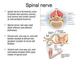
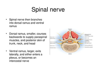
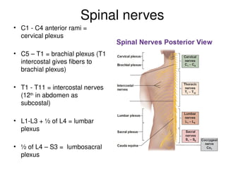
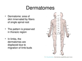
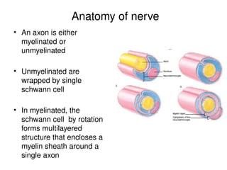
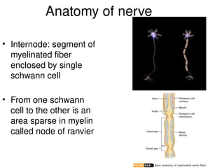
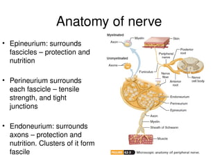
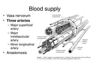
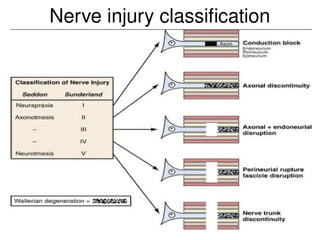
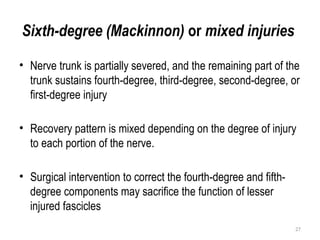
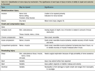
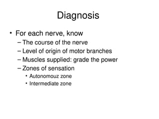
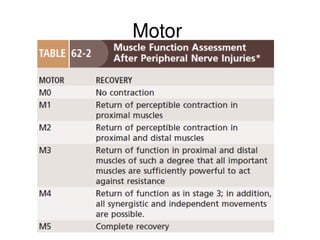
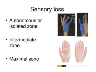
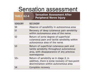
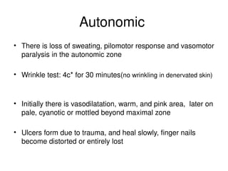
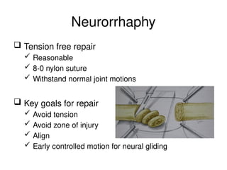
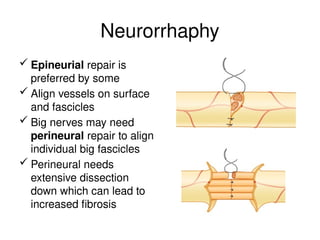
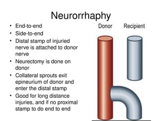
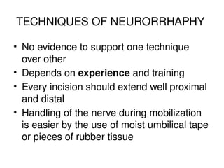
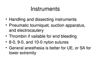
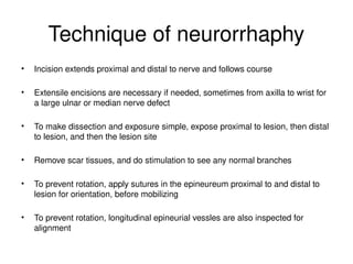
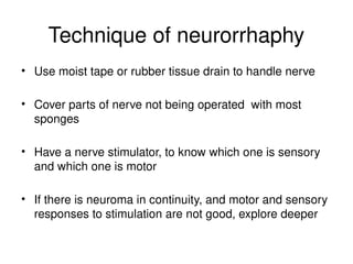
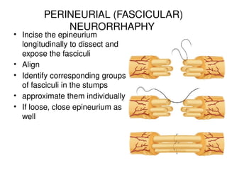
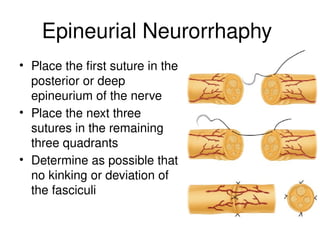
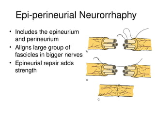
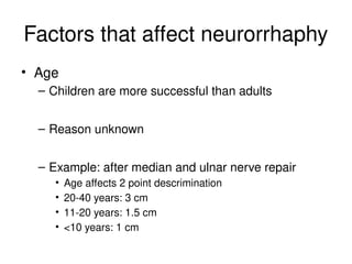
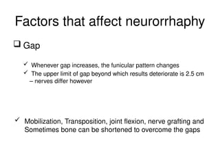
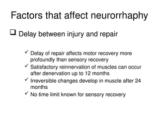
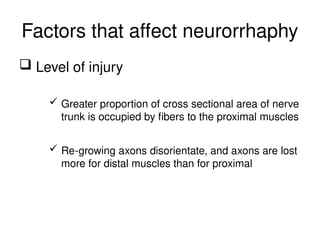
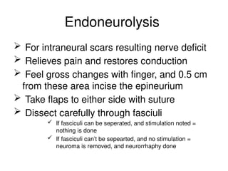
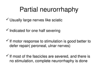
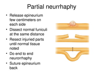
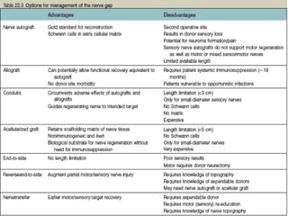
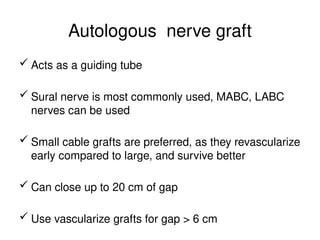
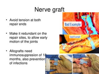
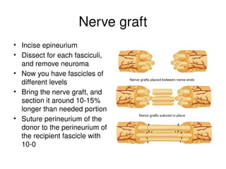
Peripheral nerve injury:
pathology, features, and treatment
Dr. Kolang (G. surgeon)
surgery dept,
JUNE.2024
Outline
• Anatomy, histology overview
• Pathology: degeneration and regeneration
• Classification of nerve injury
• Clinical feature and diagnosis
• Treatment of peripheral nerve injury
3
Organization
 The nervous system is divided into two parts
 The central nervous system (CNS)
• Brain and spinal cord
• Integrative and control centers
 The peripheral nervous system (PNS)
• Spinal and cranial nerves
• Communication lines between the CNS and the
rest of the body
Peripheral nerves – 31 pairs
• 8 cervical: C1-C7 exit above
named vertebra. C8 between C7
and T1
• 12 thoracic: exit below named
vertebra
• 5 lumbar: exit below named
vertebra
• 5 sacral: S1-S4 exit as anterior
division and posterior division,
through anterior and posterior
foramina respectively
• 1 coccygeal: S5 and Co1 exit
through sacral hiatus
Spinal nerve
• spinal nerve is formed by union
of dorsal root (sensory axons) ,
and ventral root (motor axons)
at intervertebral foramina
• Spinal nerve: two way road
(both efferent and afferent
pathways)
• Dorsal root: one way in, and cell
bodies located at dorsal root
ganglion (cluster of nerve cell
bodies)
• Ventral root: one way out, and
cell bodies located within grey
matter of spinal cord
Spinal nerve
• Spinal nerve then branches
into dorsal ramus and ventral
ramus
• Dorsal ramus, smaller, courses
backwards to supply paraspinal
muscles, and posterior skin of
trunk, neck, and head
• Ventral ramus, larger, exits
laterally, and either enters a
plexus, or becomes an
intercostal nerve
Spinal nerves
• C1 - C4 anterior rami =
cervical plexus
• C5 – T1 = brachial plexus (T1
intercostal gives fibers to
brachial plexus)
• T1 - T11 = intercostal nerves
(12th
in abdomen as
subcostal)
• L1-L3 + ½ of L4 = lumbar
plexus
• ½ of L4 – S3 = lumbosacral
plexus
Dermatomes
• Dermatome: area of
skin innervated by fibers
of single spinal root
• The pattern is preserved
in thoracic region
• In limbs, the
dermatomes are
displaced due to
migration of limb buds
Anatomy of nerve
• An axon is either
myelinated or
unmyelinated
• Unmyelinated are
wrapped by single
schwann cell
• In myelinated, the
schwann cell by rotation
forms multilayered
structure that encloses a
myelin sheath around a
single axon
Anatomy of nerve
• Internode: segment of
myelinated fiber
enclosed by single
schwann cell
• From one schwann
cell to the other is an
area sparse in myelin
called node of ranvier
Anatomy of nerve
• Epineurium: surrounds
fascicles – protection and
nutrition
• Perineurium surrounds
each fascicle – tensile
strength, and tight
junctions
• Endoneurium: surrounds
axons – protection and
nutrition. Clusters of it form
fascile
Blood supply
• Vasa nervorum
• Three arteries
– Major superficial
artery
– Major
intrafascicular
artery
– Minor longitudinal
artery
• Anastomosis
13
INTERNAL TOPOGRAPHY OF
PERIPHERAL NERVES
• Fascicular arrangement of
nerves are complex network
of branching and
intermingling fascicles that
constantly change throughout
the course of the nerve
14
• fascicular arrangement
is complex in the
proximal aspect than
the distal fascicles
Etiology
• Metabolic or collagen disease
• Malignancies
• Toxins
• Thermal
• Chemical
• Mechanical trauma
16
• Traumatic injury could be
 Primary injury
 secondary injury
 displaced osseous fragments,
 manipulation
 infection, scar, callus, or vascular
complications( hematoma, arteriovenous
fistula, ischemia, or aneurysm).
Pathogenesis
• Low grade injury can cause temporary block of hours to days
• More vigorous injury can demyelinate axons, and cause
conduction block of weeks for myelin to regrow
• Greater degrees of stretch or compression can cause
axonotmetic injury, with axon destruction, which may take
months to years
• Nerve stretch and compression injuries can disrupt vasa
nervorum  ischemia  necrosis
Pathology – degeneration & regeneration
• Pathologic events
occur at
–Cell body
–Distal stamp
–Proximal stamp
–End organ
Pathology – cell body
• Injuries nearer make
biggest changes
• Swells
• Chromotolysis, nissl
substance disperse
• Nucleus move
eccentrically
• Evident by 7 days
• In 4 – 6 weeks death or
recovery is apparent
• At recovery: edema
subside, nucleus central,
nissle reaccumulate
Pathology – proximal stamp
• Primary, traumatic or
retrograde
degeneration
• Usually one node
proximal, but can go
up to cell body
• Regenerating sprouts
noted within 24 hours
• Several day later,
sprouts emerge
Pathology – distal stamp
• Wallerian degeneration by
2 – 3 days
• Myelin detoriorates
• Axon disorganize
• Fluid loss  fragment 
shrink
• By 7 days, Phagocytes eat
myelin and axon debris
• 15 – 30 days, no debris left
• Tight junctions disrupt –
proteins act as toxins and
worsen condition (reforms
in 1 week)
Pathology – distal stamp
• Schwann cell lamina
persisted
• Bungner band
columns formed to
support sprouts
• Cone sprouts through
the band, helped by
fibronectin
• Releases proteases
to clean route
Pathology – distal stamp
• Cone survival/growth
helped by NGFs
• If endoneural tube
intact – previous
course
• Tight junctions in 01
week, complete in 03
• Schwann cells, and
endoneural tubes
degenerate 18 – 24
mo – re-growth
ceases
Pathology – muscle
• Paralyzed
• Atrophic changes 2 – 4 weeks (fibrillations)
• 30% loss in 1 month
• 50-70% loss in 2 months
• 60-80% in 4 months – hold on
• Striations and motor endplate
configurations are retained for longer
than 12 months
• Complete fibrosis not complete until 3 years
Pathology – sensory
• Only small area of sensory loss
(autonomous zone)
• Can have anesthetic intermediate zone
• 30-40% of small diameter sensory nerves
apoptose
• Sensory receptors degenerate longer
Nerve injury classification
Sixth-degree (Mackinnon) or mixed injuries
• Nerve trunk is partially severed, and the remaining part of the
trunk sustains fourth-degree, third-degree, second-degree, or
first-degree injury
• Recovery pattern is mixed depending on the degree of injury
to each portion of the nerve.
• Surgical intervention to correct the fourth-degree and fifth-
degree components may sacrifice the function of lesser
injured fascicles
27
Diagnosis
• For each nerve, know
– The course of the nerve
– Level of origin of motor branches
– Muscles supplied: grade the power
– Zones of sensation
• Autonomouz zone
• Intermediate zone
Diagnosing a nerve injury
• Evaluate nerve injury on arrival of the traumatized
patient, as well as after manipulation, cast, or
operation
• Follow patient clinically, and electrodiagnostically, to
minimize delay in recognizing a nerve deficit that will
not resolve spontaneously.
• On arrival pain usually limits examination
• There are simple tests to conduct to detect injuries of
major nerves of extremities even if patient is in severe
pain
Diagnosis
• Loss of pain perception in tip of little finger =
ulnar nerve injury
• Loss of pain perception in tip of index finger =
median nerve injury
• Inability to extend thumb = radial nerve injury
(see if extensors are intact and look muscle
belly)
Diagnosis
• Loss of pain perception in sole of foot =
sciatic or tibial nerve
• Inability to extend great toe or foot = peroneal
or sciatic injury
• Look muscle bellies or tendon continuity to
rely on these tests – many movements are
done with trick
Diagnosis
• Thumb opposition to little finger can be
done even if OP is cut or paralyzed
• Wrist can be partially extended by finger
flexion
• Elbow can be forcefully flexed by using BR
Motor
Sensory loss
• Autonomous or
isolated zone
• Intermediate
zone
• Maximal zone
Sensation assessment
Autonomic
• There is loss of sweating, pilomotor response and vasomotor
paralysis in the autonomic zone
• Wrinkle test: 4c* for 30 minutes(no wrinkling in denervated skin)
• Initially there is vasodilatation, warm, and pink area, later on
pale, cyanotic or mottled beyond maximal zone
• Ulcers form due to trauma, and heal slowly, finger nails
become distorted or entirely lost
Sweat test
• Sympathetic fibers are resistant to mechanical trauma
• Sweating within autonomous zone shows the nerve
injury is incomplete
• +20 lens of ophthalmoscope can show sweat beads
• Sweat test, is another means of testing, where
quinizarin powder is poured to the area
– denervated area remains dry and light gray
– and innervated area becomes deep purple
Diagnostic tests
• Tinel sign
• Electrodiagnostics
– NCS
– EMG
Tinel sign
• Percuss along the course of the injuried nerve with
hammer or finger, from distal to proximal
• Patient feels transient tingling sensation (which
persists for few seconds) at the distribution of the
nerve rather than area percussed
• It’s the regenerating unmyelinated portion of the
nerve that when percussed produces the tingling
sensation
Tinel sign
• Grade 1 has no tinel sign, because there is no
degenerated axon that is regenerating
• Grade 2 and 3 have advancing tinel sign (means,
every month 1 inch of proximal portion of the
nerve will have negative tinel sign as it get
myelinated)
• Grade 4 and 5 have no advancing tinel sign, as
there is no axon regeneration
NCS/EMG
• If done in 2 weeks, conduction studies can differentiate
neuropraxia from axono/neurotmesis
• In neuropraxia, there is conduction or motor unit
potentials present in 2 weeks, but there is no
conduction or MUPS in the other two
• In 12 weeks, if there are MUPS, it shows grade 2 and
3, and recovery is expected
• If no MUPS in 12 weeks, it shows grade 4 and 5 and
need surgical intervention.
43
EARLY MANAGEMENT OF NERVE
INJURIES
 General Considerations of Treatment
 Life and limb
 Systemic antibiotics and tetanus prophylaxis
 Assess PN injury
Management of PNI
• Possible scenarios
–Clean laceration
–Contaminated laceration
–Blast or crushing
• Without a gap
• With a gap
Management of PNI
• Primary repair: within 6 to 8 hours
• Delayed primary: within 1 week
• Secondary repair: after 1 week
• These timings are arbitrary
Management of PNI
• Primary repair has two main benefits
 Shortens time of end organ denervation
 Improves fascicular alignment as there is
minimal excision of ends
• In war wounds, early secondary suture is
preferred
Management of PNI
• Clean laceration
 Clean and debride thoroughly
 Do primary repair, if patient condition allows and if
personnel and equipment are available
 If not possible at the first debridement, do it in 3 to 7
days
 Suture, dress, and follow for infection
Management of PNI
 Contaminated laceration
 Debride thoroughly
 Apply sterile dressing
 Do delayed primary repair in 7 – 18 days
 Contamination increases risk of scarring
When to operate
• For nerve lacerations, due to glass or knife,
operation can be done in 72 hours, as both
ends are healthy
• Delay results in retraction of nerve stamps,
and increased gap which necessitate grafting
– outcome is not guaranteed there on
• End-to-end neurorhaphy is operative option
Management of PNI
 Blast or crush injury
Clean and debride
If there is no gap, appose loose end-to-end for later easier
repair and prevent retraction
If there is gap, suture ends to soft tissues to prevent
retraction
Do soft tissue coverage, if necessary
 Let the soft tissues heal & do secondary repair in 3 – 6
weeks
When to operate
• If injury is uncertain, like blunt trauma, or fracture,
re-innervation is followed clinically and EMG
• Demyelination corrects in 8-12 weeks: if no re-
innervation noted then there is axonal damage
which needs intervention
• If no innervation is noted in 3 months, exploration
is indicated
When to explore
 Sharp injury nerve transection
 Avulsion or blast, to identify and tag
 No regeneration noted after blunt or closed trauma
– appropriate time passed
 No regeneration after low velocity gunshot
 Nerve deficit immediately after manipulation and
casting
 Delay exploration if there is progressive
regeneration – clinical and electrodiagnostic
Why surgical timing matters
• Muscle completely atrophies in 12-18 months, when
denervated
• The nerve must reach that muscle, before it
atrophies after that period even re-innervation has
no benefit
• You calculate how long it will take a nerve to reach
the end organ by travelling 1mm/day or 1
inch/month starting from day of operation
• 3/5 muscle function is a good outcome
Surgical options
• Neurorrhaphy
• Neurolysis
• Partial neurorrhaphy
• Methods to close the gap
Neurorrhaphy
 Done as long as nerve ends can come together
without tension
 Tension results in scarring or breakdown at repair site
 Mild tension stimulates nerve growth
 Problems with fancy joint positions
 Dehiscence of repair with early motion
 Joint stiffness
Neurorrhaphy
 Tension free repair
 Reasonable
 8-0 nylon suture
 Withstand normal joint motions
 Key goals for repair
 Avoid tension
 Avoid zone of injury
 Align
 Early controlled motion for neural gliding
Neurorrhaphy
 Epineurial repair is
preferred by some
 Align vessels on surface
and fascicles
 Big nerves may need
perineural repair to align
individual big fascicles
 Perineural needs
extensive dissection
down which can lead to
increased fibrosis
Neurorrhaphy
• End-to-end
• Side-to-end
• Distal stamp of injuried
nerve is attached to donor
nerve
• Neurectomy is done on
donor
• Collateral sprouts exit
epineurium of donor and
enter the distal stamp
• Good for long distance
injuries, and if no proximal
stamp to do end to end
TECHNIQUES OF NEURORRHAPHY
• No evidence to support one technique
over other
• Depends on experience and training
• Every incision should extend well proximal
and distal
• Handling of the nerve during mobilization
is easier by the use of moist umbilical tape
or pieces of rubber tissue
Instruments
• Handling and dissecting instruments
• Pneumatic tourniquet, suction apparatus,
and electrocautery
• Thrombin if vailable for end bleeding
• 8-0, 9-0, and 10-0 nylon sutures
• General anesthesia is better for UE, or SA for
lower extremity
Technique of neurorrhaphy
• Incision extends proximal and distal to nerve and follows course
• Extensile encisions are necessary if needed, sometimes from axilla to wrist for
a large ulnar or median nerve defect
• To make dissection and exposure simple, expose proximal to lesion, then distal
to lesion, and then the lesion site
• Remove scar tissues, and do stimulation to see any normal branches
• To prevent rotation, apply sutures in the epineureum proximal to and distal to
lesion for orientation, before mobilizing
• To prevent rotation, longitudinal epineurial vessles are also inspected for
alignment
Technique of neurorrhaphy
• Use moist tape or rubber tissue drain to handle nerve
• Cover parts of nerve not being operated with most
sponges
• Have a nerve stimulator, to know which one is sensory
and which one is motor
• If there is neuroma in continuity, and motor and sensory
responses to stimulation are not good, explore deeper
PERINEURIAL (FASCICULAR)
NEURORRHAPHY
• Incise the epineurium
longitudinally to dissect and
expose the fasciculi
• Align
• Identify corresponding groups
of fasciculi in the stumps
• approximate them individually
• If loose, close epineurium as
well
Epineurial Neurorrhaphy
• Place the first suture in the
posterior or deep
epineurium of the nerve
• Place the next three
sutures in the remaining
three quadrants
• Determine as possible that
no kinking or deviation of
the fasciculi
Epi-perineurial Neurorrhaphy
• Includes the epineurium
and perineurium
• Aligns large group of
fascicles in bigger nerves
• Epineurial repair adds
strength
Factors that affect neurorrhaphy
• Age
– Children are more successful than adults
– Reason unknown
– Example: after median and ulnar nerve repair
• Age affects 2 point descrimination
• 20-40 years: 3 cm
• 11-20 years: 1.5 cm
• <10 years: 1 cm
Factors that affect neurorrhaphy
 Gap
 Whenever gap increases, the funicular pattern changes
 The upper limit of gap beyond which results deteriorate is 2.5 cm
– nerves differ however
 Mobilization, Transposition, joint flexion, nerve grafting and
Sometimes bone can be shortened to overcome the gaps
Factors that affect neurorrhaphy
 Delay between injury and repair
 Delay of repair affects motor recovery more
profoundly than sensory recovery
 Satisfactory reinnervation of muscles can occur
after denervation up to 12 months
 Irreversible changes develop in muscle after 24
months
 No time limit known for sensory recovery
Factors that affect neurorrhaphy
 Level of injury
 Greater proportion of cross sectional area of nerve
trunk is occupied by fibers to the proximal muscles
 Re-growing axons disorientate, and axons are lost
more for distal muscles than for proximal
Endoneurolysis
 For intraneural scars resulting nerve deficit
 Relieves pain and restores conduction
 Feel gross changes with finger, and 0.5 cm
from these area incise the epineurium
 Take flaps to either side with suture
 Dissect carefully through fasciuli
 If fasciculi can be seperated, and stimulation noted =
nothing is done
 If fasciculi can’t be sepearted, and no stimulation =
neuroma is removed, and neurorrhaphy done
Partial neurorrhaphy
Usually large nerves like sciatic
Indicated for one half severing
If motor response to stimulation is good better to
defer repair( peroneal, ulnar nerves)
If most of the fascicles are severed, and there is
no stimulation, complete neurorrhaphy is done
Partial neurrhaphy
• Release epineurium
few centimeters on
each side
• Dissect normal funiculi
at the same distance
• Resect injuried parts
until normal tissue
noted
• Do end to end
neurorrhaphy
• Suture epineurium
back
Methods to close nerve gap
Mobilization
Transposition
Joint positions
 Nerve autograft
 Nerve allograft
 Conduits
 Nerve transfer
Autologous nerve graft
 Acts as a guiding tube
 Sural nerve is most commonly used, MABC, LABC
nerves can be used
 Small cable grafts are preferred, as they revascularize
early compared to large, and survive better
 Can close up to 20 cm of gap
 Use vascularize grafts for gap > 6 cm
Nerve graft
• Avoid tension at both
repair ends
• Make it redundant on the
repair sites, to allow early
motion of the joints
• Allografts need
immunosuppresion of 18
months, also prevention
of infections
Nerve graft
• Incise epineurium
• Dissect for each fasciculi,
and remove neuroma
• Now you have fascicles of
different levels
• Bring the nerve graft, and
section it around 10-15%
longer than needed portion
• Suture perineurium of the
donor to the perineurium of
the recipient fascicle with
10-0
NERVE CONDUITS
Bridging tube
As primary conduit or nerve wrap
Only for sensory gaps < 3 cm
Biologic: nerve, bone, vein, artery
Synthetic: silicon
As a wrap: if repair site dehisces still nerve can
go through the conduit canal
NERVE TRANSFER
Post operative care
Immobilize for 1-2 weeks
Consider associated injury(tendon or bone)
Start gentle ROM in 3 days to help gliding –
depends on intra-op tolerated motion
Pain management – paresthesia and electric
shock, may need narcotics, neuroleptics
POSTOPERATIVE MANAGEMENT
In 1 – 2 week, inspect wound and remove
sutures
6 to 12 weeks after surgery, careful attention
should be paid to the avoidance of fixed
contractures
8 to 12 weeks after surgery, progressive
strengthening exercises are begun
References
• S. Terry Canale James Beaty. Campbell's Operative
Orthopaedics. Pp3346-3413
• Steve K. Lee, MD, and Scott W. Wolfe, MD. Peripheral Nerve
Injury and Repair. J Am Acad Orthop Surg 2000;8:243-252
• Deal DN et. Nerve conduits for nerve repair or reconstruction.
Al J Am Acad Orthop Surg. 2012
• Campbell, William W., "Evaluation and management of
peripheral nerve injury" (2008). Uniformed Services University
of the Health Sciences. 3.http://digitalcommons.unl.edu/usuhs/3
• S. Hall. The response to injury in the peripheral
• nervous system. Review article. Society of Bone and
• Joint Surgery. 2005

peripheral nerve injury.pptx from surgery

  • 1.
    Peripheral nerve injury: pathology,features, and treatment Dr. Kolang (G. surgeon) surgery dept, JUNE.2024
  • 2.
    Outline • Anatomy, histologyoverview • Pathology: degeneration and regeneration • Classification of nerve injury • Clinical feature and diagnosis • Treatment of peripheral nerve injury
  • 3.
    3 Organization  The nervoussystem is divided into two parts  The central nervous system (CNS) • Brain and spinal cord • Integrative and control centers  The peripheral nervous system (PNS) • Spinal and cranial nerves • Communication lines between the CNS and the rest of the body
  • 4.
    Peripheral nerves –31 pairs • 8 cervical: C1-C7 exit above named vertebra. C8 between C7 and T1 • 12 thoracic: exit below named vertebra • 5 lumbar: exit below named vertebra • 5 sacral: S1-S4 exit as anterior division and posterior division, through anterior and posterior foramina respectively • 1 coccygeal: S5 and Co1 exit through sacral hiatus
  • 5.
    Spinal nerve • spinalnerve is formed by union of dorsal root (sensory axons) , and ventral root (motor axons) at intervertebral foramina • Spinal nerve: two way road (both efferent and afferent pathways) • Dorsal root: one way in, and cell bodies located at dorsal root ganglion (cluster of nerve cell bodies) • Ventral root: one way out, and cell bodies located within grey matter of spinal cord
  • 6.
    Spinal nerve • Spinalnerve then branches into dorsal ramus and ventral ramus • Dorsal ramus, smaller, courses backwards to supply paraspinal muscles, and posterior skin of trunk, neck, and head • Ventral ramus, larger, exits laterally, and either enters a plexus, or becomes an intercostal nerve
  • 7.
    Spinal nerves • C1- C4 anterior rami = cervical plexus • C5 – T1 = brachial plexus (T1 intercostal gives fibers to brachial plexus) • T1 - T11 = intercostal nerves (12th in abdomen as subcostal) • L1-L3 + ½ of L4 = lumbar plexus • ½ of L4 – S3 = lumbosacral plexus
  • 8.
    Dermatomes • Dermatome: areaof skin innervated by fibers of single spinal root • The pattern is preserved in thoracic region • In limbs, the dermatomes are displaced due to migration of limb buds
  • 9.
    Anatomy of nerve •An axon is either myelinated or unmyelinated • Unmyelinated are wrapped by single schwann cell • In myelinated, the schwann cell by rotation forms multilayered structure that encloses a myelin sheath around a single axon
  • 10.
    Anatomy of nerve •Internode: segment of myelinated fiber enclosed by single schwann cell • From one schwann cell to the other is an area sparse in myelin called node of ranvier
  • 11.
    Anatomy of nerve •Epineurium: surrounds fascicles – protection and nutrition • Perineurium surrounds each fascicle – tensile strength, and tight junctions • Endoneurium: surrounds axons – protection and nutrition. Clusters of it form fascile
  • 12.
    Blood supply • Vasanervorum • Three arteries – Major superficial artery – Major intrafascicular artery – Minor longitudinal artery • Anastomosis
  • 13.
    13 INTERNAL TOPOGRAPHY OF PERIPHERALNERVES • Fascicular arrangement of nerves are complex network of branching and intermingling fascicles that constantly change throughout the course of the nerve
  • 14.
    14 • fascicular arrangement iscomplex in the proximal aspect than the distal fascicles
  • 15.
    Etiology • Metabolic orcollagen disease • Malignancies • Toxins • Thermal • Chemical • Mechanical trauma
  • 16.
    16 • Traumatic injurycould be  Primary injury  secondary injury  displaced osseous fragments,  manipulation  infection, scar, callus, or vascular complications( hematoma, arteriovenous fistula, ischemia, or aneurysm).
  • 17.
    Pathogenesis • Low gradeinjury can cause temporary block of hours to days • More vigorous injury can demyelinate axons, and cause conduction block of weeks for myelin to regrow • Greater degrees of stretch or compression can cause axonotmetic injury, with axon destruction, which may take months to years • Nerve stretch and compression injuries can disrupt vasa nervorum  ischemia  necrosis
  • 18.
    Pathology – degeneration& regeneration • Pathologic events occur at –Cell body –Distal stamp –Proximal stamp –End organ
  • 19.
    Pathology – cellbody • Injuries nearer make biggest changes • Swells • Chromotolysis, nissl substance disperse • Nucleus move eccentrically • Evident by 7 days • In 4 – 6 weeks death or recovery is apparent • At recovery: edema subside, nucleus central, nissle reaccumulate
  • 20.
    Pathology – proximalstamp • Primary, traumatic or retrograde degeneration • Usually one node proximal, but can go up to cell body • Regenerating sprouts noted within 24 hours • Several day later, sprouts emerge
  • 21.
    Pathology – distalstamp • Wallerian degeneration by 2 – 3 days • Myelin detoriorates • Axon disorganize • Fluid loss  fragment  shrink • By 7 days, Phagocytes eat myelin and axon debris • 15 – 30 days, no debris left • Tight junctions disrupt – proteins act as toxins and worsen condition (reforms in 1 week)
  • 22.
    Pathology – distalstamp • Schwann cell lamina persisted • Bungner band columns formed to support sprouts • Cone sprouts through the band, helped by fibronectin • Releases proteases to clean route
  • 23.
    Pathology – distalstamp • Cone survival/growth helped by NGFs • If endoneural tube intact – previous course • Tight junctions in 01 week, complete in 03 • Schwann cells, and endoneural tubes degenerate 18 – 24 mo – re-growth ceases
  • 24.
    Pathology – muscle •Paralyzed • Atrophic changes 2 – 4 weeks (fibrillations) • 30% loss in 1 month • 50-70% loss in 2 months • 60-80% in 4 months – hold on • Striations and motor endplate configurations are retained for longer than 12 months • Complete fibrosis not complete until 3 years
  • 25.
    Pathology – sensory •Only small area of sensory loss (autonomous zone) • Can have anesthetic intermediate zone • 30-40% of small diameter sensory nerves apoptose • Sensory receptors degenerate longer
  • 26.
  • 27.
    Sixth-degree (Mackinnon) ormixed injuries • Nerve trunk is partially severed, and the remaining part of the trunk sustains fourth-degree, third-degree, second-degree, or first-degree injury • Recovery pattern is mixed depending on the degree of injury to each portion of the nerve. • Surgical intervention to correct the fourth-degree and fifth- degree components may sacrifice the function of lesser injured fascicles 27
  • 29.
    Diagnosis • For eachnerve, know – The course of the nerve – Level of origin of motor branches – Muscles supplied: grade the power – Zones of sensation • Autonomouz zone • Intermediate zone
  • 30.
    Diagnosing a nerveinjury • Evaluate nerve injury on arrival of the traumatized patient, as well as after manipulation, cast, or operation • Follow patient clinically, and electrodiagnostically, to minimize delay in recognizing a nerve deficit that will not resolve spontaneously. • On arrival pain usually limits examination • There are simple tests to conduct to detect injuries of major nerves of extremities even if patient is in severe pain
  • 31.
    Diagnosis • Loss ofpain perception in tip of little finger = ulnar nerve injury • Loss of pain perception in tip of index finger = median nerve injury • Inability to extend thumb = radial nerve injury (see if extensors are intact and look muscle belly)
  • 32.
    Diagnosis • Loss ofpain perception in sole of foot = sciatic or tibial nerve • Inability to extend great toe or foot = peroneal or sciatic injury • Look muscle bellies or tendon continuity to rely on these tests – many movements are done with trick
  • 33.
    Diagnosis • Thumb oppositionto little finger can be done even if OP is cut or paralyzed • Wrist can be partially extended by finger flexion • Elbow can be forcefully flexed by using BR
  • 34.
  • 35.
    Sensory loss • Autonomousor isolated zone • Intermediate zone • Maximal zone
  • 36.
  • 37.
    Autonomic • There isloss of sweating, pilomotor response and vasomotor paralysis in the autonomic zone • Wrinkle test: 4c* for 30 minutes(no wrinkling in denervated skin) • Initially there is vasodilatation, warm, and pink area, later on pale, cyanotic or mottled beyond maximal zone • Ulcers form due to trauma, and heal slowly, finger nails become distorted or entirely lost
  • 38.
    Sweat test • Sympatheticfibers are resistant to mechanical trauma • Sweating within autonomous zone shows the nerve injury is incomplete • +20 lens of ophthalmoscope can show sweat beads • Sweat test, is another means of testing, where quinizarin powder is poured to the area – denervated area remains dry and light gray – and innervated area becomes deep purple
  • 39.
    Diagnostic tests • Tinelsign • Electrodiagnostics – NCS – EMG
  • 40.
    Tinel sign • Percussalong the course of the injuried nerve with hammer or finger, from distal to proximal • Patient feels transient tingling sensation (which persists for few seconds) at the distribution of the nerve rather than area percussed • It’s the regenerating unmyelinated portion of the nerve that when percussed produces the tingling sensation
  • 41.
    Tinel sign • Grade1 has no tinel sign, because there is no degenerated axon that is regenerating • Grade 2 and 3 have advancing tinel sign (means, every month 1 inch of proximal portion of the nerve will have negative tinel sign as it get myelinated) • Grade 4 and 5 have no advancing tinel sign, as there is no axon regeneration
  • 42.
    NCS/EMG • If donein 2 weeks, conduction studies can differentiate neuropraxia from axono/neurotmesis • In neuropraxia, there is conduction or motor unit potentials present in 2 weeks, but there is no conduction or MUPS in the other two • In 12 weeks, if there are MUPS, it shows grade 2 and 3, and recovery is expected • If no MUPS in 12 weeks, it shows grade 4 and 5 and need surgical intervention.
  • 43.
    43 EARLY MANAGEMENT OFNERVE INJURIES  General Considerations of Treatment  Life and limb  Systemic antibiotics and tetanus prophylaxis  Assess PN injury
  • 44.
    Management of PNI •Possible scenarios –Clean laceration –Contaminated laceration –Blast or crushing • Without a gap • With a gap
  • 45.
    Management of PNI •Primary repair: within 6 to 8 hours • Delayed primary: within 1 week • Secondary repair: after 1 week • These timings are arbitrary
  • 46.
    Management of PNI •Primary repair has two main benefits  Shortens time of end organ denervation  Improves fascicular alignment as there is minimal excision of ends • In war wounds, early secondary suture is preferred
  • 47.
    Management of PNI •Clean laceration  Clean and debride thoroughly  Do primary repair, if patient condition allows and if personnel and equipment are available  If not possible at the first debridement, do it in 3 to 7 days  Suture, dress, and follow for infection
  • 48.
    Management of PNI Contaminated laceration  Debride thoroughly  Apply sterile dressing  Do delayed primary repair in 7 – 18 days  Contamination increases risk of scarring
  • 49.
    When to operate •For nerve lacerations, due to glass or knife, operation can be done in 72 hours, as both ends are healthy • Delay results in retraction of nerve stamps, and increased gap which necessitate grafting – outcome is not guaranteed there on • End-to-end neurorhaphy is operative option
  • 51.
    Management of PNI Blast or crush injury Clean and debride If there is no gap, appose loose end-to-end for later easier repair and prevent retraction If there is gap, suture ends to soft tissues to prevent retraction Do soft tissue coverage, if necessary  Let the soft tissues heal & do secondary repair in 3 – 6 weeks
  • 52.
    When to operate •If injury is uncertain, like blunt trauma, or fracture, re-innervation is followed clinically and EMG • Demyelination corrects in 8-12 weeks: if no re- innervation noted then there is axonal damage which needs intervention • If no innervation is noted in 3 months, exploration is indicated
  • 54.
    When to explore Sharp injury nerve transection  Avulsion or blast, to identify and tag  No regeneration noted after blunt or closed trauma – appropriate time passed  No regeneration after low velocity gunshot  Nerve deficit immediately after manipulation and casting  Delay exploration if there is progressive regeneration – clinical and electrodiagnostic
  • 55.
    Why surgical timingmatters • Muscle completely atrophies in 12-18 months, when denervated • The nerve must reach that muscle, before it atrophies after that period even re-innervation has no benefit • You calculate how long it will take a nerve to reach the end organ by travelling 1mm/day or 1 inch/month starting from day of operation • 3/5 muscle function is a good outcome
  • 56.
    Surgical options • Neurorrhaphy •Neurolysis • Partial neurorrhaphy • Methods to close the gap
  • 57.
    Neurorrhaphy  Done aslong as nerve ends can come together without tension  Tension results in scarring or breakdown at repair site  Mild tension stimulates nerve growth  Problems with fancy joint positions  Dehiscence of repair with early motion  Joint stiffness
  • 58.
    Neurorrhaphy  Tension freerepair  Reasonable  8-0 nylon suture  Withstand normal joint motions  Key goals for repair  Avoid tension  Avoid zone of injury  Align  Early controlled motion for neural gliding
  • 59.
    Neurorrhaphy  Epineurial repairis preferred by some  Align vessels on surface and fascicles  Big nerves may need perineural repair to align individual big fascicles  Perineural needs extensive dissection down which can lead to increased fibrosis
  • 60.
    Neurorrhaphy • End-to-end • Side-to-end •Distal stamp of injuried nerve is attached to donor nerve • Neurectomy is done on donor • Collateral sprouts exit epineurium of donor and enter the distal stamp • Good for long distance injuries, and if no proximal stamp to do end to end
  • 61.
    TECHNIQUES OF NEURORRHAPHY •No evidence to support one technique over other • Depends on experience and training • Every incision should extend well proximal and distal • Handling of the nerve during mobilization is easier by the use of moist umbilical tape or pieces of rubber tissue
  • 62.
    Instruments • Handling anddissecting instruments • Pneumatic tourniquet, suction apparatus, and electrocautery • Thrombin if vailable for end bleeding • 8-0, 9-0, and 10-0 nylon sutures • General anesthesia is better for UE, or SA for lower extremity
  • 63.
    Technique of neurorrhaphy •Incision extends proximal and distal to nerve and follows course • Extensile encisions are necessary if needed, sometimes from axilla to wrist for a large ulnar or median nerve defect • To make dissection and exposure simple, expose proximal to lesion, then distal to lesion, and then the lesion site • Remove scar tissues, and do stimulation to see any normal branches • To prevent rotation, apply sutures in the epineureum proximal to and distal to lesion for orientation, before mobilizing • To prevent rotation, longitudinal epineurial vessles are also inspected for alignment
  • 64.
    Technique of neurorrhaphy •Use moist tape or rubber tissue drain to handle nerve • Cover parts of nerve not being operated with most sponges • Have a nerve stimulator, to know which one is sensory and which one is motor • If there is neuroma in continuity, and motor and sensory responses to stimulation are not good, explore deeper
  • 65.
    PERINEURIAL (FASCICULAR) NEURORRHAPHY • Incisethe epineurium longitudinally to dissect and expose the fasciculi • Align • Identify corresponding groups of fasciculi in the stumps • approximate them individually • If loose, close epineurium as well
  • 66.
    Epineurial Neurorrhaphy • Placethe first suture in the posterior or deep epineurium of the nerve • Place the next three sutures in the remaining three quadrants • Determine as possible that no kinking or deviation of the fasciculi
  • 67.
    Epi-perineurial Neurorrhaphy • Includesthe epineurium and perineurium • Aligns large group of fascicles in bigger nerves • Epineurial repair adds strength
  • 68.
    Factors that affectneurorrhaphy • Age – Children are more successful than adults – Reason unknown – Example: after median and ulnar nerve repair • Age affects 2 point descrimination • 20-40 years: 3 cm • 11-20 years: 1.5 cm • <10 years: 1 cm
  • 69.
    Factors that affectneurorrhaphy  Gap  Whenever gap increases, the funicular pattern changes  The upper limit of gap beyond which results deteriorate is 2.5 cm – nerves differ however  Mobilization, Transposition, joint flexion, nerve grafting and Sometimes bone can be shortened to overcome the gaps
  • 70.
    Factors that affectneurorrhaphy  Delay between injury and repair  Delay of repair affects motor recovery more profoundly than sensory recovery  Satisfactory reinnervation of muscles can occur after denervation up to 12 months  Irreversible changes develop in muscle after 24 months  No time limit known for sensory recovery
  • 71.
    Factors that affectneurorrhaphy  Level of injury  Greater proportion of cross sectional area of nerve trunk is occupied by fibers to the proximal muscles  Re-growing axons disorientate, and axons are lost more for distal muscles than for proximal
  • 72.
    Endoneurolysis  For intraneuralscars resulting nerve deficit  Relieves pain and restores conduction  Feel gross changes with finger, and 0.5 cm from these area incise the epineurium  Take flaps to either side with suture  Dissect carefully through fasciuli  If fasciculi can be seperated, and stimulation noted = nothing is done  If fasciculi can’t be sepearted, and no stimulation = neuroma is removed, and neurorrhaphy done
  • 73.
    Partial neurorrhaphy Usually largenerves like sciatic Indicated for one half severing If motor response to stimulation is good better to defer repair( peroneal, ulnar nerves) If most of the fascicles are severed, and there is no stimulation, complete neurorrhaphy is done
  • 74.
    Partial neurrhaphy • Releaseepineurium few centimeters on each side • Dissect normal funiculi at the same distance • Resect injuried parts until normal tissue noted • Do end to end neurorrhaphy • Suture epineurium back
  • 75.
    Methods to closenerve gap Mobilization Transposition Joint positions  Nerve autograft  Nerve allograft  Conduits  Nerve transfer
  • 77.
    Autologous nerve graft Acts as a guiding tube  Sural nerve is most commonly used, MABC, LABC nerves can be used  Small cable grafts are preferred, as they revascularize early compared to large, and survive better  Can close up to 20 cm of gap  Use vascularize grafts for gap > 6 cm
  • 78.
    Nerve graft • Avoidtension at both repair ends • Make it redundant on the repair sites, to allow early motion of the joints • Allografts need immunosuppresion of 18 months, also prevention of infections
  • 79.
    Nerve graft • Inciseepineurium • Dissect for each fasciculi, and remove neuroma • Now you have fascicles of different levels • Bring the nerve graft, and section it around 10-15% longer than needed portion • Suture perineurium of the donor to the perineurium of the recipient fascicle with 10-0
  • 80.
    NERVE CONDUITS Bridging tube Asprimary conduit or nerve wrap Only for sensory gaps < 3 cm Biologic: nerve, bone, vein, artery Synthetic: silicon As a wrap: if repair site dehisces still nerve can go through the conduit canal
  • 81.
  • 82.
    Post operative care Immobilizefor 1-2 weeks Consider associated injury(tendon or bone) Start gentle ROM in 3 days to help gliding – depends on intra-op tolerated motion Pain management – paresthesia and electric shock, may need narcotics, neuroleptics
  • 83.
    POSTOPERATIVE MANAGEMENT In 1– 2 week, inspect wound and remove sutures 6 to 12 weeks after surgery, careful attention should be paid to the avoidance of fixed contractures 8 to 12 weeks after surgery, progressive strengthening exercises are begun
  • 84.
    References • S. TerryCanale James Beaty. Campbell's Operative Orthopaedics. Pp3346-3413 • Steve K. Lee, MD, and Scott W. Wolfe, MD. Peripheral Nerve Injury and Repair. J Am Acad Orthop Surg 2000;8:243-252 • Deal DN et. Nerve conduits for nerve repair or reconstruction. Al J Am Acad Orthop Surg. 2012 • Campbell, William W., "Evaluation and management of peripheral nerve injury" (2008). Uniformed Services University of the Health Sciences. 3.http://digitalcommons.unl.edu/usuhs/3 • S. Hall. The response to injury in the peripheral • nervous system. Review article. Society of Bone and • Joint Surgery. 2005

Editor's Notes

  • #6 First three cervical dorsal ramus, are larger, because they supply large area of scalp
  • #52 For more distal nerve injuries operation can be done in 6 months
  • #65 If multiple small fasciculi, approximate several as a group
  • #82 No consensus on how long to immobilize