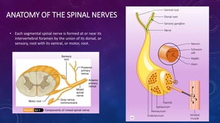
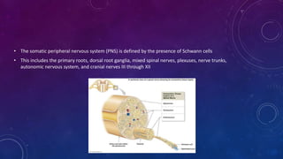
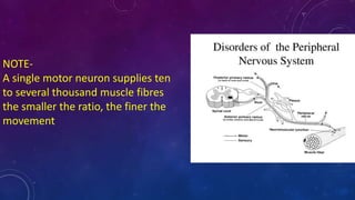
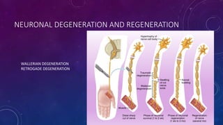
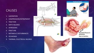
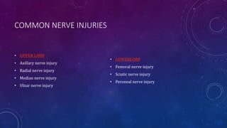
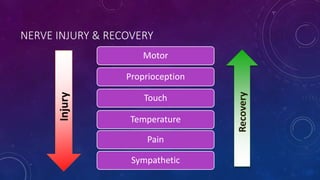
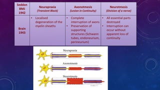
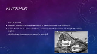
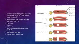
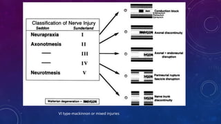
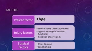
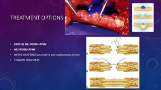
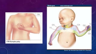
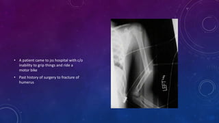
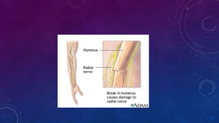
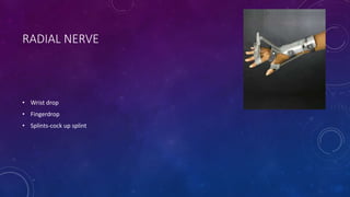
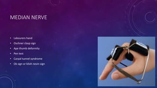
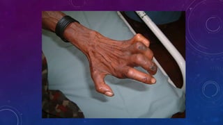
PERIPHERAL NERVE
INJURIES
DR SUPREETH
DR RAVI VARMA
ANATOMY OF THE SPINAL NERVES
• Each segmental spinal nerve is formed at or near its
intervertebral foramen by the union of its dorsal, or
sensory, root with its ventral, or motor, root.
• The somatic peripheral nervous system (PNS) is defined by the presence of Schwann cells
• This includes the primary roots, dorsal root ganglia, mixed spinal nerves, plexuses, nerve trunks,
autonomic nervous system, and cranial nerves III through XII
NOTE-
A single motor neuron supplies ten
to several thousand muscle fibres
the smaller the ratio, the finer the
movement
COMPONENTS OF SPINAL NERVE
• MOTOR
• SENSORY
• SYMPATHETIC
NEURONAL DEGENERATION AND REGENERATION
WALLERIAN DEGENERATION
RETROGADE DEGENERATION
CAUSES
1. LACERATION
2. COMPRESSION/ENTRAPMENT
3. TRACTION
4. BIRTH INJURIES
5. IATROGENIC
6. FRACTURE
7. METABOLIC DISTURBANCES
8. ISCHAEMIA
9. THERMAL /ELECTRICAL INJURIES
COMMON NERVE INJURIES
• UPPER LIMB
• Axillary nerve injury
• Radial nerve injury
• Median nerve injury
• Ulnar nerve injury
• LOWERLIMB
• Femoral nerve injury
• Sciatic nerve injury
• Peroneal nerve injury
NERVE INJURY & RECOVERY
Motor
Proprioception
Touch
Temperature
Pain
Sympathetic
Recovery
Injury
Seddon
BMJ
1942
Neurapraxia
(Transient Block)
Axonotmesis
(Lesion in Continuity)
Neurotmesis
(Division of a nerve)
Brain
1943
• Localised
degeneration of the
myelin sheaths
• Complete
interruption of axons
• Preservation of
supporting
structures (Schwann
tubes, endoneurium,
perineurium)
• All essential parts
destroyed
• Interruption can
occur without
apparent loss of
continuity
TRANSIENT ISCHAEMIA-
• Acute nerve compression causes numbness and tingling within 15 minutes
• loss of pain sensibility after 30 minutes and muscle weakness after 45 minutes
• Relief of compression is followed by intense paraesthesia lasting up to 5 minutes
• feeling is restored within 30 second
• TRANSIENT ENDONEURIAL ANOXIA AND THEY LEAVE NO TRACE OF NERVE DAMAGE.
NEURAPRAXIA
• ‘neurapraxia’ to describe a reversible block to nerve conduction
• minor contusion or compression of a peripheral nerve with preservation of the axis- cylinder
• Transmission of impulses is physiologically interrupted for a time
• recovery is complete in 3-6 weeks
• Tinel’s sign-negative
AXONOTMESIS
• designating more significant injury
• breakdown of the axon and distal wallerian degener-ation but with preservation of the Schwann cell
and endoneurial tubes.
• Spontaneous regeneration with good functional recovery can be expected
• Tinels sign-positive
NEUROTMESIS
• more severe injury
• complete anatomical severance of the nerve or extensive avulsing or crushing injury
• Axon,Schwann cell and endoneurial tubes , eperineurium and epineurium are disrupted to varying
degrees
• significant spontaneous recovery cannot be expected.
IN CLINICAL PRACTICE, HOW DO YOU DISTINGUISH?
AXONOTMESIS VERSUS NEUROTMESIS
• Nature of injury
• Serial observations
• Exploration
• Imaging
SUNDERLAND CLASSIFICATION
Sunderland
1951 I II III IV V
Focal
conduction
block
NO Wallerian
degeneration
Axonal
Disruption
Axon
+
Endoneurium
Disruption
Axon
+
Endoneurium
+
Perineurium
Disruption
Axon
+
Endoneurium
+
Perineurium
+
Epineurium
Disruption
Cross-innervation
• In this classification, peripheral nerve
injuries are arranged in ascending
order of severity
• Anatomically, the various degrees
represent injury to
• (1) myelin,
• (2) axon,
• (3) the endoneurial tube and its
contents,
• (4) perineurium, and
• (5) the entire nerve trunk
VI type-mackinnon or mixed injuries
DIAGNOSIS
• NOTE-Always test for nerve injuries following any significant trauma. And test
again after manipulation or operation, in case the nerve has been damaged
during treatment!
acute chronic
MUSCLE POWER
• Muscles supplied by the nerve
should be tested repeatedly
• Nerve regenaration at 1mm per
day
• MOTOR MARCH
SENSORY EXAMINATION
• Sensory loss usually follows a definite anatomical pattern,
• area supplied exclusively by the severed nerve and is
called the AUTONOMOUS ZONE
• Axonometesis –cross innervation
• RELEXES
• AUTONOMIC- SWEATING
• TROPHIC CHANGES
• NAIL CHANGES
• OSTEOPOROSIS
TINEL’S SIGN
• A classic sign of progressive nerve recovery is
peripheral tingling provoked by percussing
the nerve at the site of injury
• NOTE- where regenerating axons are most
sensitive
DIAGNOSTIC TESTS
• EMG-earliest indicator
• NERVE CONDUCTION STUDY
• SWEAT TEST
• MRI
TREATMENT
• Conservative
• surgical
PREREQUISITES FOR NERVE REPAIR
• Skeletal stability
• Healthy tissue bed
• Healthy nerve ends
• No undue tension
• Adequate soft tissue coverage
FACTORS
•AgePatient factor
• Level of injury (distal vs proximal)
• Type of nerve (pure vs mixed
functions)
• Condition of nerve ends
Injury factors
• Delay to repair
• Length of gap
Surgical
factors
INDICATION FOR SURGERY
• Sharp injury dividing a nerve
• Blast injury
• In a closed injury when no signs of recovery by appropriat time
TIME OF OPERATION
Open injury
Primary repair(6-8
hours)
Delayed primary
repair(7-18 days)
Secondary
repair(after 18 days)
Closed
injury
Conservative(splints)
Delayed intervention
GOOD PROGNOSTIC FACTORS
• G-growing age
• O-only motor
• O-only sensory
• D-distal lesion
• N-neuropraxia
• E-early repair
• R-radial nerve
• V-vascularity
• E-end to end repair
GOOD NERVE
TREATMENT OPTIONS
• PARTIAL NEURORRHAPHY
• NEURORRHAPHY
• NERVE GRAFTING(sural nerve and saphaneous nerve)
• TENDON TRANSFERS
CARE OF PARALYSED PARTS
While recovery is awaited,
• the skin must be protected from friction damage and burns.
• The joints should be moved through their full range twice daily to prevent stiffness and minimize the
work required of muscles when they recover.
• SPLINTS-both dynamic and static alternatively
• A newly born child, delivered by full term vaginal delivery- shoulder presentation,birth weight-4 kg,born
to a gestational diabetes mother presented with deformity of left upper limb,with hand held in
pronation of forearm and hand held close to the body
• A patient with a past history of shoulder
dislocation,which was reduced in phc
and c/o weakness in abduction and loss
of sensory supply of lateral aspect of arm
• A patient came to jss hospital with c/o
inability to grip things and ride a
motor bike
• Past history of surgery to fracture of
humerus
CRUTCH PALSY
• A jss ug student residing in hostel came to opd with c/o weakness in fingers and deformity after socials
of their batch
RADIAL NERVE
• Wrist drop
• Fingerdrop
• Splints-cock up splint
• A software engineer presented with
tingling and numbness of radial side
of hand with thenar atrophy
MEDIAN NERVE
• Labourers hand
• Oschner clasp sign
• Ape thumb deformity
• Pen test
• Carpal tunnel syndrome
• Ok sign or kiloh nevin sign
ULNAR NERVE
• Musician nerve
• Card test
• Froments sign
• Ulnar paradox
• Claw hand
Peripheral nervous system ppt

Peripheral nervous system ppt

  • 1.
  • 2.
    ANATOMY OF THESPINAL NERVES • Each segmental spinal nerve is formed at or near its intervertebral foramen by the union of its dorsal, or sensory, root with its ventral, or motor, root.
  • 3.
    • The somaticperipheral nervous system (PNS) is defined by the presence of Schwann cells • This includes the primary roots, dorsal root ganglia, mixed spinal nerves, plexuses, nerve trunks, autonomic nervous system, and cranial nerves III through XII
  • 4.
    NOTE- A single motorneuron supplies ten to several thousand muscle fibres the smaller the ratio, the finer the movement
  • 5.
    COMPONENTS OF SPINALNERVE • MOTOR • SENSORY • SYMPATHETIC
  • 6.
    NEURONAL DEGENERATION ANDREGENERATION WALLERIAN DEGENERATION RETROGADE DEGENERATION
  • 7.
    CAUSES 1. LACERATION 2. COMPRESSION/ENTRAPMENT 3.TRACTION 4. BIRTH INJURIES 5. IATROGENIC 6. FRACTURE 7. METABOLIC DISTURBANCES 8. ISCHAEMIA 9. THERMAL /ELECTRICAL INJURIES
  • 8.
    COMMON NERVE INJURIES •UPPER LIMB • Axillary nerve injury • Radial nerve injury • Median nerve injury • Ulnar nerve injury • LOWERLIMB • Femoral nerve injury • Sciatic nerve injury • Peroneal nerve injury
  • 9.
    NERVE INJURY &RECOVERY Motor Proprioception Touch Temperature Pain Sympathetic Recovery Injury
  • 10.
    Seddon BMJ 1942 Neurapraxia (Transient Block) Axonotmesis (Lesion inContinuity) Neurotmesis (Division of a nerve) Brain 1943 • Localised degeneration of the myelin sheaths • Complete interruption of axons • Preservation of supporting structures (Schwann tubes, endoneurium, perineurium) • All essential parts destroyed • Interruption can occur without apparent loss of continuity
  • 11.
    TRANSIENT ISCHAEMIA- • Acutenerve compression causes numbness and tingling within 15 minutes • loss of pain sensibility after 30 minutes and muscle weakness after 45 minutes • Relief of compression is followed by intense paraesthesia lasting up to 5 minutes • feeling is restored within 30 second • TRANSIENT ENDONEURIAL ANOXIA AND THEY LEAVE NO TRACE OF NERVE DAMAGE.
  • 12.
    NEURAPRAXIA • ‘neurapraxia’ todescribe a reversible block to nerve conduction • minor contusion or compression of a peripheral nerve with preservation of the axis- cylinder • Transmission of impulses is physiologically interrupted for a time • recovery is complete in 3-6 weeks • Tinel’s sign-negative
  • 13.
    AXONOTMESIS • designating moresignificant injury • breakdown of the axon and distal wallerian degener-ation but with preservation of the Schwann cell and endoneurial tubes. • Spontaneous regeneration with good functional recovery can be expected • Tinels sign-positive
  • 14.
    NEUROTMESIS • more severeinjury • complete anatomical severance of the nerve or extensive avulsing or crushing injury • Axon,Schwann cell and endoneurial tubes , eperineurium and epineurium are disrupted to varying degrees • significant spontaneous recovery cannot be expected.
  • 15.
    IN CLINICAL PRACTICE,HOW DO YOU DISTINGUISH? AXONOTMESIS VERSUS NEUROTMESIS • Nature of injury • Serial observations • Exploration • Imaging
  • 16.
    SUNDERLAND CLASSIFICATION Sunderland 1951 III III IV V Focal conduction block NO Wallerian degeneration Axonal Disruption Axon + Endoneurium Disruption Axon + Endoneurium + Perineurium Disruption Axon + Endoneurium + Perineurium + Epineurium Disruption Cross-innervation
  • 17.
    • In thisclassification, peripheral nerve injuries are arranged in ascending order of severity • Anatomically, the various degrees represent injury to • (1) myelin, • (2) axon, • (3) the endoneurial tube and its contents, • (4) perineurium, and • (5) the entire nerve trunk
  • 18.
    VI type-mackinnon ormixed injuries
  • 19.
    DIAGNOSIS • NOTE-Always testfor nerve injuries following any significant trauma. And test again after manipulation or operation, in case the nerve has been damaged during treatment! acute chronic
  • 21.
    MUSCLE POWER • Musclessupplied by the nerve should be tested repeatedly • Nerve regenaration at 1mm per day • MOTOR MARCH
  • 22.
    SENSORY EXAMINATION • Sensoryloss usually follows a definite anatomical pattern, • area supplied exclusively by the severed nerve and is called the AUTONOMOUS ZONE • Axonometesis –cross innervation
  • 24.
    • RELEXES • AUTONOMIC-SWEATING • TROPHIC CHANGES • NAIL CHANGES • OSTEOPOROSIS
  • 25.
    TINEL’S SIGN • Aclassic sign of progressive nerve recovery is peripheral tingling provoked by percussing the nerve at the site of injury • NOTE- where regenerating axons are most sensitive
  • 26.
    DIAGNOSTIC TESTS • EMG-earliestindicator • NERVE CONDUCTION STUDY • SWEAT TEST • MRI
  • 27.
  • 28.
    PREREQUISITES FOR NERVEREPAIR • Skeletal stability • Healthy tissue bed • Healthy nerve ends • No undue tension • Adequate soft tissue coverage
  • 29.
    FACTORS •AgePatient factor • Levelof injury (distal vs proximal) • Type of nerve (pure vs mixed functions) • Condition of nerve ends Injury factors • Delay to repair • Length of gap Surgical factors
  • 30.
    INDICATION FOR SURGERY •Sharp injury dividing a nerve • Blast injury • In a closed injury when no signs of recovery by appropriat time
  • 31.
    TIME OF OPERATION Openinjury Primary repair(6-8 hours) Delayed primary repair(7-18 days) Secondary repair(after 18 days) Closed injury Conservative(splints) Delayed intervention
  • 32.
    GOOD PROGNOSTIC FACTORS •G-growing age • O-only motor • O-only sensory • D-distal lesion • N-neuropraxia • E-early repair • R-radial nerve • V-vascularity • E-end to end repair GOOD NERVE
  • 33.
    TREATMENT OPTIONS • PARTIALNEURORRHAPHY • NEURORRHAPHY • NERVE GRAFTING(sural nerve and saphaneous nerve) • TENDON TRANSFERS
  • 34.
    CARE OF PARALYSEDPARTS While recovery is awaited, • the skin must be protected from friction damage and burns. • The joints should be moved through their full range twice daily to prevent stiffness and minimize the work required of muscles when they recover. • SPLINTS-both dynamic and static alternatively
  • 35.
    • A newlyborn child, delivered by full term vaginal delivery- shoulder presentation,birth weight-4 kg,born to a gestational diabetes mother presented with deformity of left upper limb,with hand held in pronation of forearm and hand held close to the body
  • 40.
    • A patientwith a past history of shoulder dislocation,which was reduced in phc and c/o weakness in abduction and loss of sensory supply of lateral aspect of arm
  • 41.
    • A patientcame to jss hospital with c/o inability to grip things and ride a motor bike • Past history of surgery to fracture of humerus
  • 43.
  • 44.
    • A jssug student residing in hostel came to opd with c/o weakness in fingers and deformity after socials of their batch
  • 45.
    RADIAL NERVE • Wristdrop • Fingerdrop • Splints-cock up splint
  • 46.
    • A softwareengineer presented with tingling and numbness of radial side of hand with thenar atrophy
  • 47.
    MEDIAN NERVE • Labourershand • Oschner clasp sign • Ape thumb deformity • Pen test • Carpal tunnel syndrome • Ok sign or kiloh nevin sign
  • 53.
    ULNAR NERVE • Musiciannerve • Card test • Froments sign • Ulnar paradox • Claw hand

Editor's Notes

  • #4 HOW MANY SPINLA NERVES PRESENT 31
  • #21 DEGREE OF INJURY OLD OR NEW