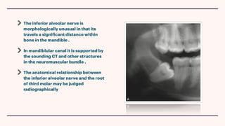
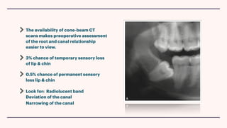
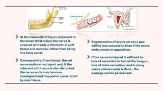
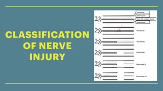
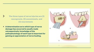
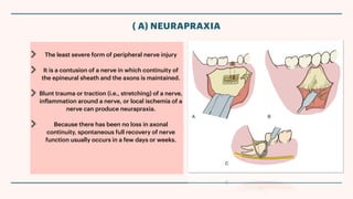
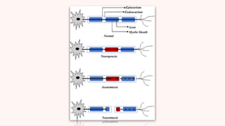
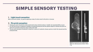
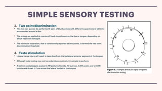
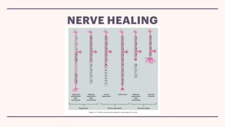
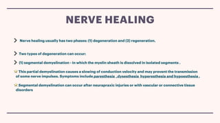
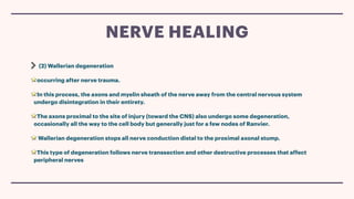
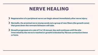
By : Fatemah Ibrahim
NERVE INJURIES IN 3RD
MOLAR EXTRACTION
Classification , clinical features , and investigations
Introduction
Nerve associated with third molar extraction
classification of nerve injury
Method used to monitor nerve recovery
Nerve healing
CONTENTS :
Injuries to sensory nerves of the maxillofacial region
occasionally occur during the treatment of impacted teeth
Fortunately, most injured nerves spontaneously recover.
However, in the past, little was done to treat persistent sensory
nerve disorders. Advances in the understanding of how nerves
heal and in the surgical means of repairing peripheral nerves
provide patients with the possibility of partially or fully
regaining normal nerve function.
INTRODUCTION :
Impacted mandibular third molar teeth are in
case proximity to the lingual , inferior alveolar
nerve , mylohyoid and buccal nerve .
The majority of injuries result in transient
sensory disturbance but in some cases ,
permanent paresthesia , hypoaesthesia, or even
worse
These sensory disturbance can be troublesome
causing problems with speech and mastication
and may adversely affect the patient’s quality of
life
INFERIOR
ALVEOLAR
NERVE
The inferior alveolar nerve is
morphologically unusual in that its
travels a significant distance within
bone in the mandible .
In mandiblular canal it is supported by
the sounding CT and other structures
in the neuromuscular bundle .
The anatomical relationship between
the inferior alveolar nerve and the root
of third molar may be judged
radiographically
The availability of cone-beam CT
scans makes preoperative assessment
of the root and canal relationship
easier to view.
3% chance of temporary sensory loss
of lip & chin
0.5% chance of permanent sensory
loss lip & chin ︎
Look for: Radiolucent band
︎Deviation of the canal
︎Narrowing of the canal
LINGUAL NERVE
At the usual site of injury (adjacent to
the lower third molar) the nerve is
covered with only a thin layer of soft
tissue and mucosa, rather than being
in a bony canal.
Consequently, if sectioned, the cut
nerve ends retract apart and, if the
adjacent soft tissue is also distorted,
the nerve ends may become
misaligned and trapped or constricted
by scar tissue.
Regeneration of axons across a gap
will be less successful than if the nerve
ends remain in apposition.
If the nerve is injured it will lead to :
loss of sensation to half of the tongue ,
loss of taste sensation ,and in many
cases unless repair is done , the
damage can be permanent .
CLASSIFICATION
OF NERVE
INJURY
The three types of nerve injuries are (1)
neurapraxia, (2) axonotmesis, and
(3) neurotmesis
• A determination as to which type of nerve
damage has occurred is usually made
retrospectively, knowledge of the
pathophysiology of each type is important for
gaining an appreciation of nerve healing.
The least severe form of peripheral nerve injury
It is a contusion of a nerve in which continuity of
the epineural sheath and the axons is maintained.
Blunt trauma or traction (i.e., stretching) of a nerve,
inflammation around a nerve, or local ischemia of a
nerve can produce neurapraxia.
Because there has been no loss in axonal
continuity, spontaneous full recovery of nerve
function usually occurs in a few days or weeks.
( A) NEURAPRAXIA
occurred when the continuity of the axons, but
not the epineural sheath, is disrupted.
Severe blunt trauma, nerve crushing, or extreme
traction of a nerve can produce this type of injury.
Because the epineural sheath is still intact,
axonal regeneration can (but does not always)
occur with a resolution of nerve dysfunction in 2
to 6 months.
(B) AXONOTMESIS
the most severe type of nerve injury,
involves a complete loss of nerve continuity.
This form of damage can be produced by badly
displaced fractures, severance by bullets or
knives during an assault, or by iatrogenic
transection.
Prognosis for spontaneous recovery of nerves
that have undergone neurotmesis is poor
except if the ends of the affected nerve have
somehow been left in approximation and properly
oriented.
(C ) NEUROTMESIS
METHODS USED TO
MONITOR NERVE RECOVER
1. Light touch sensation
Most commonly tested ; by gently applying a wisp of cotton wool to the skin or mucosa
2. Pin prick sensation
• Testing pin prick threshold is often performed using a dental probe or needle, but reproducibility is poor.
• For this test the pin is applied at steadily increasing pressures and the patient asked to indicate the point at
which the sensation becomes sharp rather than dull.
• The pin prick sensation threshold is noted for a series of randomly chosen points on both the injured and the
uninjured side.
SIMPLE SENSORY TESTING
3. Two point discrimination
• This test can quickly be performed if pairs of blunt probes with different separations (2–20 mm)
are mounted around a disc
• The probes are applied at a series of fixed sites chosen on the lips or tongue, depending on
which has been damaged.
• The minimum separation, that is consistently reported as two points, is termed the two point
discrimination threshold.
4. Taste stimulation
• Lingual nerve injury will result in taste loss from the ipsilateral anterior segment of the tongue.
• Although taste testing may not be undertaken routinely, it is simple to perform.
• A Cotton wool pledgets soaked in 1M sodium chloride, 1M sucrose, 0.4M acetic acid or 0.1M
quinine are drawn 1–2 cm across the lateral border of the tongue
SIMPLE SENSORY TESTING
NERVE HEALING
NERVE HEALING
Nerve healing usually has two phases: (1) degeneration and (2) regeneration.
Two types of degeneration can occur:
(1) segmental demyelination - in which the myelin sheath is dissolved in isolated segments .
This partial demyelination causes a slowing of conduction velocity and may prevent the transmission
of some nerve impulses. Symptoms include paresthesia ,dysesthesia hyperesthesia and hypoesthesia .
Segmental demyelination can occur after neurapraxic injuries or with vascular or connective tissue
disorders
NERVE HEALING
(2) Wallerian degeneration
occurring after nerve trauma.
In this process, the axons and myelin sheath of the nerve away from the central nervous system
undergo disintegration in their entirety.
The axons proximal to the site of injury (toward the CNS) also undergo some degeneration,
occasionally all the way to the cell body but generally just for a few nodes of Ranvier.
Wallerian degeneration stops all nerve conduction distal to the proximal axonal stump.
This type of degeneration follows nerve transsection and other destructive processes that affect
peripheral nerves
NERVE HEALING
Regeneration of a peripheral nerve can begin almost immediately after nerve injury.
Normally, the proximal nerve stump sends out a group of new fibers (the growth cone)
that grow down the remnant Schwann cell tube.
Growth progresses at a rate of 1 to 1.5 mm per day and continues until the site
innervated by the nerve is reached or growth is blocked by fibrous connective tissue
or bone.
NERVE HEALING
CONCLUSION
IT IS IMPERATIVE THAT
PATIENTS SUSTAINING A NERVE
INJURY ARE MANAGED
CORRECTLY AND THIS MUST
INCLUDE A DIAGNOSIS OF THE
TYPE OF INJURY, MONITORING
RECOVERY AND THE
TREATMENT OF APPROPRIATE
CASES.
THANK YOU FOR YOU PATIENTS !
REFERENCES :

Nerve injuries

  • 1.
    By : FatemahIbrahim NERVE INJURIES IN 3RD MOLAR EXTRACTION Classification , clinical features , and investigations
  • 2.
    Introduction Nerve associated withthird molar extraction classification of nerve injury Method used to monitor nerve recovery Nerve healing CONTENTS :
  • 3.
    Injuries to sensorynerves of the maxillofacial region occasionally occur during the treatment of impacted teeth Fortunately, most injured nerves spontaneously recover. However, in the past, little was done to treat persistent sensory nerve disorders. Advances in the understanding of how nerves heal and in the surgical means of repairing peripheral nerves provide patients with the possibility of partially or fully regaining normal nerve function. INTRODUCTION :
  • 4.
    Impacted mandibular thirdmolar teeth are in case proximity to the lingual , inferior alveolar nerve , mylohyoid and buccal nerve . The majority of injuries result in transient sensory disturbance but in some cases , permanent paresthesia , hypoaesthesia, or even worse These sensory disturbance can be troublesome causing problems with speech and mastication and may adversely affect the patient’s quality of life
  • 5.
  • 6.
    The inferior alveolarnerve is morphologically unusual in that its travels a significant distance within bone in the mandible . In mandiblular canal it is supported by the sounding CT and other structures in the neuromuscular bundle . The anatomical relationship between the inferior alveolar nerve and the root of third molar may be judged radiographically
  • 7.
    The availability ofcone-beam CT scans makes preoperative assessment of the root and canal relationship easier to view. 3% chance of temporary sensory loss of lip & chin 0.5% chance of permanent sensory loss lip & chin ︎ Look for: Radiolucent band ︎Deviation of the canal ︎Narrowing of the canal
  • 8.
  • 9.
    At the usualsite of injury (adjacent to the lower third molar) the nerve is covered with only a thin layer of soft tissue and mucosa, rather than being in a bony canal. Consequently, if sectioned, the cut nerve ends retract apart and, if the adjacent soft tissue is also distorted, the nerve ends may become misaligned and trapped or constricted by scar tissue. Regeneration of axons across a gap will be less successful than if the nerve ends remain in apposition. If the nerve is injured it will lead to : loss of sensation to half of the tongue , loss of taste sensation ,and in many cases unless repair is done , the damage can be permanent .
  • 10.
  • 11.
    The three typesof nerve injuries are (1) neurapraxia, (2) axonotmesis, and (3) neurotmesis • A determination as to which type of nerve damage has occurred is usually made retrospectively, knowledge of the pathophysiology of each type is important for gaining an appreciation of nerve healing.
  • 12.
    The least severeform of peripheral nerve injury It is a contusion of a nerve in which continuity of the epineural sheath and the axons is maintained. Blunt trauma or traction (i.e., stretching) of a nerve, inflammation around a nerve, or local ischemia of a nerve can produce neurapraxia. Because there has been no loss in axonal continuity, spontaneous full recovery of nerve function usually occurs in a few days or weeks. ( A) NEURAPRAXIA
  • 13.
    occurred when thecontinuity of the axons, but not the epineural sheath, is disrupted. Severe blunt trauma, nerve crushing, or extreme traction of a nerve can produce this type of injury. Because the epineural sheath is still intact, axonal regeneration can (but does not always) occur with a resolution of nerve dysfunction in 2 to 6 months. (B) AXONOTMESIS
  • 14.
    the most severetype of nerve injury, involves a complete loss of nerve continuity. This form of damage can be produced by badly displaced fractures, severance by bullets or knives during an assault, or by iatrogenic transection. Prognosis for spontaneous recovery of nerves that have undergone neurotmesis is poor except if the ends of the affected nerve have somehow been left in approximation and properly oriented. (C ) NEUROTMESIS
  • 16.
  • 17.
    1. Light touchsensation Most commonly tested ; by gently applying a wisp of cotton wool to the skin or mucosa 2. Pin prick sensation • Testing pin prick threshold is often performed using a dental probe or needle, but reproducibility is poor. • For this test the pin is applied at steadily increasing pressures and the patient asked to indicate the point at which the sensation becomes sharp rather than dull. • The pin prick sensation threshold is noted for a series of randomly chosen points on both the injured and the uninjured side. SIMPLE SENSORY TESTING
  • 18.
    3. Two pointdiscrimination • This test can quickly be performed if pairs of blunt probes with different separations (2–20 mm) are mounted around a disc • The probes are applied at a series of fixed sites chosen on the lips or tongue, depending on which has been damaged. • The minimum separation, that is consistently reported as two points, is termed the two point discrimination threshold. 4. Taste stimulation • Lingual nerve injury will result in taste loss from the ipsilateral anterior segment of the tongue. • Although taste testing may not be undertaken routinely, it is simple to perform. • A Cotton wool pledgets soaked in 1M sodium chloride, 1M sucrose, 0.4M acetic acid or 0.1M quinine are drawn 1–2 cm across the lateral border of the tongue SIMPLE SENSORY TESTING
  • 19.
  • 20.
  • 21.
    Nerve healing usuallyhas two phases: (1) degeneration and (2) regeneration. Two types of degeneration can occur: (1) segmental demyelination - in which the myelin sheath is dissolved in isolated segments . This partial demyelination causes a slowing of conduction velocity and may prevent the transmission of some nerve impulses. Symptoms include paresthesia ,dysesthesia hyperesthesia and hypoesthesia . Segmental demyelination can occur after neurapraxic injuries or with vascular or connective tissue disorders NERVE HEALING
  • 22.
    (2) Wallerian degeneration occurringafter nerve trauma. In this process, the axons and myelin sheath of the nerve away from the central nervous system undergo disintegration in their entirety. The axons proximal to the site of injury (toward the CNS) also undergo some degeneration, occasionally all the way to the cell body but generally just for a few nodes of Ranvier. Wallerian degeneration stops all nerve conduction distal to the proximal axonal stump. This type of degeneration follows nerve transsection and other destructive processes that affect peripheral nerves NERVE HEALING
  • 23.
    Regeneration of aperipheral nerve can begin almost immediately after nerve injury. Normally, the proximal nerve stump sends out a group of new fibers (the growth cone) that grow down the remnant Schwann cell tube. Growth progresses at a rate of 1 to 1.5 mm per day and continues until the site innervated by the nerve is reached or growth is blocked by fibrous connective tissue or bone. NERVE HEALING
  • 24.
  • 25.
    IT IS IMPERATIVETHAT PATIENTS SUSTAINING A NERVE INJURY ARE MANAGED CORRECTLY AND THIS MUST INCLUDE A DIAGNOSIS OF THE TYPE OF INJURY, MONITORING RECOVERY AND THE TREATMENT OF APPROPRIATE CASES.
  • 26.
    THANK YOU FORYOU PATIENTS !
  • 27.