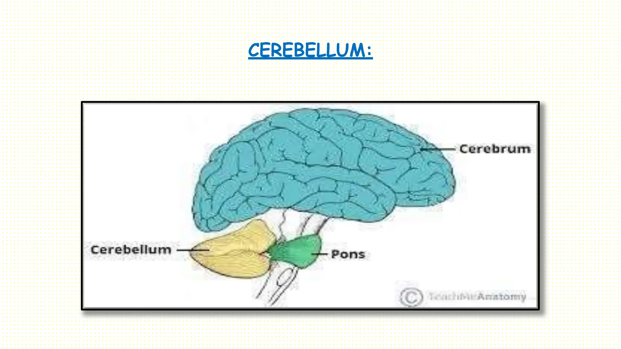
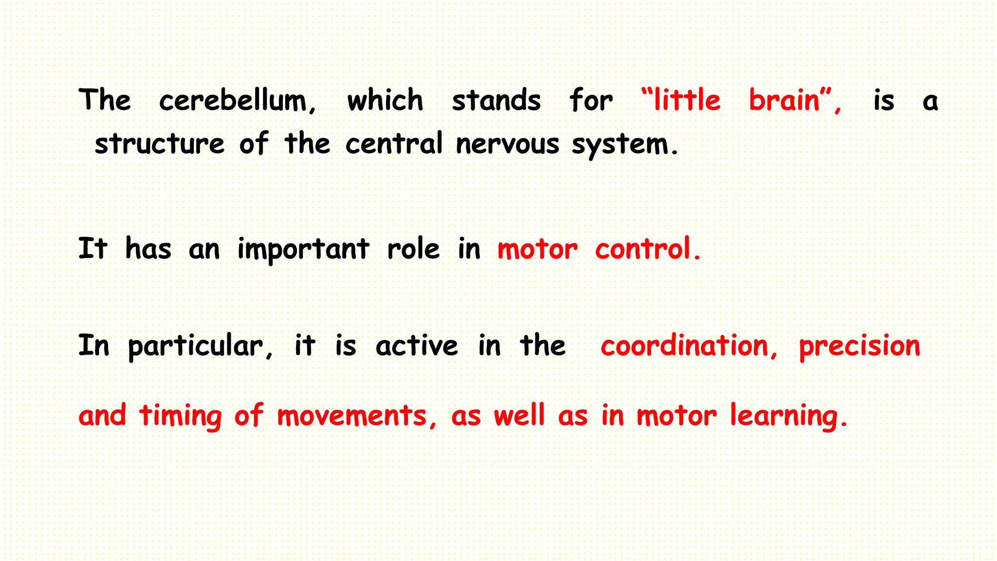
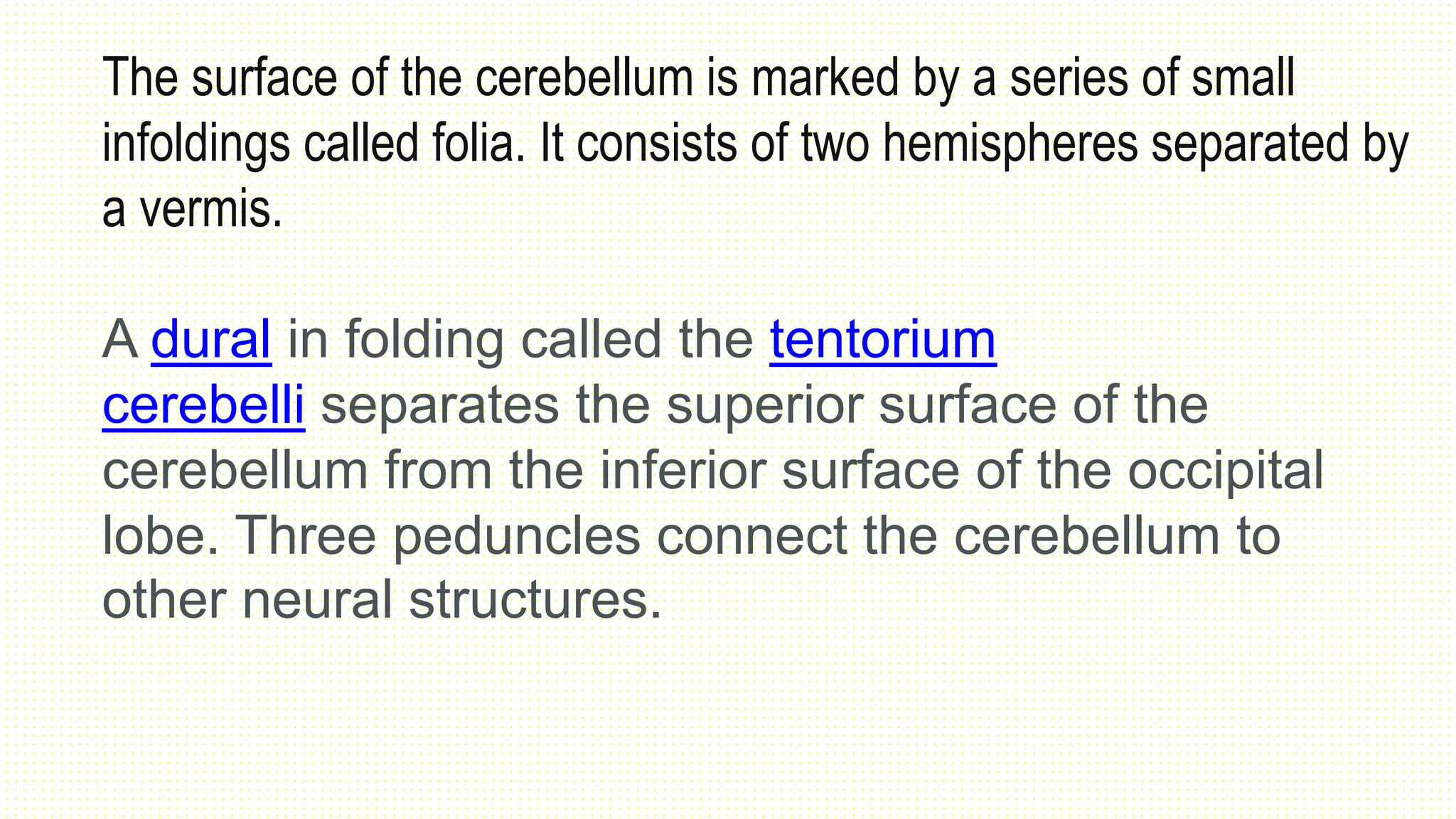
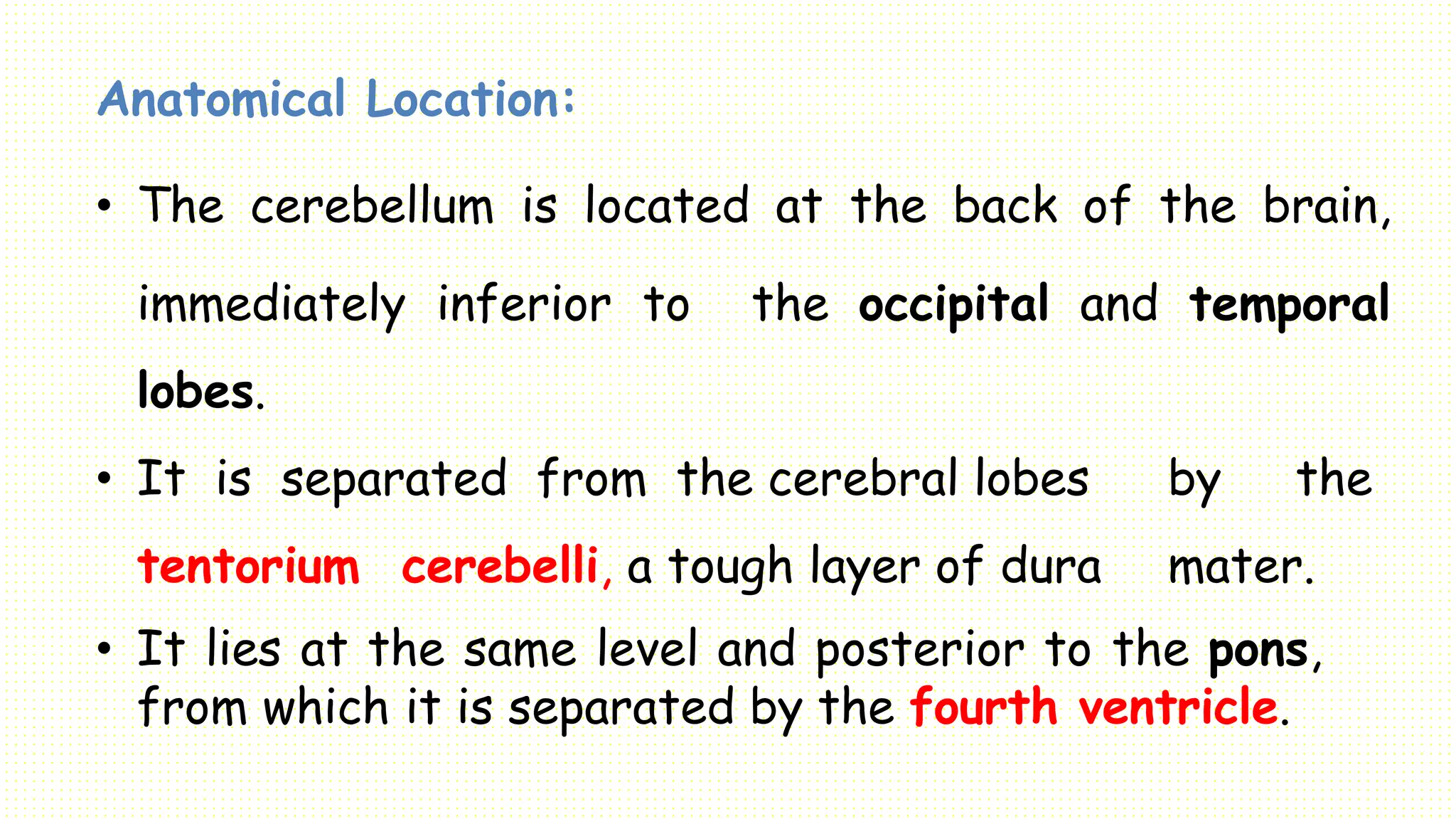
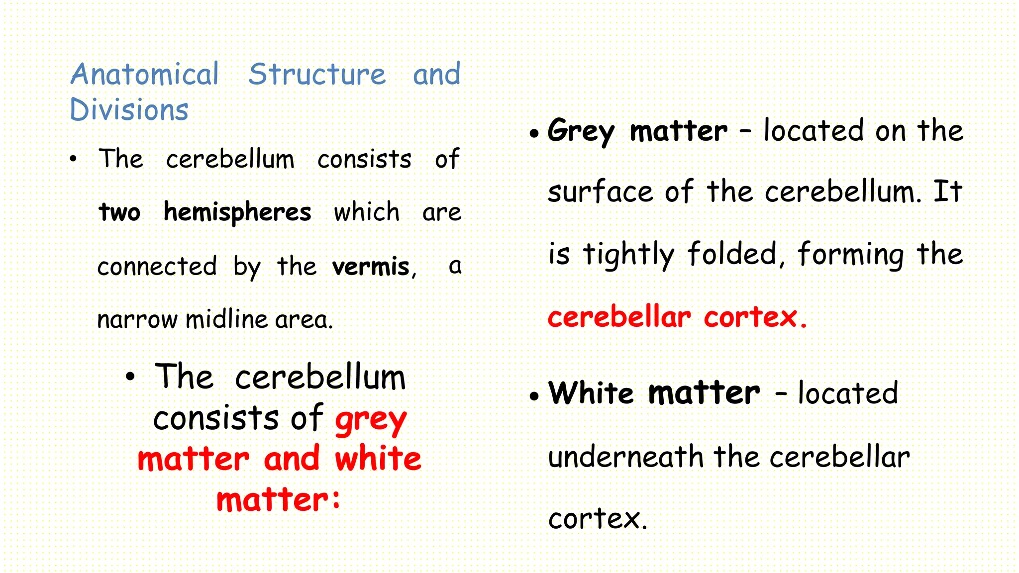
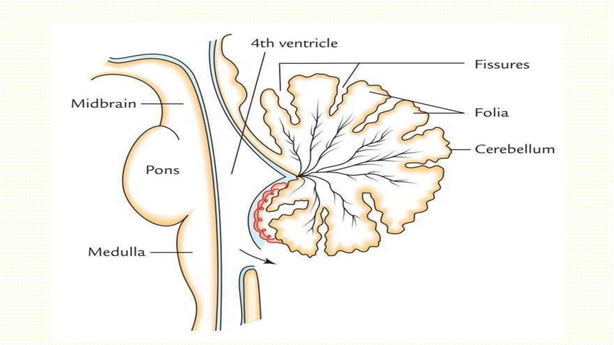
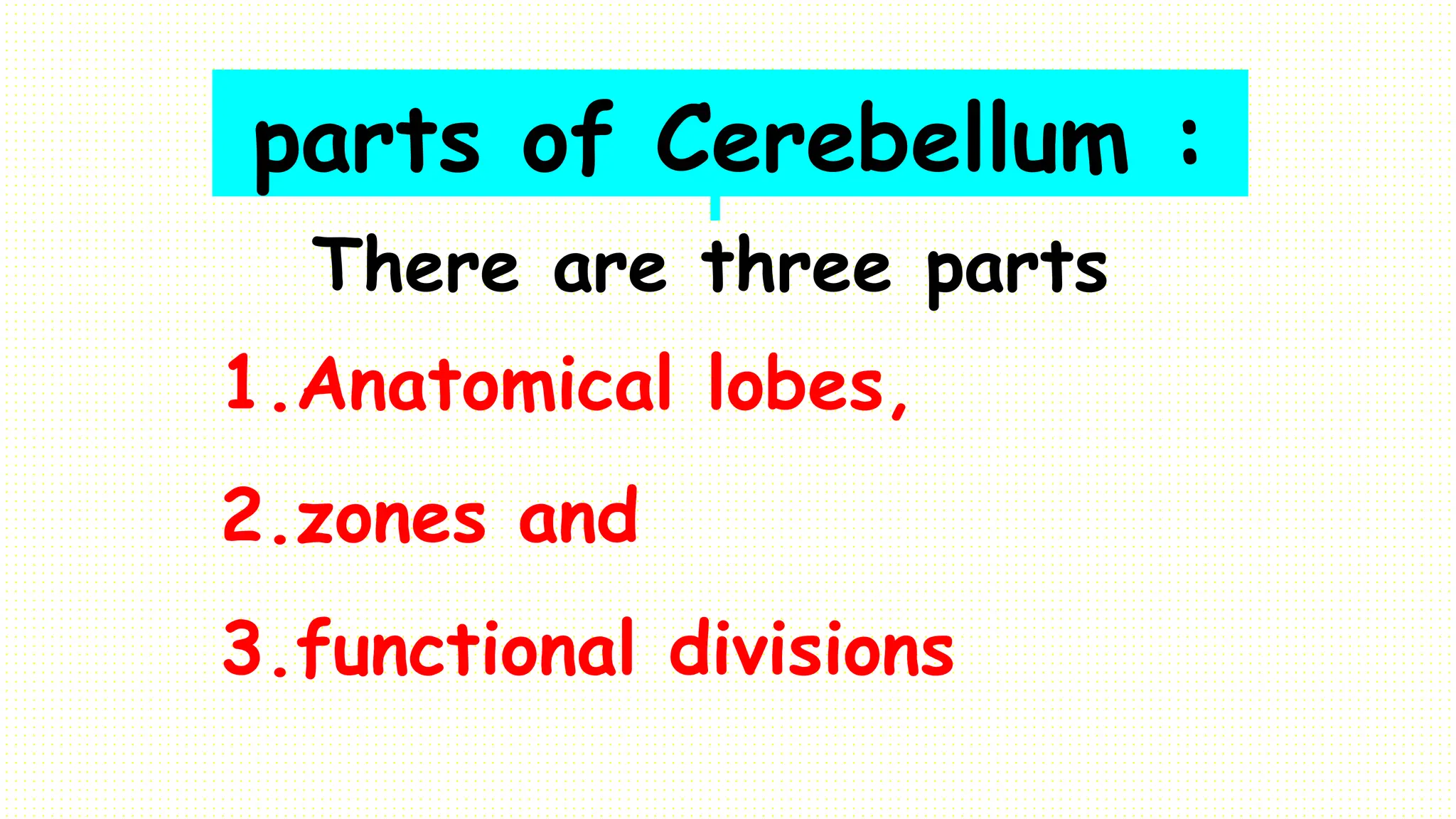
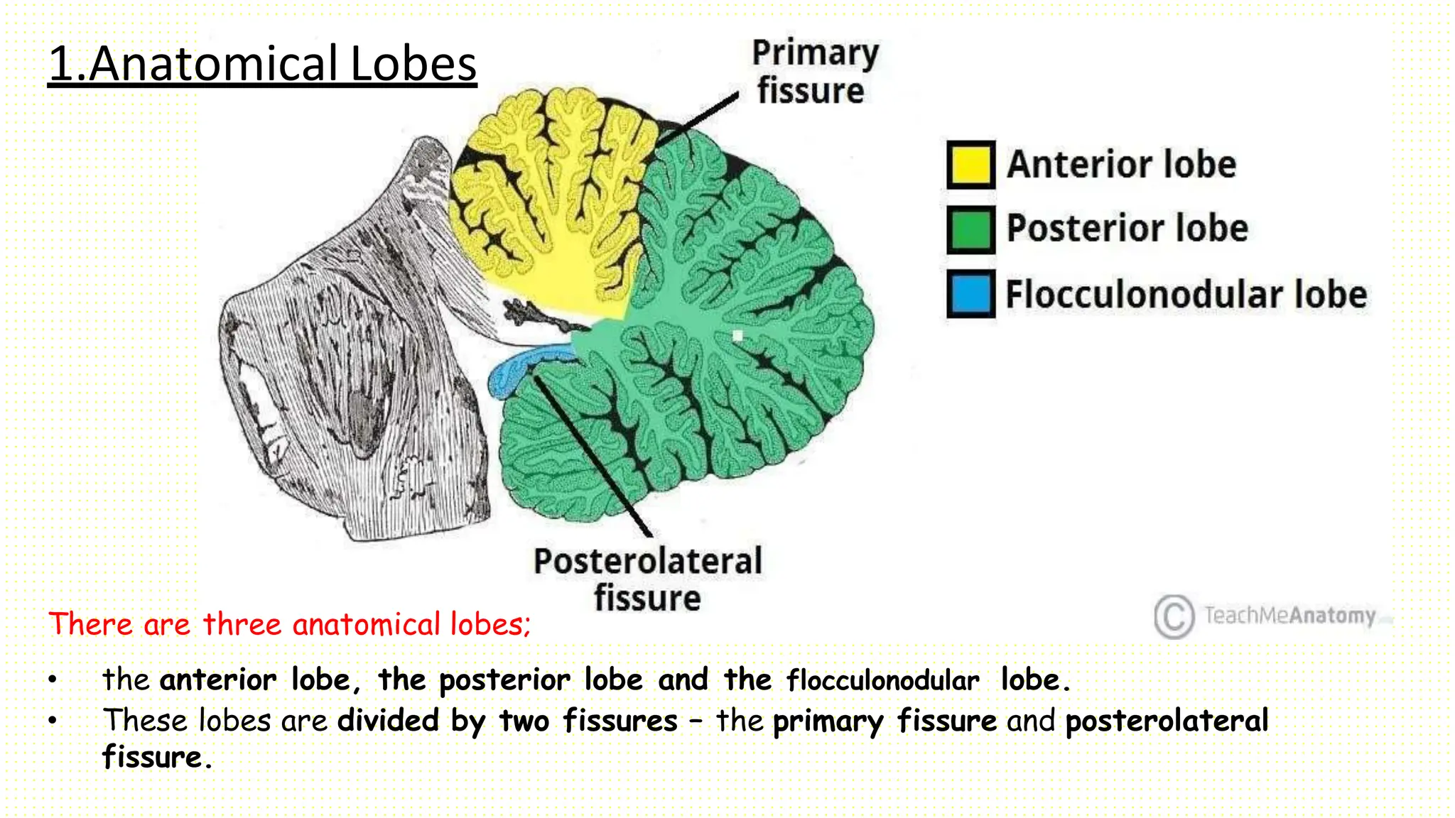
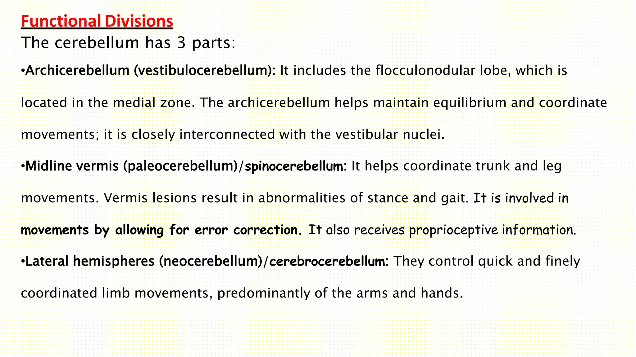
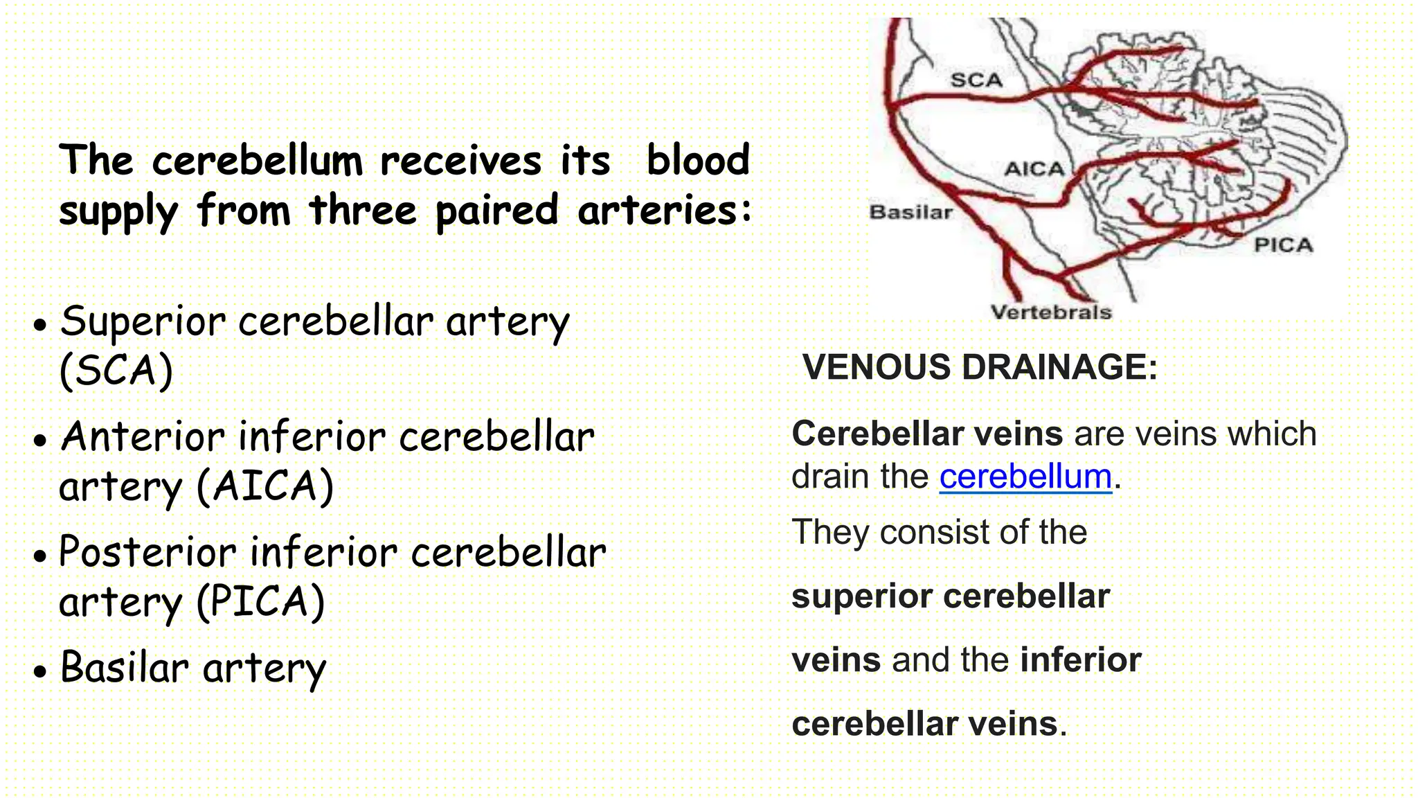
CEREBELLUM:
The cerebellum, which stands for “little brain”, is a
structure of the central nervous system.
It has an important role in motor control.
In particular, it is active in the coordination, precision
and timing of movements, as well as in motor learning.
The surface of the cerebellum is marked by a series of small
infoldings called folia. It consists of two hemispheres separated by
a vermis.
A dural in folding called the tentorium
cerebelli separates the superior surface of the
cerebellum from the inferior surface of the occipital
lobe. Three peduncles connect the cerebellum to
other neural structures.
Anatomical Location:
• The cerebellum is located at the back of the brain,
immediately inferior to the occipital and temporal
lobes.
• It is separated from the cerebral lobes by the
tentorium cerebelli, a tough layer of dura mater.
• It lies at the same level and posterior to the pons,
from which it is separated by the fourth ventricle.
Anatomical Structure and
Divisions
• The cerebellum consists of
two hemispheres which are
a
connected by the vermis,
narrow midline area.
• The cerebellum
consists of grey
matter and white
matter:
 Grey matter – located on the
surface of the cerebellum. It
is tightly folded, forming the
cerebellar cortex.
 White matter – located
underneath the cerebellar
cortex.
parts of Cerebellum :
There are three parts
1.Anatomical lobes,
2.zones and
3.functional divisions
1.Anatomical Lobes
There are three anatomical lobes;
• the anterior lobe, the posterior lobe and the flocculonodular lobe.
• These lobes are divided by two fissures – the primary fissure and posterolateral
fissure.
2. Zones
There are two cerebellar zones.
• In the midline of the cerebellum is
the vermis.
• Either side of the vermis is the intermediate zone.
• Lateral to the intermediate zone are the lateral
hemispheres/lateral zone.
Functional Divisions
The cerebellum has 3 parts:
•Archicerebellum (vestibulocerebellum): It includes the flocculonodular lobe, which is
located in the medial zone. The archicerebellum helps maintain equilibrium and coordinate
movements; it is closely interconnected with the vestibular nuclei.
•Midline vermis (paleocerebellum)/spinocerebellum: It helps coordinate trunk and leg
movements. Vermis lesions result in abnormalities of stance and gait. It is involved in
movements by allowing for error correction. It also receives proprioceptive information.
•Lateral hemispheres (neocerebellum)/cerebrocerebellum: They control quick and finely
coordinated limb movements, predominantly of the arms and hands.
The cerebellum receives its blood
supply from three paired arteries:
 Superior cerebellar artery
(SCA)
 Anterior inferior cerebellar
artery (AICA)
 Posterior inferior cerebellar
artery (PICA)
 Basilar artery
VENOUS DRAINAGE:
Cerebellar veins are veins which
drain the cerebellum.
They consist of the
superior cerebellar
veins and the inferior
cerebellar veins.
THE CEREBELLUM IS INVOLVED IN THE FOLLOWING
FUNCTIONS:
 Maintenance of balance and posture. postural adjustments
in order to maintain balance
 Coordination of voluntary movements. Most movements are
composed of a number of different muscle groups acting
together in a temporally coordinated fashion.
 Motor learning. The cerebellum is important for
motor learning. The cerebellum plays a major role in
adapting and fine-tuning motor programs to make
accurate movements through a trial-and-error process
(e.g., learning to hit a baseball).
 Cognitive functions. involved in certain cognitive
functions, such as language.
Dysdiadochokinesia (DDK) is the
medical term for an impaired ability to
perform rapid, alternating movements
(i.e., diadochokinesia).
ONLY CEREBELLUM.pptx FOR NURSING STUDENTS

ONLY CEREBELLUM.pptx FOR NURSING STUDENTS

  • 2.
  • 3.
    The cerebellum, whichstands for “little brain”, is a structure of the central nervous system. It has an important role in motor control. In particular, it is active in the coordination, precision and timing of movements, as well as in motor learning.
  • 4.
    The surface ofthe cerebellum is marked by a series of small infoldings called folia. It consists of two hemispheres separated by a vermis. A dural in folding called the tentorium cerebelli separates the superior surface of the cerebellum from the inferior surface of the occipital lobe. Three peduncles connect the cerebellum to other neural structures.
  • 5.
    Anatomical Location: • Thecerebellum is located at the back of the brain, immediately inferior to the occipital and temporal lobes. • It is separated from the cerebral lobes by the tentorium cerebelli, a tough layer of dura mater. • It lies at the same level and posterior to the pons, from which it is separated by the fourth ventricle.
  • 6.
    Anatomical Structure and Divisions •The cerebellum consists of two hemispheres which are a connected by the vermis, narrow midline area. • The cerebellum consists of grey matter and white matter:  Grey matter – located on the surface of the cerebellum. It is tightly folded, forming the cerebellar cortex.  White matter – located underneath the cerebellar cortex.
  • 8.
    parts of Cerebellum: There are three parts 1.Anatomical lobes, 2.zones and 3.functional divisions
  • 9.
    1.Anatomical Lobes There arethree anatomical lobes; • the anterior lobe, the posterior lobe and the flocculonodular lobe. • These lobes are divided by two fissures – the primary fissure and posterolateral fissure.
  • 10.
    2. Zones There aretwo cerebellar zones. • In the midline of the cerebellum is the vermis. • Either side of the vermis is the intermediate zone. • Lateral to the intermediate zone are the lateral hemispheres/lateral zone.
  • 13.
    Functional Divisions The cerebellumhas 3 parts: •Archicerebellum (vestibulocerebellum): It includes the flocculonodular lobe, which is located in the medial zone. The archicerebellum helps maintain equilibrium and coordinate movements; it is closely interconnected with the vestibular nuclei. •Midline vermis (paleocerebellum)/spinocerebellum: It helps coordinate trunk and leg movements. Vermis lesions result in abnormalities of stance and gait. It is involved in movements by allowing for error correction. It also receives proprioceptive information. •Lateral hemispheres (neocerebellum)/cerebrocerebellum: They control quick and finely coordinated limb movements, predominantly of the arms and hands.
  • 15.
    The cerebellum receivesits blood supply from three paired arteries:  Superior cerebellar artery (SCA)  Anterior inferior cerebellar artery (AICA)  Posterior inferior cerebellar artery (PICA)  Basilar artery VENOUS DRAINAGE: Cerebellar veins are veins which drain the cerebellum. They consist of the superior cerebellar veins and the inferior cerebellar veins.
  • 16.
    THE CEREBELLUM ISINVOLVED IN THE FOLLOWING FUNCTIONS:  Maintenance of balance and posture. postural adjustments in order to maintain balance  Coordination of voluntary movements. Most movements are composed of a number of different muscle groups acting together in a temporally coordinated fashion.
  • 17.
     Motor learning.The cerebellum is important for motor learning. The cerebellum plays a major role in adapting and fine-tuning motor programs to make accurate movements through a trial-and-error process (e.g., learning to hit a baseball).  Cognitive functions. involved in certain cognitive functions, such as language.
  • 18.
    Dysdiadochokinesia (DDK) isthe medical term for an impaired ability to perform rapid, alternating movements (i.e., diadochokinesia).