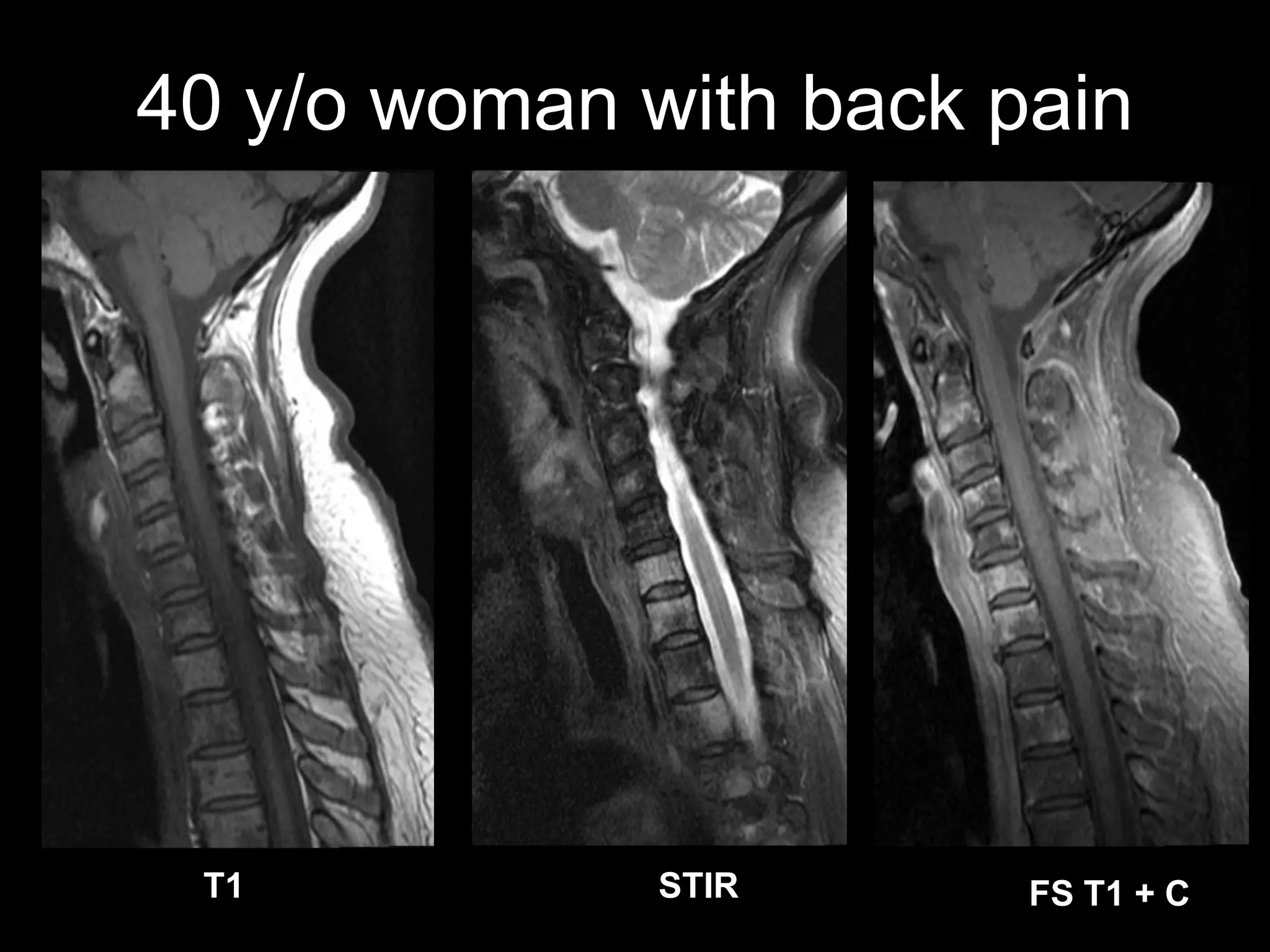
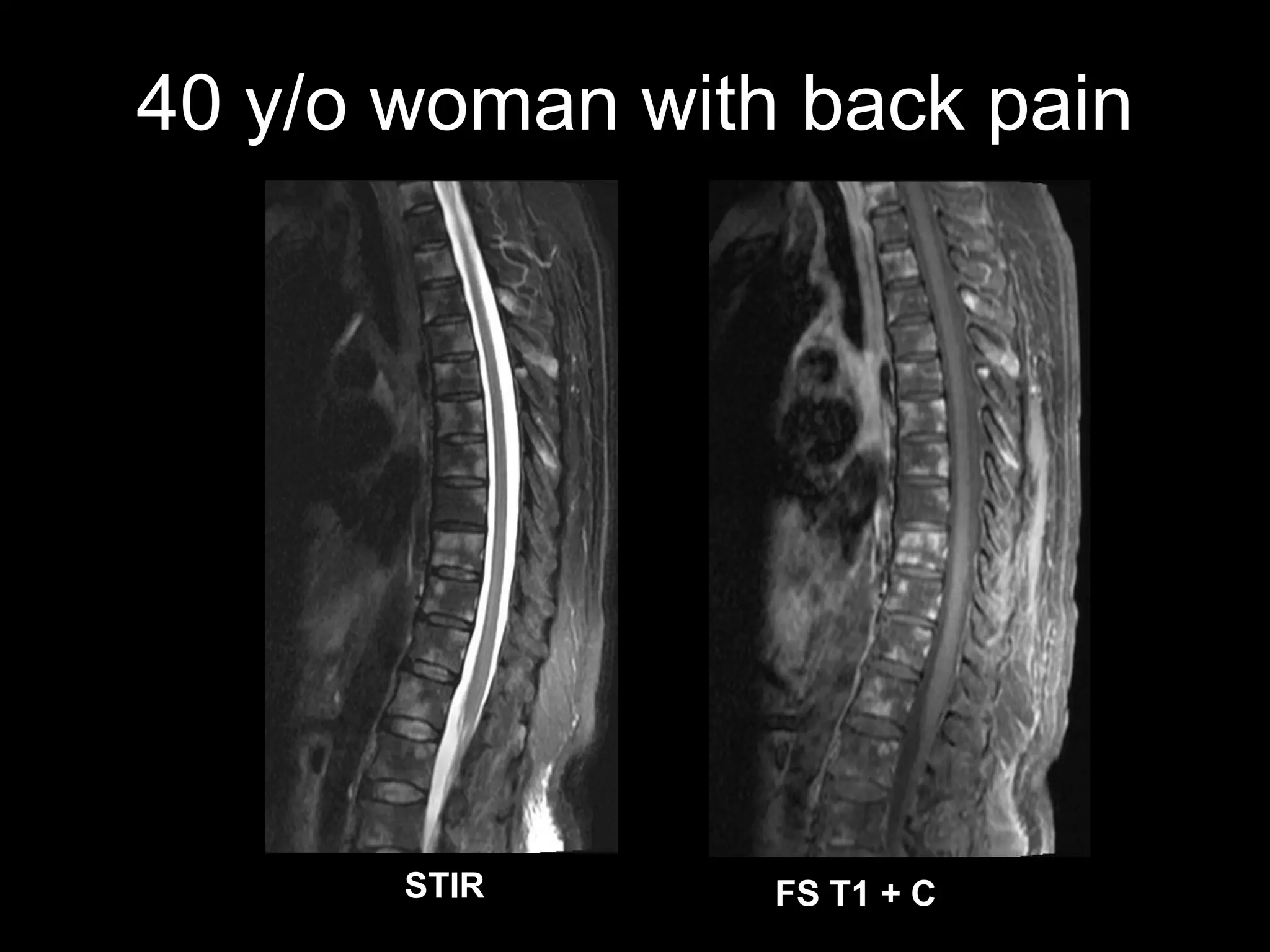
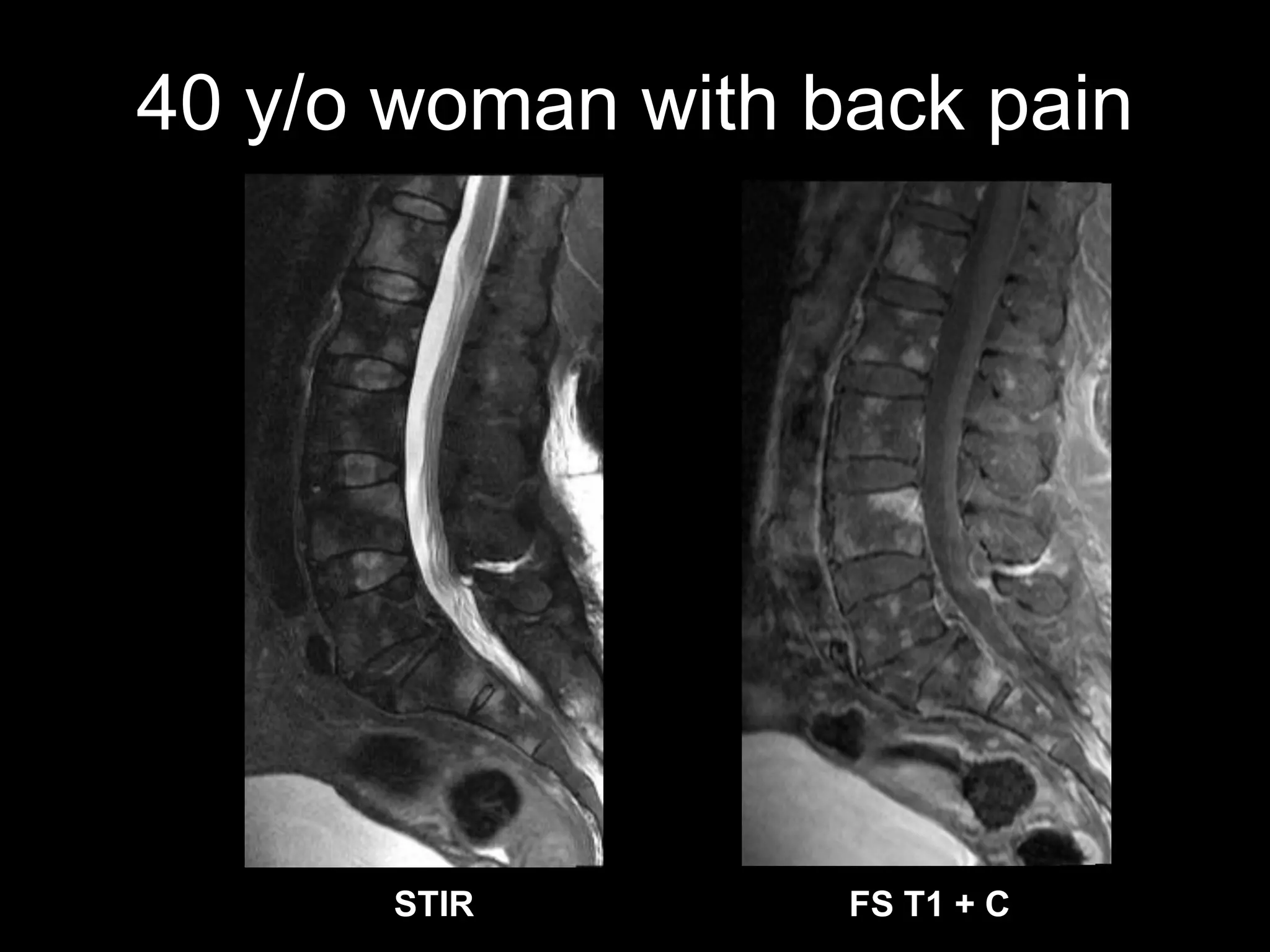
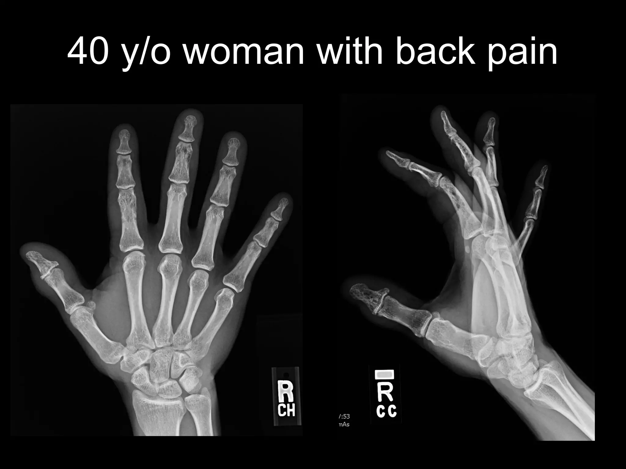
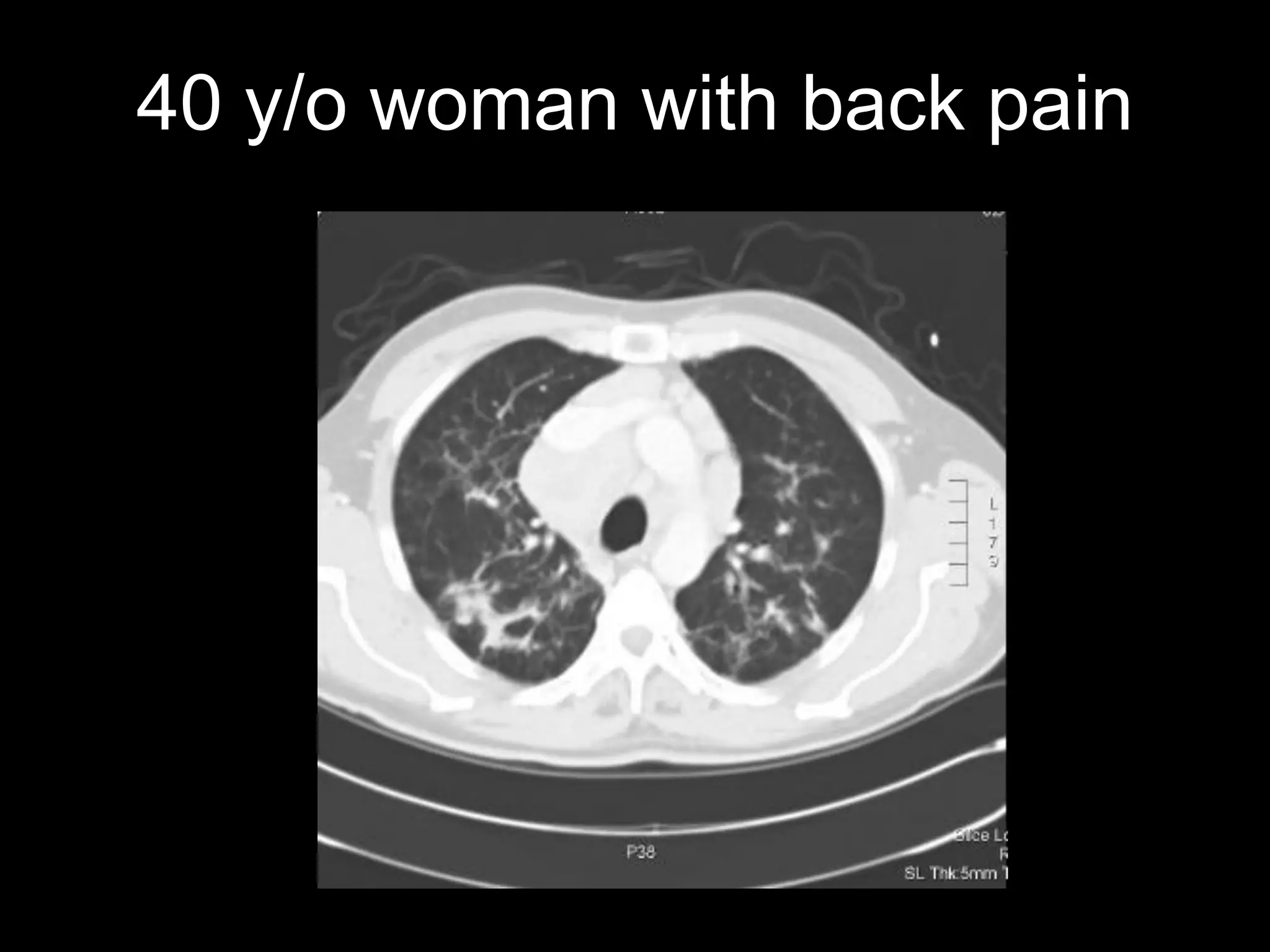
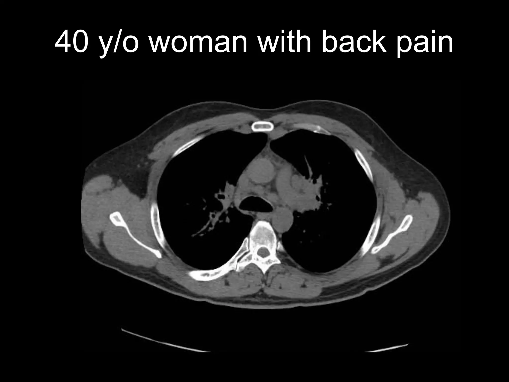
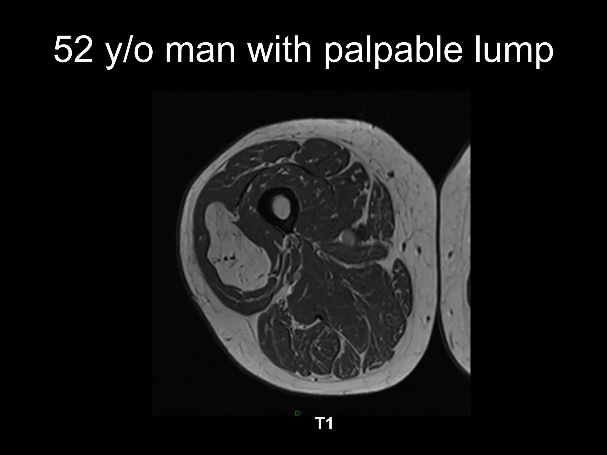
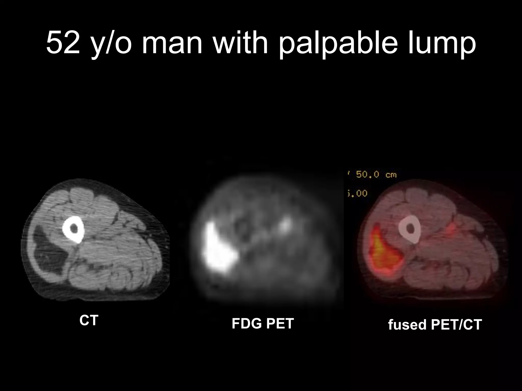
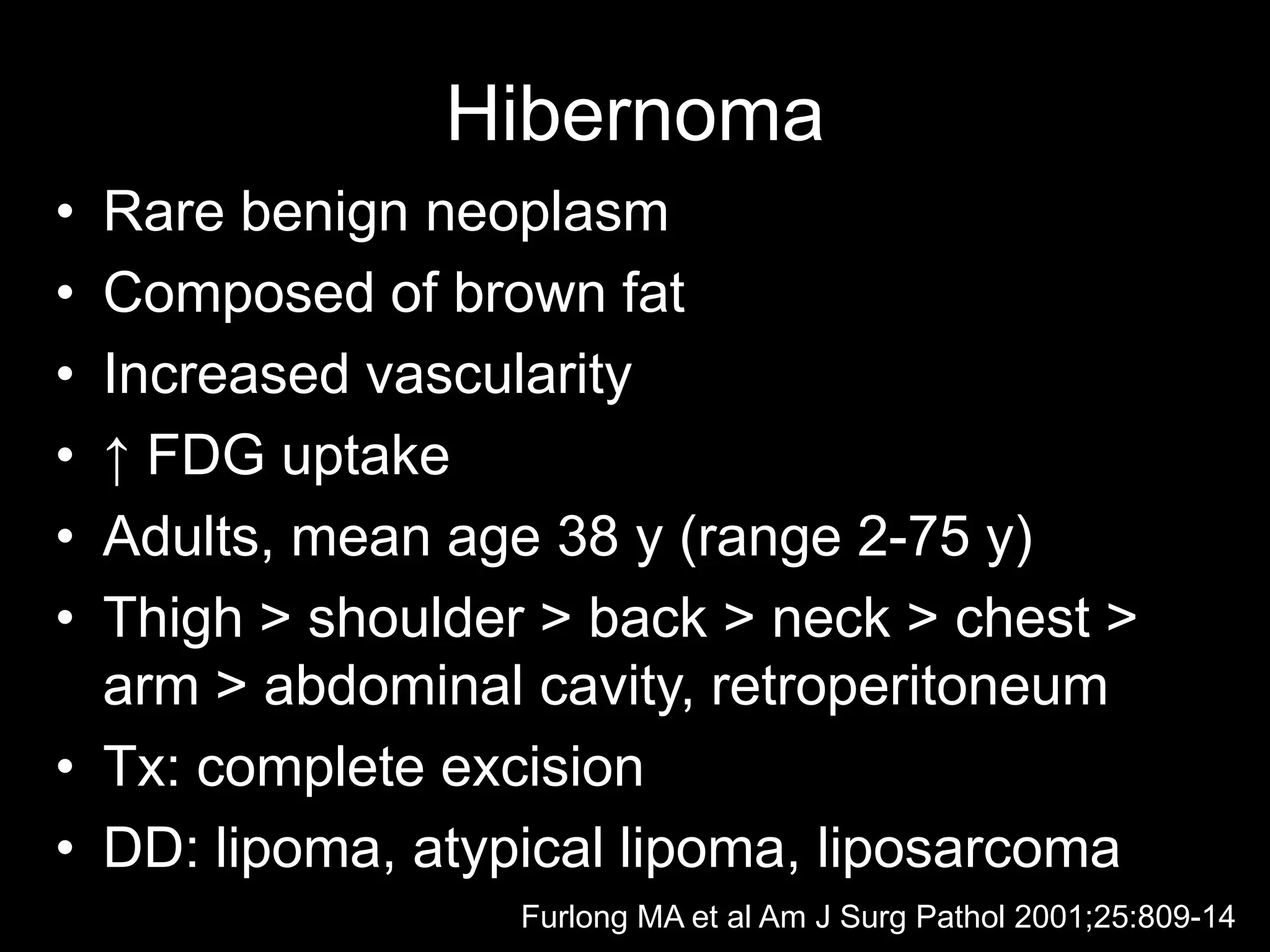
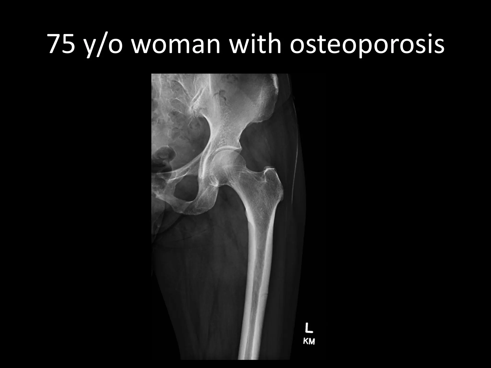
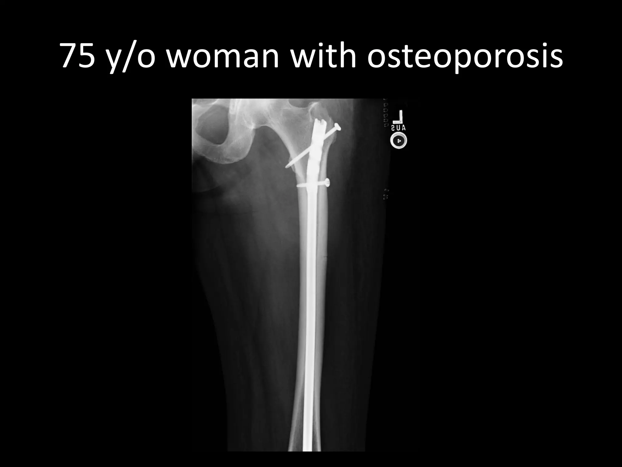
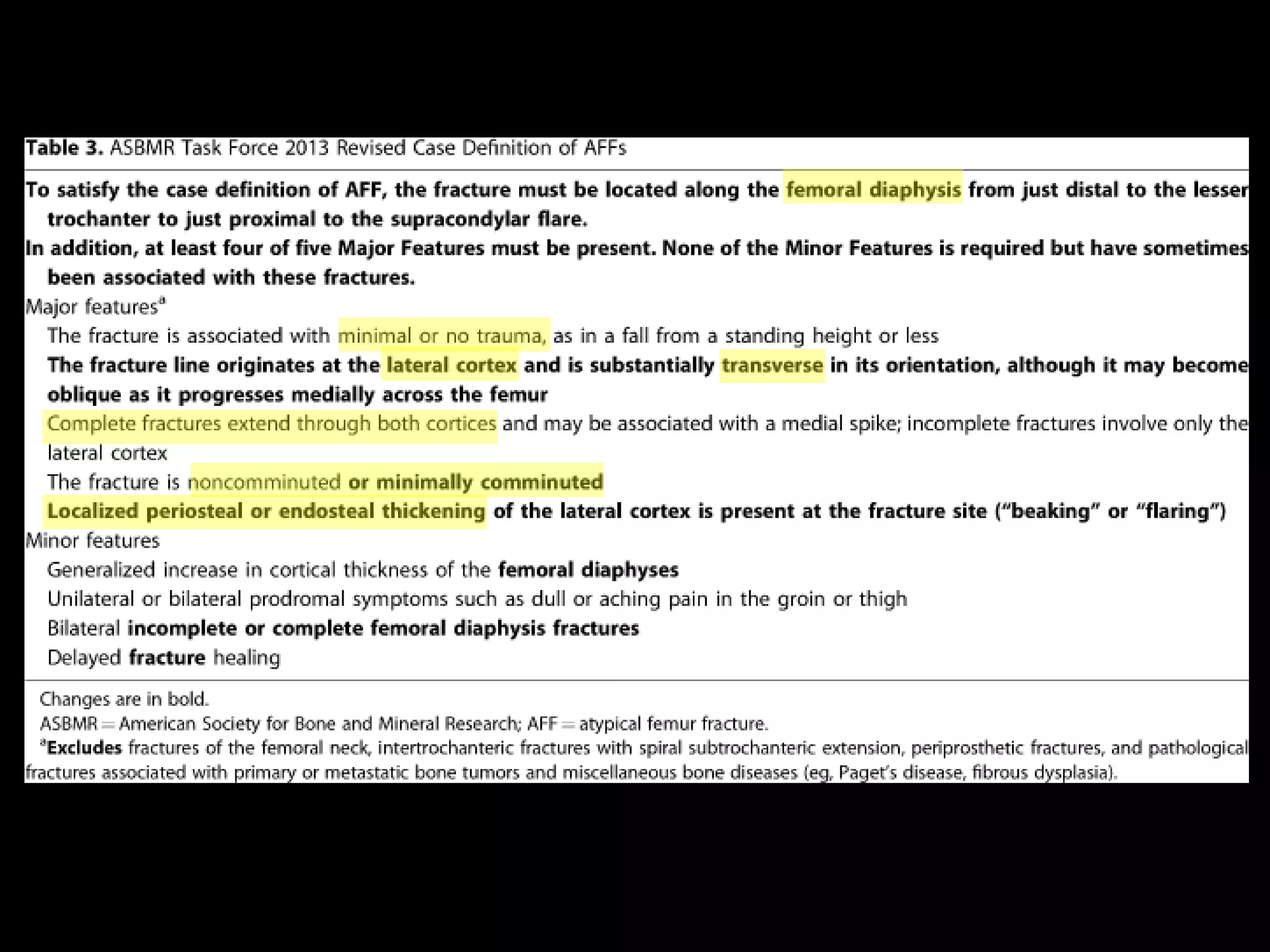
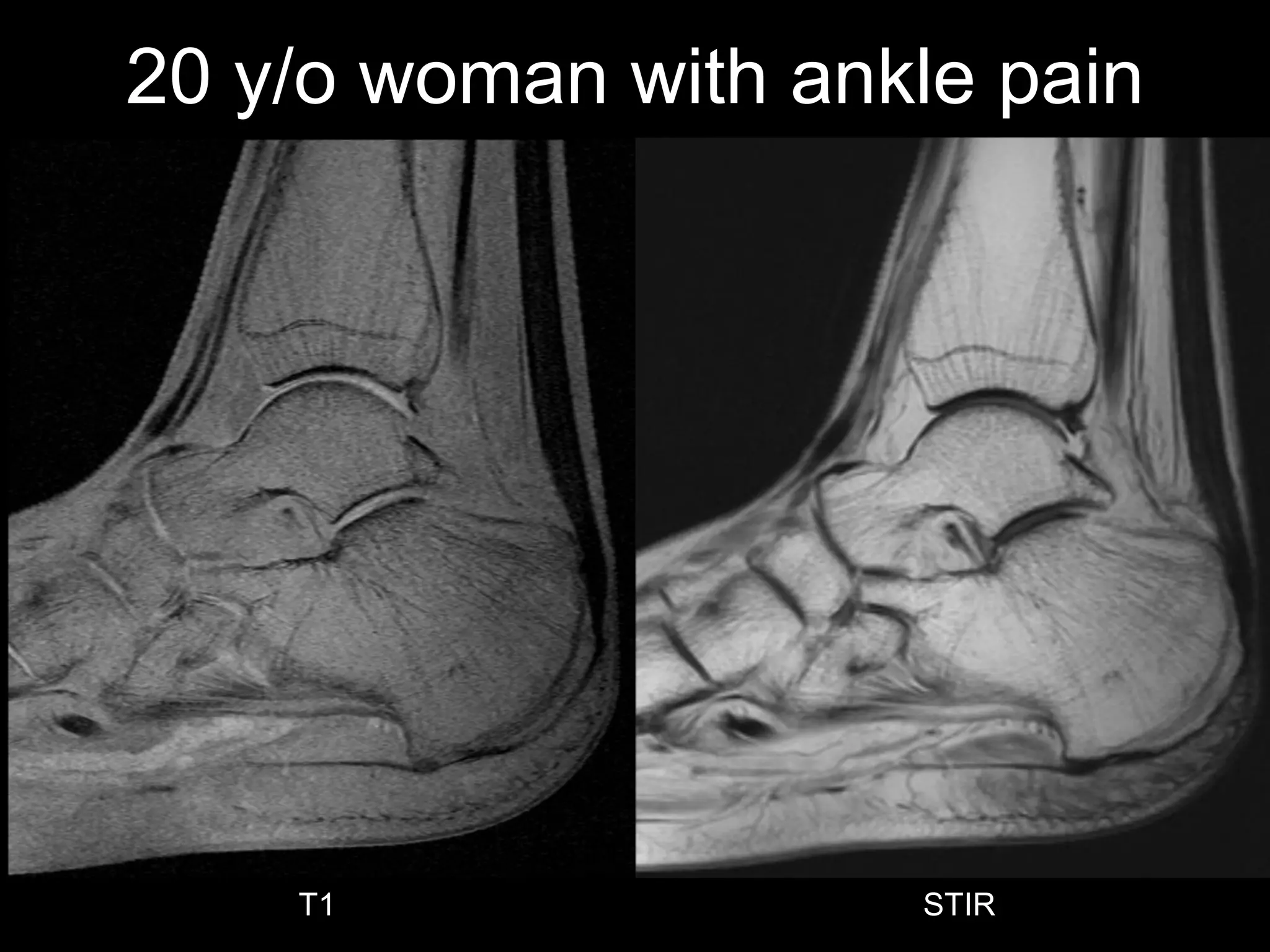
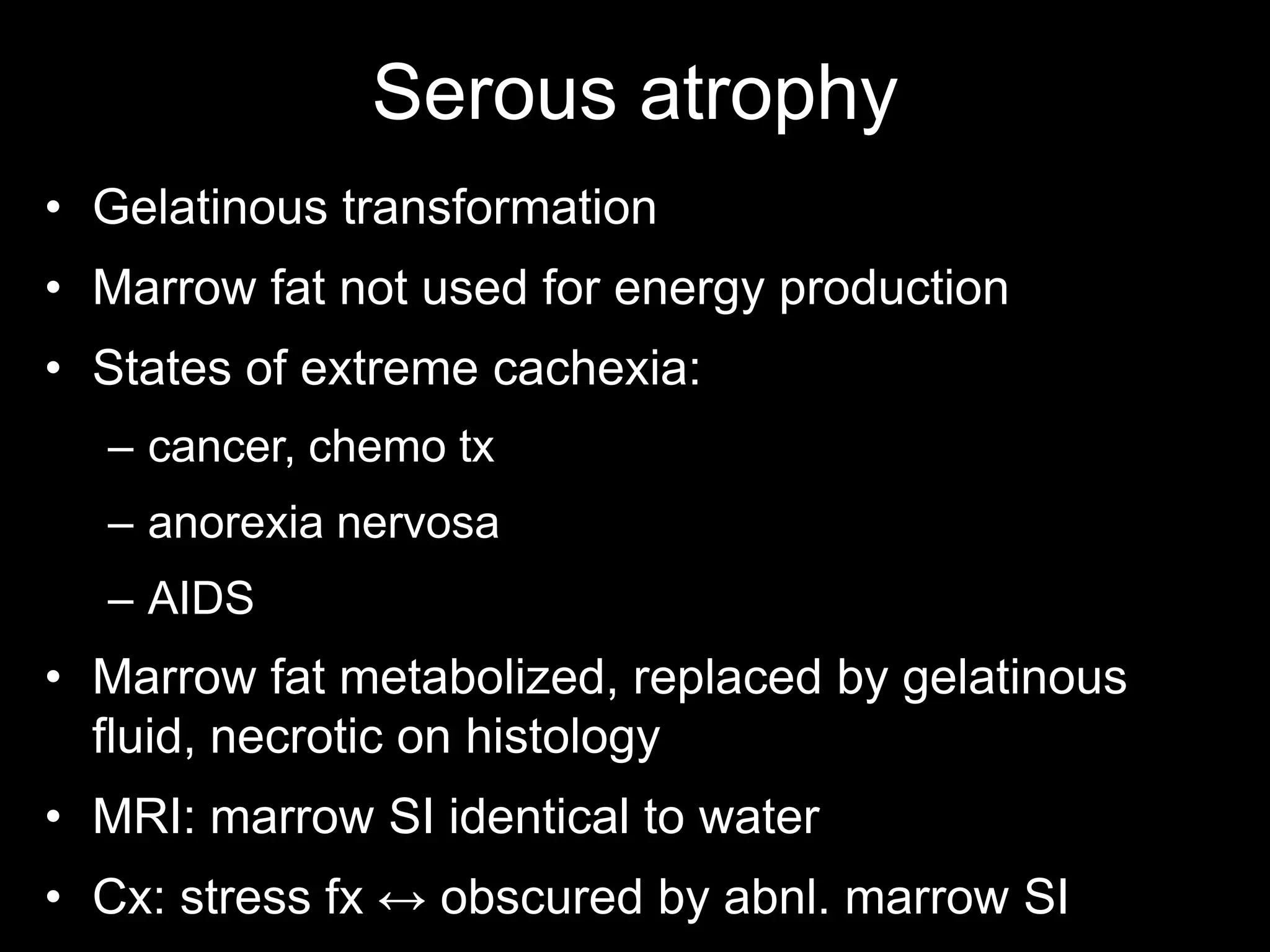
Panel of unknown cases 
Miriam A. Bredella, MD 
Associate Professor of Radiology, Massachusetts General Hospital and Harvard Medical School, Boston, MA
40 y/o woman with back pain 
T1 
STIR 
FS T1 + C
40 y/o woman with back pain 
STIR 
FS T1 + C
40 y/o woman with back pain 
STIR 
FS T1 + C
40 y/o woman with back pain
•Bone involvement rare 
•Bone involvement of axial skeleton often asymptomatic 
•MRI: intramedullary lesions 
–round large, confluent, irregular, patchy diffuse, ± enhancement 
•Lesions may involute following treatment 
•DD: metastases 
Sarcoid
52 y/o man with palpable lump 
T1
52 y/o man with palpable lump 
CT 
FDG PET 
fused PET/CT
•Rare benign neoplasm 
•Composed of brown fat 
•Increased vascularity 
•↑ FDG uptake 
•Adults, mean age 38 y (range 2-75 y) 
•Thigh > shoulder > back > neck > chest > arm > abdominal cavity, retroperitoneum 
•Tx: complete excision 
•DD: lipoma, atypical lipoma, liposarcoma 
Hibernoma 
Furlong MA et al Am J Surg Pathol 2001;25:809-14
75 y/o woman with osteoporosis
•Long term use of bisphosphonate 
•Can also occur without bisphosphonates 
•Osteoclast oversuppression → impaired bone remodeling → accumulation of microdamage 
•Decreased elasticity, increased brittleness 
Atypical femoral fracture
sag T1 
20 y/o woman with ankle pain 
T1 
STIR
Serous atrophy 
•Gelatinous transformation 
•Marrow fat not used for energy production 
•States of extreme cachexia: 
–cancer, chemo tx 
–anorexia nervosa 
–AIDS 
•Marrow fat metabolized, replaced by gelatinous fluid, necrotic on histology 
•MRI: marrow SI identical to water 
•Cx: stress fx ↔ obscured by abnl. marrow SI

2014-15 NERRS MSK Case Answers

  • 1.
    Panel of unknowncases Miriam A. Bredella, MD Associate Professor of Radiology, Massachusetts General Hospital and Harvard Medical School, Boston, MA
  • 2.
    40 y/o womanwith back pain T1 STIR FS T1 + C
  • 3.
    40 y/o womanwith back pain STIR FS T1 + C
  • 4.
    40 y/o womanwith back pain STIR FS T1 + C
  • 5.
    40 y/o womanwith back pain
  • 6.
    40 y/o womanwith back pain
  • 7.
    40 y/o womanwith back pain
  • 8.
    •Bone involvement rare •Bone involvement of axial skeleton often asymptomatic •MRI: intramedullary lesions –round large, confluent, irregular, patchy diffuse, ± enhancement •Lesions may involute following treatment •DD: metastases Sarcoid
  • 9.
    52 y/o manwith palpable lump T1
  • 10.
    52 y/o manwith palpable lump CT FDG PET fused PET/CT
  • 11.
    •Rare benign neoplasm •Composed of brown fat •Increased vascularity •↑ FDG uptake •Adults, mean age 38 y (range 2-75 y) •Thigh > shoulder > back > neck > chest > arm > abdominal cavity, retroperitoneum •Tx: complete excision •DD: lipoma, atypical lipoma, liposarcoma Hibernoma Furlong MA et al Am J Surg Pathol 2001;25:809-14
  • 12.
    75 y/o womanwith osteoporosis
  • 13.
    75 y/o womanwith osteoporosis
  • 14.
    •Long term useof bisphosphonate •Can also occur without bisphosphonates •Osteoclast oversuppression → impaired bone remodeling → accumulation of microdamage •Decreased elasticity, increased brittleness Atypical femoral fracture
  • 16.
    sag T1 20y/o woman with ankle pain T1 STIR
  • 17.
    Serous atrophy •Gelatinoustransformation •Marrow fat not used for energy production •States of extreme cachexia: –cancer, chemo tx –anorexia nervosa –AIDS •Marrow fat metabolized, replaced by gelatinous fluid, necrotic on histology •MRI: marrow SI identical to water •Cx: stress fx ↔ obscured by abnl. marrow SI