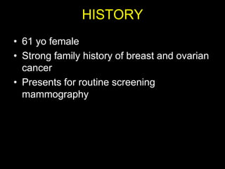
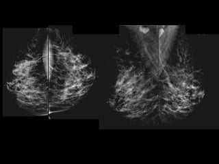
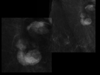
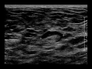
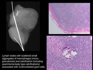
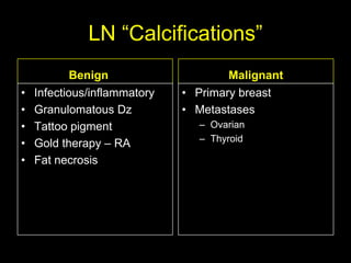
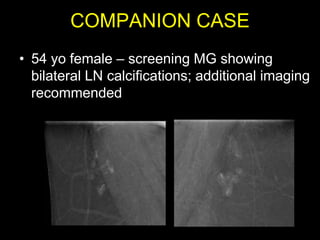
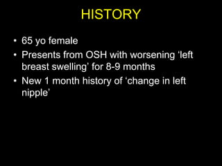
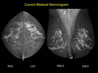
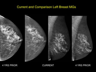
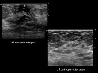
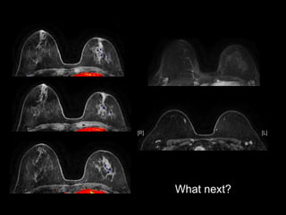
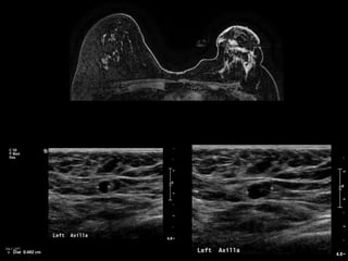
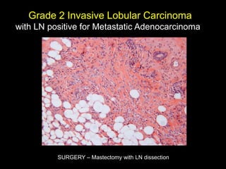
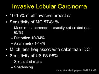
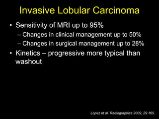
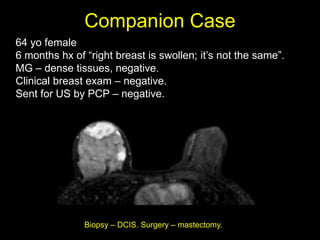
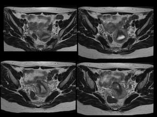
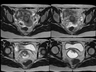
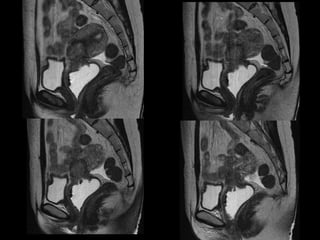
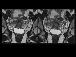
NERRS: Women‟s Imaging
        April 5, 2013


Panel of Unknown Cases
        Tejas S. Mehta MD MPH
     Assistant Professor of Radiology
          Chief, Breast Imaging
Dr. Yiming Gao
HISTORY
• 61 yo female
• Strong family history of breast and ovarian
  cancer
• Presents for routine screening
  mammography
• What would you do next?
FNA Cytology
• NEGATIVE FOR
  CARCINOMA.
  Polymorphic
  population of
  lymphocytes,
  consistent with
  lymph node sampling.

• Concordant?
• Recommend surgical
  excision
Lymph nodes with scattered small
aggregates of macrophages (micro-
granulomas) and calcification (including
psammoma-body type calcifications)
associated with multinucleated giant cells.
LN “Calcifications”
            Benign                    Malignant
•   Infectious/inflammatory   • Primary breast
•   Granulomatous Dz          • Metastases
•   Tattoo pigment              – Ovarian
                                – Thyroid
•   Gold therapy – RA
•   Fat necrosis
COMPANION CASE
• 54 yo female – screening MG showing
  bilateral LN calcifications; additional imaging
  recommended
Gold Therapy for RA
Dr. Vandana Dialani
HISTORY
• 65 yo female
• Presents from OSH with worsening „left
  breast swelling‟ for 8-9 months
• New 1 month history of „change in left
  nipple‟
Current Bilateral Mammogram


RCC      LCC             RMLO       LMLO
Current and Comparison Left Breast MGs


4 YRS PRIOR               CURRENT              4 YRS PRIOR
US retroareolar region


                         US Left upper outer breast
• What would you do next?
What next?
Grade 2 Invasive Lobular Carcinoma
with LN positive for Metastatic Adenocarcinoma


          SURGERY – Mastectomy with LN dissection
Invasive Lobular Carcinoma
• 10-15% of all invasive breast ca
• Sensitivity of MG 57-81%
  – Mass most common – usually spiculated (44-
    65%)
  – Distortion 10-34%
  – Asymmetry 1-14%
• Much less freq assoc with calcs than IDC
• Sensitivity of US 68-98%
  – Spiculated mass
  – Shadowing
                          Lopez et al. Radiographics 2009; 29:165.
Invasive Lobular Carcinoma
• Sensitivity of MRI up to 95%
  – Changes in clinical management up to 50%
  – Changes in surgical management up to 28%
• Kinetics – progressive more typical than
  washout


                          Lopez et al. Radiographics 2009; 29:165.
Companion Case
64 yo female
6 months hx of “right breast is swollen; it‟s not the same”.
MG – dense tissues, negative.
Clinical breast exam – negative.
Sent for US by PCP – negative.


               Biopsy – DCIS. Surgery – mastectomy.
Always Listen to your Patients
Dr. Ana Lourenco
HISTORY
• 36 yo female 30.5 weeks pregnant
• Referred with „multiple fetal anomalies‟ for
  fetal ultrasound
LUS – Cephalic
POSTNATAL BABYGRAM
-Cephalic
-Heart - right side
-Stomach – right side
-Liver – left side
-Des colon – right side
Esophageal Pouch
Situs Inversus Totalis
• 3-5% with cardiac abnormalities
• 25% with primary ciliary dyskinesia (PCD)
  – Kartagener‟s Syndrome
  – Chronic sinus infections; respiratory infections;
    infertility
Terminology
• Situs solitus – normal anatomy
• Situs inversus – mirror image of normal
• Situs ambiguous – heterotaxy
  – Disordered arrangement of organs in chest and
    abdomen
  – Asplenia – right atrial isomerism
  – Polyslenia – left atrial isomerism
Esophageal Atresia
      • Polyhydramnios
      • small / absent stomach if no
        fistula
      • Often other assoc anomalies
        –   Imperforate anus
        –   Duodenal atresia/stenosis
        –   Vertebral/rib anomalies
        –   VACTERL spectrum
Dr. Anne Silas
HISTORY
• 40 yo female 8.5 weeks pregant
• S/P D&C for pregnancy implanted at site of
  c-section scar
• 6 weeks post D&C patient presents with
  heavy persistent bleeding
• For Pelvic US
• HCG = 451 mIU/mL
D&C complicated by Perforation,
  AVM w RPOC at Site of Perforation
• AVM – Acquired or Congenital
• Acquired
  – Traumatic – D&C, TAB, uterine surgery
  – Less common: Endometrial / Cervical CA, GTD
  – Clue on US: numerous tortuous vessels, high velocities
• Tx:
  – transcatheter arterial embolization
      • Potential to preserve fertility
  – UA ligation, hysterectomy
Angio after MRI
POST PROCEDURE


S/P embolization of right UA and left UA due to
            cross collateralization
1 month follow up

HCG < 5 mIU/mL


                    No flow in area – smaller in size
                    Felt residual hematoma
                    Will continue US follow up
Dr. Hedvig Hricak
HISTORY
•   42 yo female
•   Recent immigrant from China
•   C/O postcoital and intermenstrual bleeding
•   Abnormal GYN exam
•   Abnormal biopsy
•   Further imaging performed
MRI findings - Stage IIB
with PET/CT - Stage IIIB
    Cervical Cancer
Stage I – Carcinoma Confined to Cervix


                           <= 4 cm greatest dimension   > 4 cm greatest dimension


    <= 7 mm wide

    A1 < =3mm deep
    A2 >3 but < 5mm deep
Stage II – Carcinoma Invades Beyond Uterus
  But not to Pelvic Wall or Lower 1/3 Vagina


                  With(A) or without (B)
                   parametrial invasion
Stage III – Carcinoma to Pelvic Wall and/or Lower 1/3 of
Vagina and/or Causes Hydronephrosis or Nonfxing Kidneys


                Lower 1/3 Vagina
                No pelvic wall


                                   Extends to pelvic wall
                                   and/or renal issues
Stage IV – Carcinoma beyond True Pelvis or Involvement
      Of Mucosa of Bladder or Rectum (bx proven)


                                                   Distant Mets
        Spread to adj organs
Role of MR Imaging in Tx Stratification
                of GYN (Cervical) Cancer
• Cervical Cancer – 2nd most common ca in women
  worldwide
   – Developing countries; pk 30-40 yrs
• FIGO classification – revised in 2009 from just clinical to
  incorporate cross-sectional imaging (CT, MR)
• Staging accuracy of MR 85-96%
   – Best test to assess tumor size and location; invasion into
     parametria, pelvic side wall, adjacent organs; local nodal enlgment
• PET/CT helpful in staging advanced disease
   – Demonstrates unexpected sites beyond pelvis
• Treatment options
   – Radical surgery - early stage (IA, IB1, IIA1)
   – Primary Chemo and Radiation – bulky IB2 or IIA2, or local
     advanced (IIB or greater)
                                              Sala et al. Radiology 2013; 266: 717.
Thank you!

NERRS WI answers 2013

  • 1.
    NERRS: Women‟s Imaging April 5, 2013 Panel of Unknown Cases Tejas S. Mehta MD MPH Assistant Professor of Radiology Chief, Breast Imaging
  • 2.
  • 3.
    HISTORY • 61 yofemale • Strong family history of breast and ovarian cancer • Presents for routine screening mammography
  • 7.
    • What wouldyou do next?
  • 8.
    FNA Cytology • NEGATIVEFOR CARCINOMA. Polymorphic population of lymphocytes, consistent with lymph node sampling. • Concordant? • Recommend surgical excision
  • 9.
    Lymph nodes withscattered small aggregates of macrophages (micro- granulomas) and calcification (including psammoma-body type calcifications) associated with multinucleated giant cells.
  • 10.
    LN “Calcifications” Benign Malignant • Infectious/inflammatory • Primary breast • Granulomatous Dz • Metastases • Tattoo pigment – Ovarian – Thyroid • Gold therapy – RA • Fat necrosis
  • 11.
    COMPANION CASE • 54yo female – screening MG showing bilateral LN calcifications; additional imaging recommended
  • 12.
  • 13.
  • 14.
    HISTORY • 65 yofemale • Presents from OSH with worsening „left breast swelling‟ for 8-9 months • New 1 month history of „change in left nipple‟
  • 15.
  • 16.
    Current and ComparisonLeft Breast MGs 4 YRS PRIOR CURRENT 4 YRS PRIOR
  • 17.
    US retroareolar region US Left upper outer breast
  • 18.
    • What wouldyou do next?
  • 19.
  • 21.
    Grade 2 InvasiveLobular Carcinoma with LN positive for Metastatic Adenocarcinoma SURGERY – Mastectomy with LN dissection
  • 22.
    Invasive Lobular Carcinoma •10-15% of all invasive breast ca • Sensitivity of MG 57-81% – Mass most common – usually spiculated (44- 65%) – Distortion 10-34% – Asymmetry 1-14% • Much less freq assoc with calcs than IDC • Sensitivity of US 68-98% – Spiculated mass – Shadowing Lopez et al. Radiographics 2009; 29:165.
  • 23.
    Invasive Lobular Carcinoma •Sensitivity of MRI up to 95% – Changes in clinical management up to 50% – Changes in surgical management up to 28% • Kinetics – progressive more typical than washout Lopez et al. Radiographics 2009; 29:165.
  • 24.
    Companion Case 64 yofemale 6 months hx of “right breast is swollen; it‟s not the same”. MG – dense tissues, negative. Clinical breast exam – negative. Sent for US by PCP – negative. Biopsy – DCIS. Surgery – mastectomy.
  • 25.
    Always Listen toyour Patients
  • 26.
  • 27.
    HISTORY • 36 yofemale 30.5 weeks pregnant • Referred with „multiple fetal anomalies‟ for fetal ultrasound
  • 28.
  • 32.
  • 34.
    -Cephalic -Heart - rightside -Stomach – right side -Liver – left side -Des colon – right side
  • 35.
  • 36.
    Situs Inversus Totalis •3-5% with cardiac abnormalities • 25% with primary ciliary dyskinesia (PCD) – Kartagener‟s Syndrome – Chronic sinus infections; respiratory infections; infertility
  • 37.
    Terminology • Situs solitus– normal anatomy • Situs inversus – mirror image of normal • Situs ambiguous – heterotaxy – Disordered arrangement of organs in chest and abdomen – Asplenia – right atrial isomerism – Polyslenia – left atrial isomerism
  • 38.
    Esophageal Atresia • Polyhydramnios • small / absent stomach if no fistula • Often other assoc anomalies – Imperforate anus – Duodenal atresia/stenosis – Vertebral/rib anomalies – VACTERL spectrum
  • 39.
  • 40.
    HISTORY • 40 yofemale 8.5 weeks pregant • S/P D&C for pregnancy implanted at site of c-section scar
  • 42.
    • 6 weekspost D&C patient presents with heavy persistent bleeding • For Pelvic US • HCG = 451 mIU/mL
  • 50.
    D&C complicated byPerforation, AVM w RPOC at Site of Perforation • AVM – Acquired or Congenital • Acquired – Traumatic – D&C, TAB, uterine surgery – Less common: Endometrial / Cervical CA, GTD – Clue on US: numerous tortuous vessels, high velocities • Tx: – transcatheter arterial embolization • Potential to preserve fertility – UA ligation, hysterectomy
  • 51.
  • 52.
    POST PROCEDURE S/P embolizationof right UA and left UA due to cross collateralization
  • 53.
    1 month followup HCG < 5 mIU/mL No flow in area – smaller in size Felt residual hematoma Will continue US follow up
  • 54.
  • 55.
    HISTORY • 42 yo female • Recent immigrant from China • C/O postcoital and intermenstrual bleeding • Abnormal GYN exam • Abnormal biopsy • Further imaging performed
  • 63.
    MRI findings -Stage IIB with PET/CT - Stage IIIB Cervical Cancer
  • 64.
    Stage I –Carcinoma Confined to Cervix <= 4 cm greatest dimension > 4 cm greatest dimension <= 7 mm wide A1 < =3mm deep A2 >3 but < 5mm deep
  • 65.
    Stage II –Carcinoma Invades Beyond Uterus But not to Pelvic Wall or Lower 1/3 Vagina With(A) or without (B) parametrial invasion
  • 66.
    Stage III –Carcinoma to Pelvic Wall and/or Lower 1/3 of Vagina and/or Causes Hydronephrosis or Nonfxing Kidneys Lower 1/3 Vagina No pelvic wall Extends to pelvic wall and/or renal issues
  • 67.
    Stage IV –Carcinoma beyond True Pelvis or Involvement Of Mucosa of Bladder or Rectum (bx proven) Distant Mets Spread to adj organs
  • 68.
    Role of MRImaging in Tx Stratification of GYN (Cervical) Cancer • Cervical Cancer – 2nd most common ca in women worldwide – Developing countries; pk 30-40 yrs • FIGO classification – revised in 2009 from just clinical to incorporate cross-sectional imaging (CT, MR) • Staging accuracy of MR 85-96% – Best test to assess tumor size and location; invasion into parametria, pelvic side wall, adjacent organs; local nodal enlgment • PET/CT helpful in staging advanced disease – Demonstrates unexpected sites beyond pelvis • Treatment options – Radical surgery - early stage (IA, IB1, IIA1) – Primary Chemo and Radiation – bulky IB2 or IIA2, or local advanced (IIB or greater) Sala et al. Radiology 2013; 266: 717.
  • 69.