The document discusses various primary retroperitoneal neoplasms. It describes the location of the retroperitoneum and divides primary retroperitoneal neoplasms into solid neoplastic masses including mesodermal neoplasms (such as liposarcomas, leiomyosarcomas, and malignant fibrous histiocytomas), neurogenic tumors, and germ cell/sex cord/stromal tumors. For each type of neoplasm, it provides details on characteristics such as common subtypes, demographic and imaging appearances, and clinical implications.
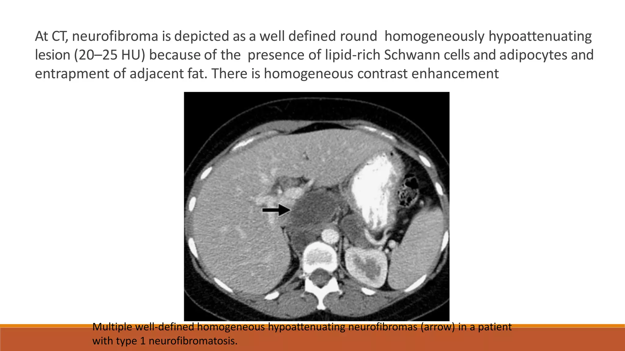
The document discusses primary retroperitoneal neoplasms. It notes that 70-80% of primary retroperitoneal neoplasms are malignant in nature. The retroperitoneum contains mesodermal neoplasms, neurogenic tumors, germ cell and sex cord tumors, and lymphoid neoplasms. The most common primary retroperitoneal sarcomas are liposarcoma, leiomyosarcoma, and malignant fibrous histiocytoma. Neurogenic tumors such as schwannomas and neurofibromas are usually benign and occur in a younger age group. Teratomas are germ cell tumors that may contain fat, calcium, or sebum levels on imaging.
The document discusses various types of primary retroperitoneal masses. It describes the anatomy of the retroperitoneum and states that 70-80% of primary retroperitoneal neoplasms are malignant. The masses are divided into solid neoplastic masses including mesodermal neoplasms (such as liposarcomas, leiomyosarcomas, and malignant fibrous histiocytomas), neurogenic tumors, and germ cell/sex cord/stromal tumors. Characteristics of common subtypes such as liposarcoma, leiomyosarcoma, and schwannoma are provided. Imaging features on CT and MRI to identify and characterize these masses are also discussed.
The document discusses retroperitoneal masses. It notes that primary retroperitoneal masses originate in the retroperitoneum outside major organs. CT and MRI are important imaging modalities. Retroperitoneal masses can be solid or cystic neoplastic or non-neoplastic tumors. The majority are malignant. Common solid neoplastic masses include mesodermal tumors like liposarcomas, neurogenic tumors, and germ cell tumors. Imaging findings of common tumor types are described.
This document provides an overview of sarcomas, including:
- Sarcomas are rare tumors accounting for less than 1% of cancers. They arise from embryonic mesoderm tissue.
- The document discusses the epidemiology, types, clinical presentation, investigation, and histopathology of sarcomas. It focuses on some of the most common subtypes like malignant fibrous histiocytoma, liposarcoma, and leiomyosarcoma.
- Evaluation of sarcoma involves clinical examination, imaging such as MRI or CT, and biopsy for histopathological diagnosis. Proper investigation is important for staging and determining the best treatment approach.
This document discusses radiological imaging modalities for assessing salivary gland diseases. It describes several imaging techniques including ultrasound, CT, MRI, nuclear scintigraphy and their roles in evaluating patients with salivary gland symptoms. Ultrasound can assess gland vascularity and guide biopsies. CT and MRI help determine lesion extent and involvement of surrounding structures. Nuclear scintigraphy uses radioactive tracers to identify tumors. The document also discusses common benign and malignant salivary gland tumors seen on imaging, including Warthin's tumor, oncocytoma, mucoepidermoid carcinoma and adenoid cystic carcinoma. Imaging features vary depending on tumor type and grade. In summary, various radiological imaging techniques play an important role in
Imaging in small bowel tumors Dr. Muhammad Bin Zulfiqar
Here we will discuss CT and MR enterography. We will further discuss the use of negative contrast.
Four important tumors will be discussed.
This document discusses primary retroperitoneal masses and provides an overview of mesodermal tumors, which constitute 47-57% of primary retroperitoneal tumors. It describes key imaging findings and pathological characteristics of common mesodermal tumors including liposarcoma, leiomyosarcoma, and malignant fibrous histiocytoma. Well-differentiated liposarcoma, dedifferentiated liposarcoma, and myxoid liposarcoma are subtypes of liposarcoma discussed in detail, outlining their imaging appearance on CT, MRI, and PET scans as well as histological features. Complete surgical excision is important but recurrence is common, necessitating long-term imaging follow-up.
Testicular tumors are rare.
1 – 2 % of all malignant tumors.
Most common malignancy in men in the 15 to 35 year age group.
Benign lesions represent a greater percentage of cases in children than in adults.
Most curable solid neoplasm
This document discusses the characteristics of benign and malignant neoplasms. It describes how malignant tumors are less differentiated, grow faster, are more locally invasive, and can metastasize to other parts of the body compared to benign tumors. The document also discusses epidemiological factors that affect cancer incidence such as geographic location, age, heredity, and preexisting conditions. Common cancer types and causes of cancer death worldwide are also presented.
The document discusses salivary gland diseases, focusing on sialadenitis (inflammation of the salivary glands) and salivary gland tumors. Sialadenitis can be caused by viruses, bacteria, or autoimmune disorders. The most common viral cause is mumps. Bacterial sialadenitis often results from ductal obstruction. Chronic sialadenitis is usually caused by Sjögren's syndrome. Common benign salivary gland tumors include pleomorphic adenoma, Warthin's tumor, and monomorphic adenomas. Malignant tumors include mucoepidermoid carcinoma and adenoid cystic carcinoma.
Soft tissue sarcomas are a rare and heterogeneous group of tumors that arise from mesenchymal tissues. They account for less than 1% of adult cancers and 7% of childhood cancers. The most common types in adults are malignant fibrous histiocytoma, leiomyosarcoma, and liposarcoma. Treatment involves surgical resection with negative margins, often combined with radiation therapy. For high-risk localized or metastatic disease, chemotherapy may also be used. Accurate diagnosis and treatment planning by a multidisciplinary team is important for managing these rare tumors.
The document discusses approaches to diagnosing benign retroperitoneal masses, noting that primary retroperitoneal neoplasms make up 0.1-0.2% of all malignancies and can be either benign or malignant. It describes methods for determining if a mass is retroperitoneal versus intra-abdominal, identifying its organ of origin, and recognizing characteristics like fat, cysts, necrosis and vascularity that provide diagnostic clues. Learning objectives cover evaluating tumor location, organ of origin, and specific imaging findings for retroperitoneal tumors.
Primary retroperitoneal tumors are rare neoplasms that arise in the retroperitoneum and pelvis. Liposarcoma is the most common type of primary retroperitoneal tumor, while lymphoma is the most common retroperitoneal malignancy overall. These tumors often grow extensively before causing symptoms. Diagnostic imaging includes CT or MRI to evaluate the tumor characteristics and relationship to surrounding structures. Surgical resection with negative margins is the standard treatment for localized primary retroperitoneal sarcomas, while chemotherapy or radiation may be used in certain settings. Prognosis depends on tumor grade, stage, and ability to achieve a complete resection.
This document discusses the management of rhabdomyosarcoma, a type of soft tissue sarcoma that is most common in children. It begins by covering the epidemiology, risk factors, clinical presentation and symptoms. Diagnostic workup involves tumor biopsy along with imaging like CT, MRI and PET scans to determine tumor location and spread. Staging uses the Children's Oncology Group system which considers tumor site, size, margins after surgery, and metastasis. There are various histologic subtypes with differing prognoses - embryonal has intermediate prognosis while alveolar and undifferentiated subtypes have poorer outcomes. Treatment involves chemotherapy, surgery and radiation therapy tailored to stage and risk group.
This document provides an overview of rhabdomyosarcoma (RMS), a rare soft tissue cancer that occurs mostly in children. It discusses the epidemiology, risk factors, clinical presentation, diagnostic workup, staging, histologic subtypes and prognosis of RMS. RMS most commonly occurs in the head, neck, genitourinary tract and extremities. Diagnostic testing includes biopsy, imaging like CT/MRI and bone marrow aspiration. Staging involves tumor size, location, margins and metastasis. The prognosis depends on the histologic subtype, with botryoid and spindle cell having the best outcomes and alveolar/undifferentiated having poorer outcomes.
Cancer is predominantly a disease of middle age and elderly. Environment and genetics influence cancer risk. The global cancer burden is estimated at 10 million new cases per year, predicted to rise to 15 million by 2020. Cancer is the second most common cause of death in developed countries. The major cancers vary by sex, with lung cancer most common in men and breast cancer in women. Cancer risk depends on factors like age, sex, geography, occupation, diet, and smoking. Cancer spreads locally, through lymphatics, blood vessels, body cavities, and along epithelial surfaces. Premalignant conditions include changes in benign tumors, intraepithelial neoplasia, and malignancies from chronic inflammation.
Renal pathology lecture 4 Tumors of kidney and urinary tract. Sufia Husain 2020Sufia Husain
This document provides an overview of tumors of the kidney and urinary tract. It begins by outlining the objectives and key topics to be covered, which include benign kidney tumors, renal cell carcinoma, Wilms tumor, and transitional cell and squamous carcinomas of the bladder. The document then covers these topics in detail over several sections, describing the histology, risk factors, clinical features, and characteristics of each tumor type. The major tumor types discussed are renal oncocytoma, angiomyolipoma, renal cell carcinoma (clear cell and papillary subtypes), Wilms tumor, and transitional cell neoplasms of the bladder.
The document discusses various types of adrenal masses and how they appear on different imaging modalities like CT, MRI, and ultrasound. It describes the normal anatomy of the adrenal glands and then discusses common benign and malignant adrenal masses. Some key masses mentioned include adrenal adenomas, pheochromocytomas, adrenocortical carcinomas, myelolipomas, and neuroblastomas. Imaging features discussed help differentiate between these different adrenal lesions.
Thymic tumors arise from the thymus gland in the anterior mediastinum. Thymomas are the most common type of thymic tumor and generally have an indolent growth pattern. Complete surgical resection is the primary treatment for thymomas. Adjuvant radiation and chemotherapy may be considered depending on the stage and completeness of resection. Thymic carcinomas are more aggressive and have a poorer prognosis than thymomas. Treatment involves surgical resection when possible along with chemotherapy and radiation.
This document discusses various types of spinal tumors, including their location, characteristics, and MRI appearances. It covers primary spinal tumors such as schwannomas, neurofibromas, meningiomas, ependymomas, astrocytomas, and hemangioblastomas. It describes these tumors' demographics, symptoms, classification as interdural exteramedullary or interdural intramedullary, and features on T1-, T2-, and contrast-enhanced MRI sequences. Rare tumors like lipomas, paragangliomas, and subependymomas are also outlined briefly.
The document discusses retroperitoneal anatomy and various etiologies of retroperitoneal masses including both neoplastic and non-neoplastic causes. It describes the clinical presentation of retroperitoneal masses as often asymptomatic initially, with later development of compressive symptoms. A variety of investigations are outlined to evaluate retroperitoneal masses including blood tests, imaging modalities, and biopsy. Common retroperitoneal masses discussed in more detail include retroperitoneal sarcoma, lymphoma, teratoma, hematoma, urinoma, and retroperitoneal fibrosis.
This document discusses spinal tumors, including:
1. Spinal tumors can be primary or secondary, extradural or intradural. The most common symptoms are pain and neurological deficits.
2. Common primary tumors include hemangiomas, aneurysmal bone cysts, and chordomas. Metastatic tumors are most frequently from breast, prostate and lung cancers.
3. Imaging like CT, MRI and bone scans are used to identify the location and extent of tumors. Tissue biopsy is needed to confirm diagnosis.
4. Treatment involves a multidisciplinary approach with surgery, radiation, chemotherapy and steroid/medical management. The goal of surgery is wide or en bloc resection with spinal reconstruction and stabilization
Soft tissue tumors are a heterogeneous group of lesions that arise from nonepithelial, extraskeletal tissues including adipose tissue, muscles, tendons, blood vessels and lymphatics. Imaging plays an important role in evaluating these tumors. There are nine major groups of soft tissue tumors including adipocytic tumors, vascular tumors, fibroblastic tumors, fibrohistiocytic tumors, smooth muscle tumors, skeletal muscle tumors, pericytic tumors, chondro-osseous tumors and tumors of uncertain differentiation. Lipoma is the most common benign adipocytic tumor while liposarcoma is a malignant adipocytic tumor. Hemangioma is a common benign vascular tumor and angiosarcoma is the most common
Cardiac tumors can be primary tumors originating from the heart or secondary tumors from metastases. Primary tumors are more commonly benign myxomas found in the left atrium of middle aged patients, while secondary tumors are more prevalent. Imaging with echocardiography, CT, and MRI can characterize tumors and determine if they are resectable. MRI provides the best evaluation of tumor extent, involvement of surrounding structures, and differentiation of tumor types.
This document provides information on testicular tumors, including:
1. It describes the two major categories of testicular tumors - germ cell tumors (95% of cases) and sex cord-stromal tumors. Germ cell tumors are aggressive cancers capable of rapid dissemination but most can now be cured.
2. It covers the various types of germ cell tumors - seminoma, embryonal carcinoma, yolk sac tumor, choriocarcinoma, and teratoma - discussing their characteristics, risk factors, pathogenesis, morphology, and microscopy.
3. Seminoma is the most common germ cell tumor, occurring most often in the third decade. Embryonal carcinoma is more
This document discusses the diagnostic workup for mediastinal tumors. It begins by describing the different types of tumors that can occur in the anterior, middle, and posterior mediastinum. Signs and symptoms are non-specific but may include cough, chest pain, and weight loss. Chest x-ray is often the initial test, while CT scan provides more detail on location and tissue characteristics. MRI and nuclear imaging can further characterize masses. Tissue sampling methods include needle biopsy, mediastinoscopy, and thoracoscopy. Surgical procedures like mediastinotomy or thoracotomy may be needed in some cases. Laboratory tests help identify certain tumor types. A multidisciplinary approach is typically needed for diagnosis and treatment planning of
This document discusses giant cell tumor (GCT), a rare type of bone tumor characterized by abnormally large multinucleated giant cells. Key points include:
- GCT most commonly affects people aged 20-40 and has a female predominance. The most common sites are the distal femur, proximal tibia, and distal radius.
- Radiographically, GCT appears as an eccentric, expansile, lytic lesion without marginal sclerosis. CT and MRI can help delineate cortical destruction and soft tissue involvement.
- Histologically, GCT contains giant cells with many nuclei and mononuclear stromal cells. Malignant degeneration is rare but can occur spontaneously or after treatment.
The vertebral column consists of 33 vertebrae separated by intervertebral discs. A typical vertebra has a vertebral body and arch enclosing the vertebral foramen through which the spinal cord passes. The spinal cord has 31 pairs of spinal nerves and is composed of gray and white matter. It transmits sensory information up the posterior columns and motor commands down tracts like the corticospinal tract. Injuries can cause syndromes like complete transection with bilateral deficits or Brown-Sequard with unilateral deficits depending on the location and extent of damage.
The document describes the various cerebrospinal fluid (CSF) filled spaces, or cisterns, within the subarachnoid space. It details both supra-tentorial and infra-tentorial cisterns, providing their locations, contents such as vessels and cranial nerves, and anatomical relationships. Key cisterns mentioned include the cistern of the lamina terminalis, chiasmatic cistern, interpeduncular cistern, prepontine cistern, cisterna magna, and cerebellopontine angle cistern. The cisterns form a interconnected network facilitating CSF circulation within the subarachnoid space.
This document provides an overview of the gross anatomy of the brain as seen on MR imaging. It describes the central sulcus, ventricular system, limbic system, and white matter. It then details the axial, sagittal, and coronal views of the brain and lists over 100 structures and their 3D localization within the brain.
The document discusses various congenital anomalies of the pancreas including annular pancreas, pancreas divisum, ectopic pancreatic tissue, horseshoe pancreas, and variations in pancreatic ductal anatomy. It describes the embryological development of the pancreas and defines important anatomical structures such as the pancreatic ducts. Imaging features of different pancreatic anomalies on modalities like CT, MRI, ERCP, and ultrasound are provided.
CT guided FNAC is a simple and minimally invasive technique for obtaining tissue samples from complex lung lesions for diagnosis. A study of 28 patients found CT guided FNAC to have a sensitivity of 80% and specificity of 100% for diagnosing malignancy. Complications occurred in 3 patients (12.5%) and were minor and self-resolving. CT guided FNAC is shown to be an effective and safe outpatient procedure for evaluating pulmonary nodules and masses.
CT guided FNAC is a simple and effective technique for diagnosing complex pulmonary lesions. In a study of 28 patients, CT guided FNAC had a sensitivity of 80% and specificity of 100% for diagnosing malignancy. CT scanning alone had sensitivity of 75% and specificity of 83.3% for malignancy. Complications occurred in 3 patients (12.5%) and were minor and resolved with conservative treatment. The study concluded that CT guided FNAC is a highly sensitive and specific technique for characterizing pulmonary lesions.
The document discusses various presacral lesions that can be seen on imaging. It describes the anatomy of the presacral space and then covers conditions with osteochondral origin like giant cell tumor and Ewing sarcoma. Neurogenic conditions such as neurofibromas, schwannomas, and perineural cysts are also discussed. Other lesions mentioned include dural ectasia and anterior myelomeningoceles. For each condition, the document provides details on clinical features, imaging appearance on modalities like CT and MRI, and examples of imaging findings.
Sarcoidosis is a multisystem granulomatous disease of unknown etiology characterized by non-caseating granulomas. It most commonly affects the lungs, presenting radiographically as bilateral hilar lymphadenopathy in 50% of cases and pulmonary nodules in 30-50% of cases. Other involved organs include the eyes, skin, and heart. On CT, it demonstrates enlarged lymph nodes and pulmonary nodules distributed along the bronchovascular bundles. Late stage disease can develop pulmonary fibrosis. Sarcoidosis can also involve bones, presenting as cystic lesions in the hands. Neurosarcoidosis manifests as leptomeningeal enhancement or intracranial masses.
The document describes various signs seen on imaging of the respiratory system. It defines signs such as the signet ring sign seen on CT scans of the lungs, the finger-in-glove appearance seen in allergic broncho-pulmonary aspergillosis, and the continuous diaphragm sign seen in pneumomediastinum where air outlines the entire diaphragm. It also provides details on other signs like the halo sign associated with hemorrhagic nodules, the reversed halo sign, and tree-in-bud appearance seen in conditions like tuberculosis.
1. The document defines and describes solitary pulmonary nodules, providing details on measurements, characteristics, and imaging features that help determine if a nodule is benign or malignant.
2. Malignant nodules are more likely to be larger in size, irregular or spiculated in shape, located in the upper lobes, and demonstrate rapid growth. Benign nodules often have fat, calcification, or show long-term stability.
3. Guidelines are provided for follow-up of solid versus subsolid nodules based on size, with smaller or stable nodules requiring less frequent follow-up, and suspicious nodules warranting further evaluation including PET scans or biopsy.
Esophageal webs are thin mucosal membranes that project into the esophageal lumen, causing constriction. They more commonly occur in the cervical esophagus near the cricopharyngeus muscle. Associations include Plummer-Vinson syndrome, graft-versus-host disease, and gastroesophageal reflux disease. On barium swallows, esophageal webs appear as smooth tapered concentric narrowing in the cervical esophagus.
The parathyroid glands are located posterior to the thyroid gland in the neck. Parathyroid adenomas, the most common cause of primary hyperparathyroidism, enhance vividly on arterial phase CT then wash out rapidly on delayed phase with low attenuation on non-contrast images. Localizing the adenoma precisely with 4D CT guides focused surgical treatment through a small incision. The characteristic enhancement pattern and morphology help identify ectopic adenomas located during fetal development in the mediastinum.
This document provides an overview of brain anatomy including:
1. It describes the MRI appearance of different brain tissues and structures including white matter, fat, CSF, and gray matter on different sequences.
2. It then covers the sulcal and gyral anatomy of the brain, describing the lobes, major sulci like the central sulcus and sylvian fissure, and how they can be identified.
3. The anatomy of each lobe is then covered in more detail including the surfaces and sulci that make up the frontal, parietal, occipital, and temporal lobes.
Osteomyelitis is an infection of bone that is usually caused by bacteria entering through the bloodstream or direct inoculation via injury. It can be acute, subacute, or chronic. Common symptoms include fever, pain, and swelling near the infected bone. Diagnosis involves blood tests, imaging like x-rays, MRI, and bone scans, and bone/blood cultures. Treatment consists of antibiotics tailored to the identified bacteria as well as possible surgical drainage of abscesses.
This document discusses primary retroperitoneal neoplasms, which arise outside of major retroperitoneal organs. It notes that 70-80% of retroperitoneal masses are malignant in nature. The document then categorizes and describes several specific types of solid neoplastic masses that can occur in the retroperitoneum, including mesodermal neoplasms (such as liposarcomas and leiomyosarcomas), neurogenic tumors, and others. For each type of mass, it provides details on prevalence, appearance on CT and MRI scans, characteristics, associated syndromes, and other relevant clinical information.
1. The goals of first trimester ultrasound include visualization of the gestational sac, identification of embryonic demise, determination of gestational age, and early diagnosis of fetal anomalies.
2. A normal intrauterine gestation will demonstrate a gestational sac, yolk sac, embryo, amnion, and cardiac activity on ultrasound. Measurement of the mean sac diameter, crown-rump length, and biometric measurements can be used to estimate gestational age.
3. Absence of cardiac activity along with signs of bleeding have a high probability of embryonic demise. Criteria such as large sac size without visualizing fetal structures indicate a poor pregnancy outcome.
This document discusses Legg-Calve-Perthes disease, which is avascular necrosis of the femoral head that occurs in children. It begins by describing the etiology as an ischemic episode affecting the capital femoral epiphysis, though the exact cause is unknown. The stages of the disease are then outlined based on radiographic appearance, from initial avascular necrosis to revascularization and bone remodeling. Complications including deformities of the femoral head and neck are also summarized. The document provides detailed information on the radiographic signs and classifications systems used to evaluate the progression and prognosis of Legg-Calve-Perthes disease.
X-ray grids are devices used to remove scattered radiation from radiographic images. They consist of alternating strips of lead and transparent material. Grids work by absorbing most of the multidirectional scattered radiation while allowing the directional primary radiation to pass through. Grid performance is evaluated based on primary transmission, Bucky factor, and contrast improvement factor. Proper grid selection and positioning are important to avoid grid cutoff and increased patient radiation dose. Moving grids eliminate grid line artifacts but have some disadvantages.
This document discusses fluoroscopy, including conventional fluoroscopy units and modern fluoroscopic units. It describes the key components of a fluoroscopic unit, including the image intensifier, vidicon camera, and TV monitor. It also discusses factors that influence fluoroscopic image quality such as radiation dose rates, image resolution both vertically and horizontally, and techniques to reduce image noise like frame averaging.
A fluoroscope uses x-rays and a fluorescent screen to enable direct observation of internal organs. It consists of an x-ray tube, table, and image intensifier. The image intensifier converts x-rays into visible light images and amplifies them for viewing. It works by accelerating photoelectrons emitted from a photocathode onto a phosphor screen, producing light photons and gaining brightness. Newer generations of image intensifiers use additional electron multiplication for higher sensitivity. Fluoroscopy provides real-time moving images for procedures while fluorography captures still diagnostic images.
Embracing Deep Variability For Reproducibility and Replicability
Abstract: Reproducibility (aka determinism in some cases) constitutes a fundamental aspect in various fields of computer science, such as floating-point computations in numerical analysis and simulation, concurrency models in parallelism, reproducible builds for third parties integration and packaging, and containerization for execution environments. These concepts, while pervasive across diverse concerns, often exhibit intricate inter-dependencies, making it challenging to achieve a comprehensive understanding. In this short and vision paper we delve into the application of software engineering techniques, specifically variability management, to systematically identify and explicit points of variability that may give rise to reproducibility issues (eg language, libraries, compiler, virtual machine, OS, environment variables, etc). The primary objectives are: i) gaining insights into the variability layers and their possible interactions, ii) capturing and documenting configurations for the sake of reproducibility, and iii) exploring diverse configurations to replicate, and hence validate and ensure the robustness of results. By adopting these methodologies, we aim to address the complexities associated with reproducibility and replicability in modern software systems and environments, facilitating a more comprehensive and nuanced perspective on these critical aspects.
https://hal.science/hal-04582287
Mechanisms and Applications of Antiviral Neutralizing Antibodies - Creative B...Creative-Biolabs
Neutralizing antibodies, pivotal in immune defense, specifically bind and inhibit viral pathogens, thereby playing a crucial role in protecting against and mitigating infectious diseases. In this slide, we will introduce what antibodies and neutralizing antibodies are, the production and regulation of neutralizing antibodies, their mechanisms of action, classification and applications, as well as the challenges they face.
Microbial interaction
Microorganisms interacts with each other and can be physically associated with another organisms in a variety of ways.
One organism can be located on the surface of another organism as an ectobiont or located within another organism as endobiont.
Microbial interaction may be positive such as mutualism, proto-cooperation, commensalism or may be negative such as parasitism, predation or competition
Types of microbial interaction
Positive interaction: mutualism, proto-cooperation, commensalism
Negative interaction: Ammensalism (antagonism), parasitism, predation, competition
I. Mutualism:
It is defined as the relationship in which each organism in interaction gets benefits from association. It is an obligatory relationship in which mutualist and host are metabolically dependent on each other.
Mutualistic relationship is very specific where one member of association cannot be replaced by another species.
Mutualism require close physical contact between interacting organisms.
Relationship of mutualism allows organisms to exist in habitat that could not occupied by either species alone.
Mutualistic relationship between organisms allows them to act as a single organism.
Examples of mutualism:
i. Lichens:
Lichens are excellent example of mutualism.
They are the association of specific fungi and certain genus of algae. In lichen, fungal partner is called mycobiont and algal partner is called
II. Syntrophism:
It is an association in which the growth of one organism either depends on or improved by the substrate provided by another organism.
In syntrophism both organism in association gets benefits.
Compound A
Utilized by population 1
Compound B
Utilized by population 2
Compound C
utilized by both Population 1+2
Products
In this theoretical example of syntrophism, population 1 is able to utilize and metabolize compound A, forming compound B but cannot metabolize beyond compound B without co-operation of population 2. Population 2is unable to utilize compound A but it can metabolize compound B forming compound C. Then both population 1 and 2 are able to carry out metabolic reaction which leads to formation of end product that neither population could produce alone.
Examples of syntrophism:
i. Methanogenic ecosystem in sludge digester
Methane produced by methanogenic bacteria depends upon interspecies hydrogen transfer by other fermentative bacteria.
Anaerobic fermentative bacteria generate CO2 and H2 utilizing carbohydrates which is then utilized by methanogenic bacteria (Methanobacter) to produce methane.
ii. Lactobacillus arobinosus and Enterococcus faecalis:
In the minimal media, Lactobacillus arobinosus and Enterococcus faecalis are able to grow together but not alone.
The synergistic relationship between E. faecalis and L. arobinosus occurs in which E. faecalis require folic acid
Candidate young stellar objects in the S-cluster: Kinematic analysis of a sub...Sérgio Sacani
Context. The observation of several L-band emission sources in the S cluster has led to a rich discussion of their nature. However, a definitive answer to the classification of the dusty objects requires an explanation for the detection of compact Doppler-shifted Brγ emission. The ionized hydrogen in combination with the observation of mid-infrared L-band continuum emission suggests that most of these sources are embedded in a dusty envelope. These embedded sources are part of the S-cluster, and their relationship to the S-stars is still under debate. To date, the question of the origin of these two populations has been vague, although all explanations favor migration processes for the individual cluster members. Aims. This work revisits the S-cluster and its dusty members orbiting the supermassive black hole SgrA* on bound Keplerian orbits from a kinematic perspective. The aim is to explore the Keplerian parameters for patterns that might imply a nonrandom distribution of the sample. Additionally, various analytical aspects are considered to address the nature of the dusty sources. Methods. Based on the photometric analysis, we estimated the individual H−K and K−L colors for the source sample and compared the results to known cluster members. The classification revealed a noticeable contrast between the S-stars and the dusty sources. To fit the flux-density distribution, we utilized the radiative transfer code HYPERION and implemented a young stellar object Class I model. We obtained the position angle from the Keplerian fit results; additionally, we analyzed the distribution of the inclinations and the longitudes of the ascending node. Results. The colors of the dusty sources suggest a stellar nature consistent with the spectral energy distribution in the near and midinfrared domains. Furthermore, the evaporation timescales of dusty and gaseous clumps in the vicinity of SgrA* are much shorter ( 2yr) than the epochs covered by the observations (≈15yr). In addition to the strong evidence for the stellar classification of the D-sources, we also find a clear disk-like pattern following the arrangements of S-stars proposed in the literature. Furthermore, we find a global intrinsic inclination for all dusty sources of 60 ± 20◦, implying a common formation process. Conclusions. The pattern of the dusty sources manifested in the distribution of the position angles, inclinations, and longitudes of the ascending node strongly suggests two different scenarios: the main-sequence stars and the dusty stellar S-cluster sources share a common formation history or migrated with a similar formation channel in the vicinity of SgrA*. Alternatively, the gravitational influence of SgrA* in combination with a massive perturber, such as a putative intermediate mass black hole in the IRS 13 cluster, forces the dusty objects and S-stars to follow a particular orbital arrangement. Key words. stars: black holes– stars: formation– Galaxy: center– galaxies: star formation
Sexuality - Issues, Attitude and Behaviour - Applied Social Psychology - Psyc...PsychoTech Services
A proprietary approach developed by bringing together the best of learning theories from Psychology, design principles from the world of visualization, and pedagogical methods from over a decade of training experience, that enables you to: Learn better, faster!
Anti-Universe And Emergent Gravity and the Dark UniverseSérgio Sacani
Recent theoretical progress indicates that spacetime and gravity emerge together from the entanglement structure of an underlying microscopic theory. These ideas are best understood in Anti-de Sitter space, where they rely on the area law for entanglement entropy. The extension to de Sitter space requires taking into account the entropy and temperature associated with the cosmological horizon. Using insights from string theory, black hole physics and quantum information theory we argue that the positive dark energy leads to a thermal volume law contribution to the entropy that overtakes the area law precisely at the cosmological horizon. Due to the competition between area and volume law entanglement the microscopic de Sitter states do not thermalise at sub-Hubble scales: they exhibit memory effects in the form of an entropy displacement caused by matter. The emergent laws of gravity contain an additional ‘dark’ gravitational force describing the ‘elastic’ response due to the entropy displacement. We derive an estimate of the strength of this extra force in terms of the baryonic mass, Newton’s constant and the Hubble acceleration scale a0 = cH0, and provide evidence for the fact that this additional ‘dark gravity force’ explains the observed phenomena in galaxies and clusters currently attributed to dark matter.
Anti-Universe And Emergent Gravity and the Dark Universe
retroperitoneal masses.pptx
1. RETROPERITONEAL MASSES
DR. SABHILASH SUGATHAN
Imaging of Uncommon Retroperitoneal Masses. Prabhakar Rajiah et al.
Primary Retroperitoneal Neoplasms: CT and MR Imaging Findings with Anatomic and
Pathologic Diagnostic Clues. Mizuki Nishino et al.
2. PRIMARY RETROPERITONEAL NEOPLASMS
(Retroperitoneal Masses not arising from major retroperitoneal organs are- outside
the major organs)
• 70%–80% are malignant in nature, and these account for 0.1%–0.2% of all
malignancies in the body.
• The retroperitoneum extends from the diaphragm superiorly to the pelvis inferiorly
and is situated between the posterior parietal peritoneum anteriorly and the
transversalis fascia posteriorly.
• The retroperitoneum is broadly divided into the
• anterior and posterior pararenal, perirenal, and great vessel spaces.
7. SOLID NEOPLASTIC MASSES
Solid neoplasms in the retroperitoneum can be broadly divided into four groups:
(a)mesodermal neoplasms;
(b)neurogenic tumors;
(c)germ cell, sex cord, and stromal tumors; and
(d)lymphoid and hematologic neoplasms.
8. MESODERMAL NEOPLASMS
• Most of the retroperitoneal neoplasms are of mesodermal origin, with liposarcomas,
leiomyosarcomas, and malignant fibrous histiocytomas making up more than 80% of
these tumors.
• Retroperitoneal sarcomas are commonly seen in the 5th and 6th decades of life.
• These tumors are large at the time of clinical presentation and often involve adjacent
structures.
• Compression of adjacent organs causes formation of a pseudocapsule.
• The recurrence rates are high, and metastases to liver, lung, bones, and brain may be seen
9. LIPOSARCOMA
Liposarcoma is the most common (33%) primary retroperitoneal sarcoma.
Liposarcoma is usually large (average diameter, >20 cm) and is a slow- growing tumor.
It is a predominantly hypoattenuating lesion on CT because of its fat content. At MR
imaging, it follows fat signal(high T1 intermediateT2 , loss of fat in fat suprsd MR).
The appearance of liposarcoma may be similar to that of a lipoma, but liposarcoma has
thicker, irregular, and nodular septa that show enhancement after contrast material
administration.
11. .
Occasionally, a portion of the well-differentiated liposarcoma undergoes histologic
dedifferentiation and becomes more aggressive and metastatic and then carries a worse
prognosis.
At CT and MR imaging, these dedifferentiated tumors are depicted as heterogeneous
tumors with both fat and solid components and show a lack of clear delineation
between solid and fat components
Calcification is seen in as many as 30% of cases and is
an important sign of dedifferentiation.
Variable signal intensity and enhancement of the
solid portion may be seen.
12. LEIOMYOSARCOMA
Leiomyosarcoma is the second most common (28%) primary retroperitoneal sarcoma.
It arises from retroperitoneal smooth muscle tissue, blood vessels, or wolffian duct
remnants and can grow to a large size (>10 cm) before compromising adjacent organs and
precipitating clinical symptoms such as venous thrombosis.
It is more common in women, in the 5th to 6th decades.
Leiomyosarcoma can be predominantly extravascular (62%) or intravascular (5%) in the
retroperitoneum or can have
a combination of extra- and intravascular components (33%).
13. At CT, small tumors may be
homogeneously solid, but large
tumors have extensive areas of
necrosis and occasional
hemorrhage.
14. At MR imaging, these tumors have intermediate to low signal intensity on T1-weighted
images and intermediate to high signal intensity on T2-weighted images, depending on the
amount of necrosis.
Mixed signal intensity and a fluid-debris level can be seen in hemorrhagic lesions.
The presence of extensive necrosis in a retroperitoneal mass, with contiguous involvement of
a vessel, is highly suggestive of leiomyosarcoma.
Metastasis to the liver, lungs, or lymph nodes occurs late in the course of the disease
15. Approximately 6% of leiomyosarcomas arise from the IVC.
Most of these tumors have a large extravascular component that makes it difficult to
distinguish them from a secondary involvement of the IVC with an extrinsic tumor.
16. The most commonly affected location is the segment between the diaphragm and
renal veins.
Tumors involving the superior segment of the IVC (above the hepatic veins) may
manifest with Budd-Chiari syndrome,
those of the middle segment (between the hepatic and renal veins) manifest with
right upper quadrant pain or tenderness or the nephrotic syndrome, and
tumors of the inferior segment (below the renal veins) manifest with
pain and lower extremity edema.
17. MR imaging typically shows an intraluminal intermediate-signal- intensity mass
on T1- and T2-weighted images, with contrast enhancement.
The differential diagnosis includes benign thrombus, angiosarcoma, and a tumor
extending to the IVC from adjacent organs.
While benign bland thrombus does not show any contrast enhancement,
leiomyosaroma of the IVC enhances on both early and delayed (10-minute)
contrast-enhanced images and causes more expansion of the IVC.
Tumors from the middle segment of the IVC have a better prognosis than
those from other IVC segments.
18. MALIGNANT FIBROUS HISTIOCYTOMA
It is the third most common retroperitoneal sarcoma (19%) and overall is the most
common soft-tissue sarcoma in the body.
Malignant fibrous histiocytoma arises from primitive mesenchymal elements, and 15% of
these tumors occur in the retroperitoneum.
This tumor is more common in males (3:1), particularly in the 50–60- year age group.
CT and MR imaging appearances are nonspecific and demonstrate a large, infiltrating, and
heterogeneously enhancing soft-tissue mass with areas of necrosis and hemorrhage and
with invasion
of adjacent organs.
19. Variable patterns of calcification can be seen (7%–20% of
cases) in the peripheral portions of these tumors.
The presence of calcification may help to distinguish malignant
fibrous histiocytoma from leiomyosarcoma.
20. Less Common Sarcomas.—
RHABDOMYOSARCOMA
Rhabdomyosarcoma has a bimodal distribution in the pediatric population, with peaks in
occurrence at 7 years and at adolescence.
CT or MR imaging shows a mass lesion with areas of calcification, necrosis, and
heterogeneous enhancement. Occasionally, high-flow blood vessels may be seen.
Metastases occur in 10%–20% of
rhabdomyosarcoma cases because of lymphatic or
hematogenous spread.
21. ANGIOMYOLIPOMA
It has varying amounts of blood vessels, smooth muscle cells, and adipose tissue.
It is more common in females and can be associated with tuberous sclerosis.
It occurs in a younger age group (25–35 years).
It is usually located in the kidneys but occasionally can be seen in the retroperitoneum, solid
organs, skin, or gynecologic tract.
At CT and MR imaging of angiomyolipomas, small tumors are homogeneous and larger
tumors are heterogeneous soft tissue masses that typically contain a large amount of
macroscopic fat and hyperenhancing vascular soft tissue.
22. angiomyolipoma from liposarcoma.
Axial contrast enhanced T1-weighted fat-suppressed gradient-echo MR image shows a
retroperitoneal mass that contains intensely enhancing vessels (arrows)
23. MYELOLIPOMA
It is a benign tumor composed of hematopoietic cells and mature adipose tissue.
Common in females, particularly in middle to old age.
The adrenal glands are the most common location for myelolipoma.
It is rarely seen in the retroperitoneum or presacral region.
At CT, myelolipoma is depicted as a heterogeneous mass with areas of fat and enhancing
soft tissue.
The attenuation value of fat
in myelolipoma is higher
than that of normal retroperitoneal fat because of the
mixed hematopoietic tissue.
24. Extraadrenal myelolipomas are difficult to distinguish from liposarcoma or other fat-
containing tumors.
Liposarcomas are less well defined, are unencapsulated, and are infiltrative without
hemorrhage, compared with myelolipomas. Technetium 99m (99mTc) sulfur colloid
scintigraphy can be used to confirm the presence of erythroid elements in myelolipomas.
25. NEUROGENIC TUMORS
Neurogenic tumors constitute 10%–20% of primary retroperitoneal
tumors.
Compared with the mesenchymal tumors, neurogenic tumors occur in a
younger age group and are more likely to be benign and have a better
prognosis.
Neurogenic tumors can originate from the
1. nerve sheath (schwannoma, neurofibroma, neurofibromatosis,
malignant nerve sheath tumors [malignant schwannoma,
Neurofibrosarcoma]),
2. ganglionic cells (ganglioneuroma, ganglioneuroblastoma,
neuroblastoma)
3. paraganglionic cells (paraganglioma, pheochromocytoma).
26. Neurogenic tumors are seen commonly
1. along the sympathetic ganglia, which are located in the
paraspinal region, and
2. in the adrenal medulla or the organs of Zuckerkandl (paraaortic
bodies)
27. SCHWANNOMA
Schwannoma, or neurilemoma, is a benign tumor that arises from the perineural
sheath of Schwann (neurilemma).
Schwannoma accounts for 6% of retroperitoneal neoplasms and is more common
than neurofibroma.
• asymptomatic
• females (2:1)
• 20–50-year age group
28. At CT, small schwannomas are round, well defined, and homogeneous, but large schwannomas
may be heterogeneous in appearance.
Calcification can be punctate, mottled, or curvilinear. The nerve of origin is
often difficult to identify.
After contrast enhancement, schwannoma demonstrates variable homogeneous or
heterogeneous enhancement
29. NEUROFIBROMA
It is a benign nerve sheath tumor that can occur as an isolated tumor (90%) or as part of
type 1 neurofibromatosis.
Approximately 30% of solitary tumors and 100% of multiple tumors or plexiform
neurofibromas are associated with type 1 neurofibromatosis.
• Common in Men
• 20–40-year age
30. At CT, neurofibroma is depicted as a well defined round homogeneously hypoattenuating
lesion (20–25 HU) because of the presence of lipid-rich Schwann cells and adipocytes and
entrapment of adjacent fat. There is homogeneous contrast enhancement
Multiple well-defined homogeneous hypoattenuating neurofibromas (arrow) in a patient
with type 1 neurofibromatosis.
31. Tumors involving the neural foramen have a dumbbell shape with expansion of the bone
foramina or vertebral body scalloping.
Plexiform neurofibroma is seen as a large extensive infiltrating
mass.
32. MALIGNANT NERVE SHEATH TUMOR
Malignant nerve sheath tumors include malignant schwannoma,
neurogenic sarcoma, and neurofibrosarcoma.
Progressive enlargement, pain, irregular margins, a heterogeneous nature, and infiltration
into adjacent soft tissues are suggestive of malignancy, especially when associated with type
1 neurofibromatosis
33. GANGLIONEUROMA
It is a rare benign tumor that arises from the sympathetic ganglia.
The retroperitoneum (32%–52% of cases) and mediastinum (39%– 43% of cases) are the
most common sites for ganglioneuroma, followed by the cervical region (8%–9% of
cases).
In the retroperitoneum, the tumor is commonly seen along the paravertebral sympathetic
ganglia (59% of cases) or, less commonly, in the adrenal medulla.
CT- circumscribed lobulated hypoattenuating mass that may surround a blood vessel without
narrowing the lumen.
Discrete punctate calcifications are seen in 20%–30%
34. GANGLIONEUROBLASTOMA AND
NEUROBLASTOMA
Ganglioneuroblastomais an intermediate-grade tumor that has elements of benign
ganglioneuroma and malignant neuroblastoma.
Ganglioneuroblastoma is a pediatric tumor occurring in the 2–4-year
age group.
Imaging appearances vary, and the tumor could be solid or cystic with solid components.
35. NEUROBLASTOMAmore commonly
seen in males and in the 1st decade of life.
Two-thirds of neuroblastomas are located in the adrenal gland, and the remaining
neuroblastomas occur along the paravertebral sympathetic chain.
At CT and MR imaging, neuroblastoma is irregular, lobulated, and heterogeneous and
demonstrates coarse amorphous calcifications and variable contrast enhancement, as
well as invasion of adjacent organs and encasement of vessels with luminal compression.
As many as 70% of patients have metastatic disease at the time of diagnosis
36. PARAGANGLIOMA
(EXTRAADRENAL PHEOCHROMOCYTOMA)
Tumors that arise from the chromaffin cells of the adrenal medulla are called pheochromocytomas,
and those that arise in an extraadrenal location (10%) are referred to as paragangliomas.
Paraganglioma is commonly seen in the 3rd to 4th decades, with no sex predilection.
In the retroperitoneum, the most common site for a
paraganglioma is the organs of Zuckerkandl, which are
located anterior to the aorta at the level of the origin of
the inferior mesenteric artery.
37. CT
• large well-defined lobular tumor with areas of hemorrhage and necrosis.
• Punctate calcification is seen in 15% of cases, and a fluid-fluid level
can be seen that is due to hemorrhage.
• Because of the hypervascular nature of paraganglioma, intense contrast enhancement is
seen.
MR
• Signal voids can be seen with T1-weighted spin-echo sequences.
• Variable signal intensity is seen on T2-weighted images.
• Although paraganglioma may be “bright” (Fig 16), the tumor is usually complex and
heterogeneous (because of hemorrhage)
38.
39. GERM CELL, SEX CORD, AND STROMAL CELL
TUMORS
Primary Extragonadal Germ Cell Tumors
Histopathologically, these tumors can be seminomas or nonseminomatous germ cell tumors, which include
embryonal carcinoma, yolk sac tumor, choriocarcinoma, teratoma, and mixed
germ cell tumors.
The retroperitoneum is the second most common site of extra-gonadal germ cell tumor after the
mediastinum.
Extragonadal germ cell tumor is often seen in or near the midline, especially
between the T6 and S2 vertebrae.
A midline mass is more suggestive of a primary extragonadal germ cell tumor than of metastasis.
At CT and MR imaging, the findings for primary extragonadal germ cell tumors are nonspecific.
40. Nonseminomatous germ cell tumors are depicted as heterogeneous tumors with
areas of hemorrhage, necrosis, and heterogeneous enhancement.
41. TERATOMA
It is a germ cell tumor that originates from pluripotent germ cells that have been
interrupted in their normal migration to the genital ridges.
Less than 10% of teratomas are found in the retroperitoneum.
It is third most common tumor in the retroperitoneum in children, after neuroblastoma
and Wilms tumor.
Common in females, with a bimodal age distribution (<6 months and
early adulthood).
42. Mature teratomas are predominantly cystic. Calcification and fat can be seen. A fat-
fluid (sebum) level and chemical shift between fat and fluid are pathognomonic.
Mature teratoma.
Axial CT image of a 21-year-old woman with a mature teratoma shows a well-defined
heterogeneous mass that demonstrates fat (arrow) and teeth (arrowhead).
43. PRIMARY SEX CORD STROMAL TUMORS
Extraovarian primary sex cord stromal tumor is more commonly seen in the pelvis
along the broad ligament or fallopian tubes and is less commonly seen in the
retroperitoneum or adrenal glands.
Most of these tumors are granulosa cell tumors.
The imaging findings are nonspecific.
CT and MR images show heterogeneous solid tumors, with heterogeneous
enhancement
44.
45. LYMPHOID NEOPLASMS
Lymphoma
It is the most common retroperitoneal malignancy, accounting for 33% of all of these
cases.
CT
Lymphoma is seen as a well-defined homogeneous mass, with mild homogeneous contrast
enhancement, that spreads between normal structures without compressing them.
The aorta and IVC can be anteriorly displaced, producing the “floating aorta” or “CT
angiogram” sign.
Calcification and necrosis are unusual before therapy.
48. LIPOMATOSIS
Lipomatosis is a benign metaplastic overgrowth of mature unencapsulated
white fat.
Lipomatosis is seen commonly in the pelvis and along the perirectal and perivesicular
spaces and is seen less commonly in the abdominal retroperitoneum.
CT and MR imaging
show excess fat in the pelvis crowding the anatomic structures, with a few fibrous tissue
strands but no soft-tissue mass or enhancement.
49. A pear-shaped bladder is produced by symmetric compression and displacement of the
bladder caused by pelvic lipomatosis.
The lower portions of the ureters may be pinched medially,
with resultant hydroureteronephrosis.
50. RETROPERITONEAL FIBROSIS
Retroperitoneal fibrosis is an uncommon collagen vascular disease
of unknown cause that can mimic a retroperitoneal tumor.
Retroperitoneal fibrosis is typically idiopathic (>70% of cases) and is likely
autoimmune in origin.
It can be secondary to drugs, malignancy, hemorrhage, inflammatory conditions, infection,
radiation, chemotherapy, renal trauma, and amyloidosis.
More common in males (3:1), 40–60-year age group
51. CT - irregular plaque like soft-tissue mass in the retroperitoneum, located around the
aortic bifurcation and extending along the iliac arteries and involving the ureters,
duodenum, pancreas, and spleen.
It does not displace the aorta and IVC anteriorly, as lymphoma or metastatic nodes often
do, but causes tethering of these structures to the underlying vertebrae.
52. Avid enhancement is seen in the active stages of retroperitoneal fibrosis with little or no
enhancement in the chronic phase.
MR-
high signal intensity on T2 WI in the acute phase of the disease, with early contrast
enhancement, and
shows low signal intensity in the chronic fibrosing phase, with delayed enhancement.
53. ERDHEIM-CHESTER DISEASE
It is a rare non-Langerhans form of histiocytosis of unknown origin.
Extraskeletal lesions are seen in 50% of cases, with retroperitoneal involvement seen in
one-third.
Radiography shows characteristic bilateral symmetric
osteosclerosis of the metaphyseal-diaphyseal region of
the long bones, with sparing of the epiphysis and lesser
involvement of the flat bones and axial skeleton.
54. Retroperitoneal involvement with Erdheim-Chester disease characteristically produces a soft-
tissue rind of fibrous perinephritis surrounding the kidneys and ureters, which can result in
renal failure.
55. EXTRAMEDULLARY HEMATOPOIESIS
It is seen in hemoglobinopathies, myelofibrosis, leukemia, lymphoma, and carcinomas.
The typical CT appearance is hyper- or isoattenuating round or lobulated masses in the
paravertebral region, with or without macroscopic fat.
56. The MR imaging appearance is variable.
Low signal intensity can be seen on T1- and T2-weighted
images because of the red marrow or hemosiderin content.
High signal intensity may be found on T1- and T2-weighted images because of fatty
tissue.
Enhancement is variable and often mild and there is no associated bone destruction or
calcification.
57.
58. CYSTIC NEOPLASTIC MASSES
Cystic Change in Solid Neoplasms
Cystic changes may develop in solid lesions such as paragangliomas
and neurilemomas
Cystic Teratoma
CT shows a complex cystic mass, with areas of fat and calcification.
Lymphangioma
Lymphangioma is a developmental malformation that is caused by failure of
communication of retroperitoneal lymphatic tissue with the main lymphatic vessels.
59. LYMPHANGIOMA
It can be seen in perirenal, pararenal, or pelvic extraperitoneal spaces
and can involve more than one compartment.
CT - large thin-walled unilocular or multilocular cystic mass with attenuation values
ranging from that of fat (caused by chyle) to that of fluid.
Calcification is rarely seen.
60. An elongated shape and the involvement of multiple compartments are salient features
of lymphangiomas, particularly those prone to recurrence.
The occurrence of septa, compression of intestinal loops, and a lack of fluid in
dependent recesses and mesenteric leaves differentiate lymphangioma from ascites.
61. MUCINOUS CYSTADENOMA
CT and MR imaging show a well-
defined unilocular homogeneous cystic
mass.
Serous Cystadenocarcinoma
CT and MR imaging findings are
nonspecific and show a well- defined
cystic lesion with solid intramural
nodules,
62. CYSTIC NONNEOPLASTIC MASSES HEMATOMA
Retroperitoneal hematoma can have a variable imaging appearance, depending on
its stage.
Acute and subacute hematomas have heterogeneous high attenuation at CT and may be
hyperintense on T1- and T2- weighted images.
Chronic hematomas have
low attenuation on CT images and low signal intensity
on MR images because of hemosiderin deposition.
63. URINOMA
A well-defined cystic lesion is seen in the retroperitoneum, more commonly in the
perirenal space.
CT shows a well-defined fluid collection , with progressively increasing attenuation
caused by contrast-enhanced urine entering the urinoma .
64. PANCREATIC PSEUDOCYST
Pancreatic pseudocyst is a collection of pancreatic fluid that occurs secondary to acute
pancreatitis.
CT shows a well-defined round or oval fluid collection with enhancing walls.
79. ?
Name the Retroperitoneal masses that grow and
extend into spaces between preexisting structures and
surround vessels without compressing their lumina
????