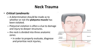
Neck Trauma
Neck Trauma
• Approximately 5-10% of traumatic injuries involve the neck.
• The most critical and immediately life-threatening problem is
– airway obstruction and massive hemorrhage.
• Approximately 3,500 people die each year from neck trauma
due to
– suicides, hangings and accidents.
• Most blunt trauma is caused by MVA
– driver hitting the steering wheel or dashboard
– commonly resulting in laryngotracheal injuries.
Neck Trauma
• Critical Landmarks
– A determination should be made as to
whether or not the platysma muscle has
been violated.
– Platysmal violation is often a clue to damage
and injury to deeper structures.
– the neck is divided into three anatomic
zones
• In order to properly evaluate, diagnose
and prioritize neck injuries,
• Zone I
– is bounded by the clavicles inferiorly and the cricoid cartilage
superiorly.
– contains many crucial structures including:
• the lung apices, trachea, aortic arch, the great vessels,
esophagus, cervical spine and spinal cord.
– Injuries in this zone are often
• difficult to access and repair and
• carry the highest morbidity and mortality risk.
• Zone II
– is bounded by the cricoid cartilage inferiorly and the angle of the
mandible superiorly.
– Crucial structures in this zone include:
• the trachea, larynx, esophagus, carotid arteries, jugular veins,
vertebral vessels, cervical spine and cord.
• This zone is easily surgically accessible, and an oblique neck
incision can often be used with minimal morbidity.
• Zone III
– is bounded by the angle of the mandible inferiorly and the base of
the skull superiorly.
– Structures in this zone include:
• the trachea, pharynx, esophagus, vertebral arteries, internal
carotid arteries and cranial nerves.
• Injuries in this region are surgically difficult secondary to
problems with obtaining proper exposure.
• Evaluation of Neck Trauma
– Early intubation is preferred for proper airway mgt.
– An expanding hematoma or laryngeal edema can occur quickly
causing airway compromise or occlusion leading to significant
morbidity.
– Neck wounds should NOT be explored in the ED due to the risk of
dislodging a clot and disrupting hemostasis.
• In patients with neck trauma, evaluation for arterial injury should be performed.
– The majority of injuries due to penetrating neck trauma are vascular injuries.
– The internal jugular vein and common and internal carotid artery are the vessels most frequently
injured with frequencies of 9% and 6.7%, respectively.
– Vertebral artery injury is rare. Typically, this lesion is clinically unsuspected and incidentally identified on
angiography.
– In hemodynamically stable patients, supportive and expectant mgt is advocated.
– Definitive Tx is required in patients with
• persistent bleeding,
• arteriovenous fistula formation or
• pseudoaneurysm.
– Clues to arterial injury include:
• expanding hematoma,
• pulsatile bleeding,
• shock unresponsive to fluids,
• presence of a new bruit or thrill,
• diminished distal pulses.
**Patients with the above findings often require immediate surgical intervention**
• A high index of suspicion should be maintained for esophageal injury secondary to blunt trauma.
• Diagnostic Studies
– Most patients require a three-view cervical spine series.
– Plain films can demonstrate
• SQ emphysema,
• fractures,
• tracheal deviation, and
• foreign bodies (e.g., bullet fragments).
– A chest X-ray is especially important to evaluate Zone I injuries since this region includes the
lung apices.
• A pneumothorax, hemothorax,
• SQ emphysema (due to an associated pneumothorax or injury to the larynx, trachea or esophagus),
• widened mediastinum (due to injury to a major mediastinal vessel) or foreign body may be visualized.
– Angiography is especially useful in evaluating Zone I and III injuries in hemodynamically stable
patients with platysmal violation.
• The use of color flow doppler is increasing since it is noninvasive and relatively inexpensive.
– However, this technique is operator dependent and its role in
assessing vascular injuries is still unclear.
– The CT scan is an important tool for diagnosing laryngeal injuries.
– The role of MRI in penetrating neck injuries is still being evaluated.
– Endoscopic evaluation of the trachea and/or esophagus should be
performed in patients at risk for injuries to these structures.
Esophagography (70-80% sensitivity) is important for evaluating
esophageal injuries; there is a 17% mortality rate after a 12 h delay
in diagnosis of esophageal injuries.
• ED Management
– Hemodynamic stability:
• Hemodynamically stable patients may undergo a diagnostic evaluation
depending on the zone involved and may not require surgical evaluation.
• However, hemodynamically unstable patients with neck injuries in any zone
require immediate surgical intervention.
– Zone I:
• angiography, esophagography or endoscopy (laryngoscopy or
bronchoscopy).
• If the result of any of the above studies is negative the patient is observed
and managed medically.
• If a study result is positive, surgical evaluation should be obtained.
– Zone II:
• There are two alternatives for evaluating penetrating Zone II injuries in a
hemodynamically stable patient.
• In the past, any injury with platysmal violation necessitated mandatory surgical
intervention.
• However, this approach is losing favor due to the high negative exploration rate;
many centers now favor selective mgt involving
• endoscopy, esophagography and angiography as indicated to determine the need
for surgical intervention.
– Zone III:
• Injuries in this zone are most commonly evaluated by a thorough oropharyngeal
examination, as well as laryngoscopy and angiography as indicated.

Neck Trauma.pptx

  • 1.
  • 2.
    Neck Trauma • Approximately5-10% of traumatic injuries involve the neck. • The most critical and immediately life-threatening problem is – airway obstruction and massive hemorrhage. • Approximately 3,500 people die each year from neck trauma due to – suicides, hangings and accidents. • Most blunt trauma is caused by MVA – driver hitting the steering wheel or dashboard – commonly resulting in laryngotracheal injuries.
  • 3.
    Neck Trauma • CriticalLandmarks – A determination should be made as to whether or not the platysma muscle has been violated. – Platysmal violation is often a clue to damage and injury to deeper structures. – the neck is divided into three anatomic zones • In order to properly evaluate, diagnose and prioritize neck injuries,
  • 4.
    • Zone I –is bounded by the clavicles inferiorly and the cricoid cartilage superiorly. – contains many crucial structures including: • the lung apices, trachea, aortic arch, the great vessels, esophagus, cervical spine and spinal cord. – Injuries in this zone are often • difficult to access and repair and • carry the highest morbidity and mortality risk.
  • 5.
    • Zone II –is bounded by the cricoid cartilage inferiorly and the angle of the mandible superiorly. – Crucial structures in this zone include: • the trachea, larynx, esophagus, carotid arteries, jugular veins, vertebral vessels, cervical spine and cord. • This zone is easily surgically accessible, and an oblique neck incision can often be used with minimal morbidity.
  • 6.
    • Zone III –is bounded by the angle of the mandible inferiorly and the base of the skull superiorly. – Structures in this zone include: • the trachea, pharynx, esophagus, vertebral arteries, internal carotid arteries and cranial nerves. • Injuries in this region are surgically difficult secondary to problems with obtaining proper exposure.
  • 7.
    • Evaluation ofNeck Trauma – Early intubation is preferred for proper airway mgt. – An expanding hematoma or laryngeal edema can occur quickly causing airway compromise or occlusion leading to significant morbidity. – Neck wounds should NOT be explored in the ED due to the risk of dislodging a clot and disrupting hemostasis.
  • 8.
    • In patientswith neck trauma, evaluation for arterial injury should be performed. – The majority of injuries due to penetrating neck trauma are vascular injuries. – The internal jugular vein and common and internal carotid artery are the vessels most frequently injured with frequencies of 9% and 6.7%, respectively. – Vertebral artery injury is rare. Typically, this lesion is clinically unsuspected and incidentally identified on angiography. – In hemodynamically stable patients, supportive and expectant mgt is advocated. – Definitive Tx is required in patients with • persistent bleeding, • arteriovenous fistula formation or • pseudoaneurysm. – Clues to arterial injury include: • expanding hematoma, • pulsatile bleeding, • shock unresponsive to fluids, • presence of a new bruit or thrill, • diminished distal pulses. **Patients with the above findings often require immediate surgical intervention** • A high index of suspicion should be maintained for esophageal injury secondary to blunt trauma.
  • 9.
    • Diagnostic Studies –Most patients require a three-view cervical spine series. – Plain films can demonstrate • SQ emphysema, • fractures, • tracheal deviation, and • foreign bodies (e.g., bullet fragments). – A chest X-ray is especially important to evaluate Zone I injuries since this region includes the lung apices. • A pneumothorax, hemothorax, • SQ emphysema (due to an associated pneumothorax or injury to the larynx, trachea or esophagus), • widened mediastinum (due to injury to a major mediastinal vessel) or foreign body may be visualized. – Angiography is especially useful in evaluating Zone I and III injuries in hemodynamically stable patients with platysmal violation. • The use of color flow doppler is increasing since it is noninvasive and relatively inexpensive.
  • 10.
    – However, thistechnique is operator dependent and its role in assessing vascular injuries is still unclear. – The CT scan is an important tool for diagnosing laryngeal injuries. – The role of MRI in penetrating neck injuries is still being evaluated. – Endoscopic evaluation of the trachea and/or esophagus should be performed in patients at risk for injuries to these structures. Esophagography (70-80% sensitivity) is important for evaluating esophageal injuries; there is a 17% mortality rate after a 12 h delay in diagnosis of esophageal injuries.
  • 11.
    • ED Management –Hemodynamic stability: • Hemodynamically stable patients may undergo a diagnostic evaluation depending on the zone involved and may not require surgical evaluation. • However, hemodynamically unstable patients with neck injuries in any zone require immediate surgical intervention. – Zone I: • angiography, esophagography or endoscopy (laryngoscopy or bronchoscopy). • If the result of any of the above studies is negative the patient is observed and managed medically. • If a study result is positive, surgical evaluation should be obtained.
  • 12.
    – Zone II: •There are two alternatives for evaluating penetrating Zone II injuries in a hemodynamically stable patient. • In the past, any injury with platysmal violation necessitated mandatory surgical intervention. • However, this approach is losing favor due to the high negative exploration rate; many centers now favor selective mgt involving • endoscopy, esophagography and angiography as indicated to determine the need for surgical intervention. – Zone III: • Injuries in this zone are most commonly evaluated by a thorough oropharyngeal examination, as well as laryngoscopy and angiography as indicated.