SlideShare a Scribd company logo
1 of 53
Dr. NDAYISABA CORNEILLE
CEO of CHG
MBChB,DCM,BCSIT,CCNA
Supported BY
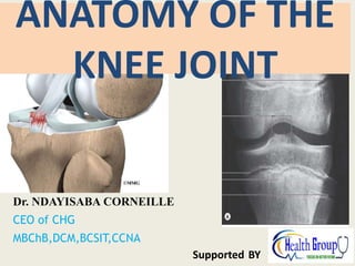
ANATOMY OF THE
KNEE JOINT
PRESENTATION OBJECTIVES
• To discuss the knee joint: At the end of the presentation we should be able to note the following
 The type of joint.
 Bones and part of the bone that forms the joints
 Type of cartilage covering the articular surface.
 Attachment of fibrous capsule.
 The attachment or lining of the synovial membrane.
 Structures found outside the fibrous capsules (Extracapsular structures).
 Structures found within the capsules (Intracapsular structures).
 Movement and muscle causing the movement.
 Blood and Nerve supply.
– Applied Anatomy.
Dr Ndayisaba Corneille
THE KNEE JOINT: Introduction
• The knee joint is the largest
synovial joint in the body
and it is of the hinge variety
• Ordinarily it would have
been a highly unstable joint
but the articulating bones
are held by strong ligaments
and further fortified by the
tendons of adjoining
muscles
Dr Ndayisaba Corneille
ARTICULATIONS, ARTICULAR
SURFACES
It is formed by articulations between the
patella, distal end of the femur and the
proximal end of the tibia.
It consist of two articulations
– Tibiofemoral and
Patellofemoral.
•Tibiofemoral – medial and lateral condyles
of the femur articulate with the tibial condyles
•Patellofemoral – anterior aspect of the
distal femur articulates with the patella. It is a
gliding joint
•The joint surfaces are lined
with hyaline cartilage
Dr Ndayisaba Corneille
Articular surfaces……….
• The medial and lateral condyles
of the tibia are slightly concave
• The femoral condyles are slightly
convex, separated posteriorly by
a deep notch but fusing
anteriorly to form the trochlear
groove for articulation with the
patella
• Patella- It articulates with
the patellar surface of the
femur. It presents articular
surfaces for the medial and
lateral condyles of the femur
Dr Ndayisaba Corneille
FIBROUS CAPSULE OF KNEE JOINT
(FCKJ)
• The joint capsule consist of an external
fibrous layer of the capsule (fibrous
capsule)
• The fibrous layer attaches to the femur
superiorly, just proximal to the articular
margins of the condyles.
• Posteriorly, the fibrous layer encloses
the condyles and the intercondylar fossa.
• it is strengthened posteriorly by the
oblique popliteal ligament, a thickening
of semimebranosus muscle
Dr Ndayisaba Corneille
FCKJ
• Inferiorly, the fibrous layer
attaches to the margin of the
superior articular surface
(tibial plateau) of the tibia
Anteriorly the fibrous layer is
continuous with the lateral and medial
margins of quadriceps tendon, patella,
and patellar ligament
Dr Ndayisaba Corneille
Synovial membrane
• The synovial membrane of the knee is
the most extensive and complex in
the body
• The synovial membrane of the
capsule lines all surfaces within the
articular cavity that is not covered by
articular cartilage
• It is attached to the periphery of
– the articular cartilage covering the
femoral and tibial condyles;
– the posterior surface of the patella;
– and the edges of the menisci,
Dr Ndayisaba Corneille
synovial membrane………………….
• The synovial membrane lines
the internal surface of the
fibrous capsule laterally and
medially,
• Centrally it becomes
separated from the fibrous
capsule by extending into the
intercondylar region, where it
covers the cruciate ligaments
and the infrapatellar fat pad,
so that they are excluded from
the articular cavity
Dr Ndayisaba Corneille
EXTRACAPSULAR LIGAMENTS OF
KNEE JOINT
• The joint capsule is strengthened
by the following extracapsular
ligaments:
– Patellar ligament,
– Fibular (lateral) collateral ligament,
– Tibial (medial) collateral ligament,
• Other extracapsular ligaments
include:
– Quadriceps tendon,
– Oblique popliteal ligament
Dr Ndayisaba Corneille
The patellar ligament
• The patellar ligament, is the
distal part of the
quadriceps femoris tendon,
it is a strong, thick fibrous
band passing from the apex
and adjoining margins of
the patella to the tibial
tuberosity
• The patellar ligament is the
anterior ligament of the
knee joint.
Patella
lig.
Dr Ndayisaba Corneille
The fibular collateral ligament (FCL;
• The fibular collateral ligament
(FCL; lateral collateral ligament),
is a strong cord-like
extracapsular ligament,
• It extends inferiorly from the
lateral epicondyle of the femur
to the lateral surface of the
fibular head
• The tendon of the popliteus
passes deep to the FCL,
separating it from the lateral
meniscus.
FCL
Dr Ndayisaba Corneille
The tibial collateral ligament (TCL
• The tibial collateral ligament
(TCL; medial collateral
ligament) is strong, flat, and
extends from the medial
epicondyle of the femur to
the medial condyle and the
superior part of the medial
surface of the tibia
• . At its midpoint, the deep
fibers of the TCL are firmly
attached to the medial
meniscus.
TCL.
Dr Ndayisaba Corneille
Quadriceps femoris tendon
Connects the quadriceps
femoris muscles to the
superior aspects of the
patella. Controls knee flexion
and extension
Dr Ndayisaba Corneille
OBLIQUE POPLITEAL LIGAMENT
A strong broad flat fibrous ligament
that pass obliquely across and
strengthens the posterior part of the
knee.
It is a reflected expansion of the
tendon of semimembranosus
Arises from the post. surface of the
medial tibial condyle and blends with
the posterior aspect of the joint
capsule
Dr Ndayisaba Corneille
INTRA-ARTICULAR LIGAMENTS OF
KNEE JOINT
• The intra-articular
ligaments within the knee
joint consist of:
– the Anterior and Posterior
cruciate ligaments and
– Medial and Lateral menisci.
• The tendon of the popliteus
is also intra-articular during
part of its course
Dr Ndayisaba Corneille
The cruciate ligaments
• The cruciate ligaments are
located in the center of the joint
and cross each other obliquely,
like the letter X.
• They crisscross within the fibrous
capsule of the joint but outside
the synovial cavity
• They described as intracapsular
and extrasynovial
Dr Ndayisaba Corneille
The anterior cruciate
ligament
• The anterior
cruciate ligament
(ACL) arises from
the anterior
intercondylar area
of the tibia, just
posterior to the
attachment of the
medial meniscus
Dr Ndayisaba Corneille
ACL
• It extends superiorly,
posteriorly, and laterally to
attach to the posterior part
of the medial side of the
lateral condyle of the femur
• It limits posterior rolling of
the femoral condyles on the
tibial plateau during flexion
AC
L
Dr Ndayisaba Corneille
ACL
• It also prevents posterior
displacement of the femur
on the tibia and
hyperextension of the knee
joint.
• When the joint is flexed at
a right angle, the tibia
cannot be pulled anteriorly
(like pulling out a drawer)
because it is held by the
ACL
Dr Ndayisaba Corneille
The posterior cruciate ligament (PCL),
• The posterior cruciate
ligament (PCL) arises
from the posterior
intercondylar area of
the tibia
Dr Ndayisaba Corneille
PCL
• The PCL passes
superiorly and
anteriorly on the
medial side of the ACL
to attach to the anterior
part of the lateral
surface of the medial
condyle of the femur
PCL
Dr Ndayisaba Corneille
PCL
• The PCL limits anterior rolling of
the femur on the tibial plateau
during extension.
• It also prevents anterior
displacement of the femur on
the tibia or posterior
displacement of the tibia on the
femur and helps prevent
hyperflexion of the knee joint
Dr Ndayisaba Corneille
The menisci of the knee
joint
• The menisci are
crescentic plates of
fibrocartilage lying on
the articular surface of
the tibia
• They are firmly attached
at their ends to the
intercondylar area of
the tibia.
Dr Ndayisaba Corneille
They comprises of the Medial and
Lateral Menisci
The main purpose of the Menisci are:
1) Equalize weight distribution across
the knee joint.
2) Act as Shock absorbers.
3) They serve to widen and deepen
the tibial articular surfaces that
receive the femoral condyles.
Dr Ndayisaba Corneille
BURSAE AROUND KNEE JOINT
• A bursa is a fluid-filled sac. It helps
cushion the muscles, tendons, and
bones around a joint.
• The bursae around the knee joint include:
• The prepatellar and infrapatellar bursae
– They are subcutaneous and lie in relation to
the patella, allowing the skin to be able to
move freely during movements of the knee
• The large suprapatellar bursa lies posterior to
the quadriceps tendon and it is especially
important because it communicates with the
joint cavity so any infection in it may spread to
the knee joint cavity
Dr Ndayisaba Corneille
BURSAE AROUND KNEE JOINT
• Other bursae around the
knee joint include:
– popliteus bursa (deep to
the popliteus tendon),
– Pes anserine bursa
(deep to the tendinous
distal attachments of the
sartorius, gracilis, and
semitendinosus), and
– Gastrocnemius bursae
– Semimembranosus
bursa
Dr Ndayisaba Corneille
Movements at the Knee Joint
• There are four main movements that the knee
joint permits:
• Extension: Produced by the quadriceps
femoris, which inserts into the tibial tuberosity.
• Flexion: Produced by the hamstrings, gracilis,
sartorius and popliteus.
• Lateral rotation: Produced by the biceps
femoris.
• Medial rotation: Produced by five muscles;
semimembranosus, semitendinosus, gracilis,
• NB: Lateral and medial rotation can only occur
when the knee is flexed (if the knee is not
flexed, the medial/lateral rotation occurs at the
hip joint).
Dr Ndayisaba Corneille
MOVEMENTS OF KNEE JOINT…………
• When the knee is fully extended with
the foot on the ground, the knee
passively “locks” because of medial
rotation of the femoral condyles on the
tibial plateau (the “screw-home
mechanism”).
• This position makes the lower limb a
solid column and more adapted for
weight-bearing.
• To unlock the knee, the popliteus
contracts, rotating the femur laterally
about 5° on the tibial plateau so that
flexion of the knee can occur.
Dr Ndayisaba Corneille
BLOOD SUPPLY OF KNEE JOINT
• The arteries supplying the
knee joint are the vessels
that form the genicular
anastomoses around the
knee. The vessels involved are:
– the superior, middle and inferior
genicular branches of the popliteal
artery;
– descending genicular branches of the
femoral artery and
– descending branch of the lateral
circumflex femoral artery;
– circumflex fibular artery; and
– anterior and posterior tibial recurrent
arteries.
Dr Ndayisaba Corneille
INNERVATION OF KNEE JOINT
• Reflecting Hilton’s law, the nerves
supplying the muscles crossing (acting on)
the knee joint also supply the joint; thus
the joint is supplied by articular branches
arising from:
• The femoral (the branches to the vasti)
– Supply its anterior aspect
• Tibial, and common fibular nerves supply
– posterior, and lateral aspects,
respectively.
• The obturator and saphenous (cutaneous)
nerves
– supply its medial aspect.
Dr Ndayisaba Corneille
APPLIED ANATOMY OF
THE KNEE JOINT
Dr Ndayisaba Corneille
Due to forced abduction of tibia on
femur. Cc: tenderness over
attachments of ligament.
Dr Ndayisaba Corneille
ANTERIOR
CRUCIATE
LIGAMENT
DAMAGE
Dr Ndayisaba Corneille
TORN ANTERIOR CRUCIATE LIG
Dr Ndayisaba Corneille
ANTERIOR
DRAWER’S
SIGN:
POSITIVE
FOR
ANTERIOR
CRUCIATE
LIGAMENT
DAMAGE
Dr Ndayisaba Corneille
POSTERIOR
CRUCIATE
LIGAMENT
DAMAGE
Dr Ndayisaba Corneille
POSTERIOR
DRAWER’S
SIGN:
POSITIVE
FOR
POSTERIOR
CRUCIATE
LIGAMENT
DAMAGE
Dr Ndayisaba Corneille
MENISCAL INJURY
Medial Meniscus:
excessive external
rotation of the
tibia.
Lateral Meniscus:
excessive flexion of
the knee.
Dr Ndayisaba Corneille
The “unhappy
triad”
• The “unhappy triad” refers
to a sprain injury that
involves three structures of
the knee. These structures
are:
– the medial collateral
ligament,
– anterior cruciate ligament,
and
– the medial meniscus.
Dr Ndayisaba Corneille
The “unhappy triad”………………………….
Dr Ndayisaba Corneille
Bursitis of the Knee Joint
• When a bursa becomes
inflamed, it’s called bursitis.
• Common symptoms are
pain, (tenderness), and
swelling that limits
movement of the joint.
• Bursitis is most often caused
by overuse of a joint due to
repeated movements.
Dr Ndayisaba Corneille
PREPATELLAR
BURSITIS
also called housemaid’s
knee or coal miner’s
knee. These names
arose because people
whose jobs require
frequent kneeling are
prone to prepatellar
bursitis.
Dr Ndayisaba Corneille
INFRAPATELLAR BURSITIS (IB):
Infrapatellar bursa is located below the
patella in relation to the patella tendon. It
is commonly associated with repetitive
jumping injury called “jumper's knee. It is
also referred to as the Vicars knee
SUPRAPATELLAR
BURSITIS:
located between
the distal femur
and the
quadriceps
tendon.
It can be irritated
by a direct blow or
from repeated
stress or motions.
(IB
)
Dr Ndayisaba Corneille
The stability of the knee joint
• The stability of the knee joint depends on (
– 1) the strength and actions of the surrounding muscles and
their tendons, and
– (2) the ligaments that connect the femur and tibia. Of
these supports, the muscles are most important;
therefore, many sport injuries are preventable through
appropriate conditioning and training.
• The most important muscle in stabilizing the knee joint
is
– the large quadriceps femoris, particularly the inferior fibers
of the vastus medialis and lateralis . The knee joint
functions surprisingly well after a ligament strain if the
quadriceps is well conditioned
Dr Ndayisaba Corneille
Knee replacement
Dr Ndayisaba Corneille
END
Dr Ndayisaba Corneille
THANKS FOR LISTENING
By
DR NDAYISABA CORNEILLE
MBChB,DCM,BCSIT,CCNA
Contact us:
amentalhealths@gmail.com/
ndayicoll@gmail.com
whatsaps :+256772497591
/+250788958241

More Related Content

What's hot

Anterior compartment of leg and Dorsum of foot
Anterior compartment  of leg and Dorsum of foot Anterior compartment  of leg and Dorsum of foot
Anterior compartment of leg and Dorsum of foot CIMS
 
Hip joint anatomy
Hip joint anatomyHip joint anatomy
Hip joint anatomyRuhul Amin
 
3.5 cervical vertebra
3.5 cervical vertebra3.5 cervical vertebra
3.5 cervical vertebraSado Anatomist
 
Anatomy of the leg
Anatomy of the legAnatomy of the leg
Anatomy of the legRandolph Tulsie
 
arches of foot.pptx
arches of foot.pptxarches of foot.pptx
arches of foot.pptxanatomysrs
 
Anatomy of the Back of thigh and popliteal fossa By DR. G Kamau.
Anatomy of the Back of thigh and popliteal fossa By DR. G Kamau.Anatomy of the Back of thigh and popliteal fossa By DR. G Kamau.
Anatomy of the Back of thigh and popliteal fossa By DR. G Kamau.MathewJude
 
Slideshow: Posterior Thigh Adductors
Slideshow: Posterior Thigh AdductorsSlideshow: Posterior Thigh Adductors
Slideshow: Posterior Thigh AdductorsThe Funky Professor
 
Sole of the foot
Sole of the footSole of the foot
Sole of the footEimaan Ktk
 
9. posterior compartment of the forearm
9. posterior compartment of the forearm9. posterior compartment of the forearm
9. posterior compartment of the forearmDr. Mohammad Mahmoud
 
Ankle and Tibiofibular Joint.pptx
Ankle and Tibiofibular Joint.pptxAnkle and Tibiofibular Joint.pptx
Ankle and Tibiofibular Joint.pptxMathew Joseph
 
Peripheral Nerves of the Lower Limb
Peripheral Nerves of the Lower LimbPeripheral Nerves of the Lower Limb
Peripheral Nerves of the Lower LimbSado Anatomist
 
Anterior & postetior tibial artery
Anterior & postetior tibial arteryAnterior & postetior tibial artery
Anterior & postetior tibial arteryIdris Siddiqui
 
Skull base : Development and anatomy.
Skull base : Development and anatomy. Skull base : Development and anatomy.
Skull base : Development and anatomy. Sarbesh Tiwari
 
Thigh - Ant.Compartment - Anatomy
Thigh - Ant.Compartment - AnatomyThigh - Ant.Compartment - Anatomy
Thigh - Ant.Compartment - AnatomyUthamalingam Murali
 
SCAPULAR REGION by Shapi. MD.pdf
SCAPULAR REGION by Shapi. MD.pdfSCAPULAR REGION by Shapi. MD.pdf
SCAPULAR REGION by Shapi. MD.pdfShapi. MD
 

What's hot (20)

Slideshow: Posterior Leg
Slideshow: Posterior LegSlideshow: Posterior Leg
Slideshow: Posterior Leg
 
Anterior compartment of leg and Dorsum of foot
Anterior compartment  of leg and Dorsum of foot Anterior compartment  of leg and Dorsum of foot
Anterior compartment of leg and Dorsum of foot
 
Anatomy of ankle joint
Anatomy of ankle jointAnatomy of ankle joint
Anatomy of ankle joint
 
Hip joint anatomy
Hip joint anatomyHip joint anatomy
Hip joint anatomy
 
Slideshow: The Foot
Slideshow: The FootSlideshow: The Foot
Slideshow: The Foot
 
3.5 cervical vertebra
3.5 cervical vertebra3.5 cervical vertebra
3.5 cervical vertebra
 
Anatomy of the leg
Anatomy of the legAnatomy of the leg
Anatomy of the leg
 
arches of foot.pptx
arches of foot.pptxarches of foot.pptx
arches of foot.pptx
 
Anatomy of the Back of thigh and popliteal fossa By DR. G Kamau.
Anatomy of the Back of thigh and popliteal fossa By DR. G Kamau.Anatomy of the Back of thigh and popliteal fossa By DR. G Kamau.
Anatomy of the Back of thigh and popliteal fossa By DR. G Kamau.
 
Slideshow: Posterior Thigh Adductors
Slideshow: Posterior Thigh AdductorsSlideshow: Posterior Thigh Adductors
Slideshow: Posterior Thigh Adductors
 
Sole of the foot
Sole of the footSole of the foot
Sole of the foot
 
9. posterior compartment of the forearm
9. posterior compartment of the forearm9. posterior compartment of the forearm
9. posterior compartment of the forearm
 
Ankle and Tibiofibular Joint.pptx
Ankle and Tibiofibular Joint.pptxAnkle and Tibiofibular Joint.pptx
Ankle and Tibiofibular Joint.pptx
 
Peripheral Nerves of the Lower Limb
Peripheral Nerves of the Lower LimbPeripheral Nerves of the Lower Limb
Peripheral Nerves of the Lower Limb
 
Anterior & postetior tibial artery
Anterior & postetior tibial arteryAnterior & postetior tibial artery
Anterior & postetior tibial artery
 
2. front of the thigh ii
2. front of the thigh ii2. front of the thigh ii
2. front of the thigh ii
 
Skull base : Development and anatomy.
Skull base : Development and anatomy. Skull base : Development and anatomy.
Skull base : Development and anatomy.
 
Thigh - Ant.Compartment - Anatomy
Thigh - Ant.Compartment - AnatomyThigh - Ant.Compartment - Anatomy
Thigh - Ant.Compartment - Anatomy
 
Hip joint anatomy
Hip joint anatomy Hip joint anatomy
Hip joint anatomy
 
SCAPULAR REGION by Shapi. MD.pdf
SCAPULAR REGION by Shapi. MD.pdfSCAPULAR REGION by Shapi. MD.pdf
SCAPULAR REGION by Shapi. MD.pdf
 

Similar to The Knee Joint.pptx

Knee joint.pptx
Knee joint.pptxKnee joint.pptx
Knee joint.pptxApoorvGarg28
 
KNEE JOINT (1).pptx ....................
KNEE JOINT (1).pptx ....................KNEE JOINT (1).pptx ....................
KNEE JOINT (1).pptx ....................vsoorajvismika
 
Knee instability
Knee instabilityKnee instability
Knee instabilitypunithpc605
 
Ligamnet around knee and injury and management
Ligamnet around knee and injury and managementLigamnet around knee and injury and management
Ligamnet around knee and injury and managementBirajkc5
 
anatomyofthekneejoint-170329185745.pdf
anatomyofthekneejoint-170329185745.pdfanatomyofthekneejoint-170329185745.pdf
anatomyofthekneejoint-170329185745.pdfUmaMaheshwariJ3
 
Anatomy of the knee joint
Anatomy of the knee jointAnatomy of the knee joint
Anatomy of the knee jointRajesh Raj
 
Anatomy Of Knee Joint.pptx
Anatomy Of Knee Joint.pptxAnatomy Of Knee Joint.pptx
Anatomy Of Knee Joint.pptxsuleymanfantahun
 
ANATOMY OF KNEE JOINT
ANATOMY OF KNEE JOINTANATOMY OF KNEE JOINT
ANATOMY OF KNEE JOINTDR. SONAL MORE
 
Anatomy and Examination of the Knee
Anatomy and Examination of the KneeAnatomy and Examination of the Knee
Anatomy and Examination of the KneeSri Harsha Gutta
 
The Knee Complex
The Knee ComplexThe Knee Complex
The Knee ComplexPhilip Keriaka
 
Complete knee joint anatomy
Complete knee joint anatomyComplete knee joint anatomy
Complete knee joint anatomyShivrajsinhJhala1
 
Anatomy of knee
Anatomy of kneeAnatomy of knee
Anatomy of kneeDibinKThomas3
 
Knee joint biomechanics
Knee joint biomechanicsKnee joint biomechanics
Knee joint biomechanicsBhavnaRajpurohit
 
Knee joint.Session 1
Knee joint.Session 1Knee joint.Session 1
Knee joint.Session 1Zinat Ashnagar
 
THE KNEE JOINT.pptx
THE KNEE JOINT.pptxTHE KNEE JOINT.pptx
THE KNEE JOINT.pptxElizabeth781016
 

Similar to The Knee Joint.pptx (20)

Knee joint.pptx
Knee joint.pptxKnee joint.pptx
Knee joint.pptx
 
KNEE JOINT (1).pptx ....................
KNEE JOINT (1).pptx ....................KNEE JOINT (1).pptx ....................
KNEE JOINT (1).pptx ....................
 
Knee instability
Knee instabilityKnee instability
Knee instability
 
Ligamnet around knee and injury and management
Ligamnet around knee and injury and managementLigamnet around knee and injury and management
Ligamnet around knee and injury and management
 
anatomyofthekneejoint-170329185745.pdf
anatomyofthekneejoint-170329185745.pdfanatomyofthekneejoint-170329185745.pdf
anatomyofthekneejoint-170329185745.pdf
 
Anatomy of the knee joint
Anatomy of the knee jointAnatomy of the knee joint
Anatomy of the knee joint
 
knee ..pptx
knee ..pptxknee ..pptx
knee ..pptx
 
Anatomy Of Knee Joint.pptx
Anatomy Of Knee Joint.pptxAnatomy Of Knee Joint.pptx
Anatomy Of Knee Joint.pptx
 
ANATOMY OF KNEE JOINT
ANATOMY OF KNEE JOINTANATOMY OF KNEE JOINT
ANATOMY OF KNEE JOINT
 
Knee joint.pptx
Knee joint.pptxKnee joint.pptx
Knee joint.pptx
 
Anatomy and Examination of the Knee
Anatomy and Examination of the KneeAnatomy and Examination of the Knee
Anatomy and Examination of the Knee
 
Knee joint
Knee jointKnee joint
Knee joint
 
The Knee Complex
The Knee ComplexThe Knee Complex
The Knee Complex
 
Complete knee joint anatomy
Complete knee joint anatomyComplete knee joint anatomy
Complete knee joint anatomy
 
Anatomy of knee
Anatomy of kneeAnatomy of knee
Anatomy of knee
 
Knee joint biomechanics
Knee joint biomechanicsKnee joint biomechanics
Knee joint biomechanics
 
GLUTEAL REGION.ppt
GLUTEAL REGION.pptGLUTEAL REGION.ppt
GLUTEAL REGION.ppt
 
Knee joint.Session 1
Knee joint.Session 1Knee joint.Session 1
Knee joint.Session 1
 
THE KNEE JOINT.pptx
THE KNEE JOINT.pptxTHE KNEE JOINT.pptx
THE KNEE JOINT.pptx
 
Bursae around knee joint
Bursae around knee jointBursae around knee joint
Bursae around knee joint
 

More from Dr Ndayisaba Corneille

ANATOMY_OF_THE_EYE_AND_ORBITS.ppt
ANATOMY_OF_THE_EYE_AND_ORBITS.pptANATOMY_OF_THE_EYE_AND_ORBITS.ppt
ANATOMY_OF_THE_EYE_AND_ORBITS.pptDr Ndayisaba Corneille
 
THE_MENINGES,CEREBRAL_SINAL_FLUID.pptx
THE_MENINGES,CEREBRAL_SINAL_FLUID.pptxTHE_MENINGES,CEREBRAL_SINAL_FLUID.pptx
THE_MENINGES,CEREBRAL_SINAL_FLUID.pptxDr Ndayisaba Corneille
 
The_nose_and_paranasal_sinuses.ppt
The_nose_and_paranasal_sinuses.pptThe_nose_and_paranasal_sinuses.ppt
The_nose_and_paranasal_sinuses.pptDr Ndayisaba Corneille
 
THE_EMBRYOLOGY_OF_HEAD_&_NECK[1].ppt
THE_EMBRYOLOGY_OF_HEAD_&_NECK[1].pptTHE_EMBRYOLOGY_OF_HEAD_&_NECK[1].ppt
THE_EMBRYOLOGY_OF_HEAD_&_NECK[1].pptDr Ndayisaba Corneille
 
Temporal_and_infratemporal_fossa.ppt
Temporal_and_infratemporal_fossa.pptTemporal_and_infratemporal_fossa.ppt
Temporal_and_infratemporal_fossa.pptDr Ndayisaba Corneille
 
Anatomy of Upper & Lower Urinary Tract.pptx
Anatomy  of Upper & Lower  Urinary Tract.pptxAnatomy  of Upper & Lower  Urinary Tract.pptx
Anatomy of Upper & Lower Urinary Tract.pptxDr Ndayisaba Corneille
 
Anatomy of Esophagus & Stomach.pptx
Anatomy of Esophagus & Stomach.pptxAnatomy of Esophagus & Stomach.pptx
Anatomy of Esophagus & Stomach.pptxDr Ndayisaba Corneille
 
Anatomy of Female internal genitalia.pptx
Anatomy of Female internal genitalia.pptxAnatomy of Female internal genitalia.pptx
Anatomy of Female internal genitalia.pptxDr Ndayisaba Corneille
 
Anatomy of Suprarenal (Adrenal) Glands.pptx
Anatomy of Suprarenal (Adrenal) Glands.pptxAnatomy of Suprarenal (Adrenal) Glands.pptx
Anatomy of Suprarenal (Adrenal) Glands.pptxDr Ndayisaba Corneille
 
Anatomy of the Male External genitalia.pptx
Anatomy of the Male External genitalia.pptxAnatomy of the Male External genitalia.pptx
Anatomy of the Male External genitalia.pptxDr Ndayisaba Corneille
 
Anatomy of the Male internal genitalia.pptx
Anatomy of the Male internal genitalia.pptxAnatomy of the Male internal genitalia.pptx
Anatomy of the Male internal genitalia.pptxDr Ndayisaba Corneille
 
Azygos ,Thoracic duct and Porto-Systemic anastomosis.pptx
Azygos ,Thoracic duct and Porto-Systemic anastomosis.pptxAzygos ,Thoracic duct and Porto-Systemic anastomosis.pptx
Azygos ,Thoracic duct and Porto-Systemic anastomosis.pptxDr Ndayisaba Corneille
 

More from Dr Ndayisaba Corneille (20)

ANATOMY_OF_THE_EYE_AND_ORBITS.ppt
ANATOMY_OF_THE_EYE_AND_ORBITS.pptANATOMY_OF_THE_EYE_AND_ORBITS.ppt
ANATOMY_OF_THE_EYE_AND_ORBITS.ppt
 
THYROID&PARATHYROID_GLAND.pdf
THYROID&PARATHYROID_GLAND.pdfTHYROID&PARATHYROID_GLAND.pdf
THYROID&PARATHYROID_GLAND.pdf
 
THE_SCALP_AND_THE_FACE.pptx
THE_SCALP_AND_THE_FACE.pptxTHE_SCALP_AND_THE_FACE.pptx
THE_SCALP_AND_THE_FACE.pptx
 
THE_MENINGES,CEREBRAL_SINAL_FLUID.pptx
THE_MENINGES,CEREBRAL_SINAL_FLUID.pptxTHE_MENINGES,CEREBRAL_SINAL_FLUID.pptx
THE_MENINGES,CEREBRAL_SINAL_FLUID.pptx
 
Temporomandibular-joint.pptx
Temporomandibular-joint.pptxTemporomandibular-joint.pptx
Temporomandibular-joint.pptx
 
Parotid_Region.ppt
Parotid_Region.pptParotid_Region.ppt
Parotid_Region.ppt
 
TRIANGLES_OF_THE_NECK.pptx
TRIANGLES_OF_THE_NECK.pptxTRIANGLES_OF_THE_NECK.pptx
TRIANGLES_OF_THE_NECK.pptx
 
The_trachea.ppt
The_trachea.pptThe_trachea.ppt
The_trachea.ppt
 
The_nose_and_paranasal_sinuses.ppt
The_nose_and_paranasal_sinuses.pptThe_nose_and_paranasal_sinuses.ppt
The_nose_and_paranasal_sinuses.ppt
 
THE_EMBRYOLOGY_OF_HEAD_&_NECK[1].ppt
THE_EMBRYOLOGY_OF_HEAD_&_NECK[1].pptTHE_EMBRYOLOGY_OF_HEAD_&_NECK[1].ppt
THE_EMBRYOLOGY_OF_HEAD_&_NECK[1].ppt
 
Temporal_and_infratemporal_fossa.ppt
Temporal_and_infratemporal_fossa.pptTemporal_and_infratemporal_fossa.ppt
Temporal_and_infratemporal_fossa.ppt
 
ANATOMY_OF_THE_LARYNX.pptx
ANATOMY_OF_THE_LARYNX.pptxANATOMY_OF_THE_LARYNX.pptx
ANATOMY_OF_THE_LARYNX.pptx
 
Anatomy of Upper & Lower Urinary Tract.pptx
Anatomy  of Upper & Lower  Urinary Tract.pptxAnatomy  of Upper & Lower  Urinary Tract.pptx
Anatomy of Upper & Lower Urinary Tract.pptx
 
Anatomy of Esophagus & Stomach.pptx
Anatomy of Esophagus & Stomach.pptxAnatomy of Esophagus & Stomach.pptx
Anatomy of Esophagus & Stomach.pptx
 
Anatomy of Female internal genitalia.pptx
Anatomy of Female internal genitalia.pptxAnatomy of Female internal genitalia.pptx
Anatomy of Female internal genitalia.pptx
 
Anatomy of Suprarenal (Adrenal) Glands.pptx
Anatomy of Suprarenal (Adrenal) Glands.pptxAnatomy of Suprarenal (Adrenal) Glands.pptx
Anatomy of Suprarenal (Adrenal) Glands.pptx
 
Anatomy of The Heart.pptx
Anatomy of The Heart.pptxAnatomy of The Heart.pptx
Anatomy of The Heart.pptx
 
Anatomy of the Male External genitalia.pptx
Anatomy of the Male External genitalia.pptxAnatomy of the Male External genitalia.pptx
Anatomy of the Male External genitalia.pptx
 
Anatomy of the Male internal genitalia.pptx
Anatomy of the Male internal genitalia.pptxAnatomy of the Male internal genitalia.pptx
Anatomy of the Male internal genitalia.pptx
 
Azygos ,Thoracic duct and Porto-Systemic anastomosis.pptx
Azygos ,Thoracic duct and Porto-Systemic anastomosis.pptxAzygos ,Thoracic duct and Porto-Systemic anastomosis.pptx
Azygos ,Thoracic duct and Porto-Systemic anastomosis.pptx
 

Recently uploaded

Bangalore Call Girls Majestic 📞 9907093804 High Profile Service 100% Safe
Bangalore Call Girls Majestic 📞 9907093804 High Profile Service 100% SafeBangalore Call Girls Majestic 📞 9907093804 High Profile Service 100% Safe
Bangalore Call Girls Majestic 📞 9907093804 High Profile Service 100% Safenarwatsonia7
 
♛VVIP Hyderabad Call Girls Chintalkunta🖕7001035870🖕Riya Kappor Top Call Girl ...
♛VVIP Hyderabad Call Girls Chintalkunta🖕7001035870🖕Riya Kappor Top Call Girl ...♛VVIP Hyderabad Call Girls Chintalkunta🖕7001035870🖕Riya Kappor Top Call Girl ...
♛VVIP Hyderabad Call Girls Chintalkunta🖕7001035870🖕Riya Kappor Top Call Girl ...astropune
 
Bangalore Call Girls Marathahalli 📞 9907093804 High Profile Service 100% Safe
Bangalore Call Girls Marathahalli 📞 9907093804 High Profile Service 100% SafeBangalore Call Girls Marathahalli 📞 9907093804 High Profile Service 100% Safe
Bangalore Call Girls Marathahalli 📞 9907093804 High Profile Service 100% Safenarwatsonia7
 
Lucknow Call girls - 8800925952 - 24x7 service with hotel room
Lucknow Call girls - 8800925952 - 24x7 service with hotel roomLucknow Call girls - 8800925952 - 24x7 service with hotel room
Lucknow Call girls - 8800925952 - 24x7 service with hotel roomdiscovermytutordmt
 
Call Girls Horamavu WhatsApp Number 7001035870 Meeting With Bangalore Escorts
Call Girls Horamavu WhatsApp Number 7001035870 Meeting With Bangalore EscortsCall Girls Horamavu WhatsApp Number 7001035870 Meeting With Bangalore Escorts
Call Girls Horamavu WhatsApp Number 7001035870 Meeting With Bangalore Escortsvidya singh
 
Call Girls Service Surat Samaira ❤️🍑 8250192130 👄 Independent Escort Service ...
Call Girls Service Surat Samaira ❤️🍑 8250192130 👄 Independent Escort Service ...Call Girls Service Surat Samaira ❤️🍑 8250192130 👄 Independent Escort Service ...
Call Girls Service Surat Samaira ❤️🍑 8250192130 👄 Independent Escort Service ...CALL GIRLS
 
💎VVIP Kolkata Call Girls Parganas🩱7001035870🩱Independent Girl ( Ac Rooms Avai...
💎VVIP Kolkata Call Girls Parganas🩱7001035870🩱Independent Girl ( Ac Rooms Avai...💎VVIP Kolkata Call Girls Parganas🩱7001035870🩱Independent Girl ( Ac Rooms Avai...
💎VVIP Kolkata Call Girls Parganas🩱7001035870🩱Independent Girl ( Ac Rooms Avai...Taniya Sharma
 
Call Girl Coimbatore Prisha☎️ 8250192130 Independent Escort Service Coimbatore
Call Girl Coimbatore Prisha☎️  8250192130 Independent Escort Service CoimbatoreCall Girl Coimbatore Prisha☎️  8250192130 Independent Escort Service Coimbatore
Call Girl Coimbatore Prisha☎️ 8250192130 Independent Escort Service Coimbatorenarwatsonia7
 
VIP Mumbai Call Girls Hiranandani Gardens Just Call 9920874524 with A/C Room ...
VIP Mumbai Call Girls Hiranandani Gardens Just Call 9920874524 with A/C Room ...VIP Mumbai Call Girls Hiranandani Gardens Just Call 9920874524 with A/C Room ...
VIP Mumbai Call Girls Hiranandani Gardens Just Call 9920874524 with A/C Room ...Garima Khatri
 
Aspirin presentation slides by Dr. Rewas Ali
Aspirin presentation slides by Dr. Rewas AliAspirin presentation slides by Dr. Rewas Ali
Aspirin presentation slides by Dr. Rewas AliRewAs ALI
 
Call Girls Service In Shyam Nagar Whatsapp 8445551418 Independent Escort Service
Call Girls Service In Shyam Nagar Whatsapp 8445551418 Independent Escort ServiceCall Girls Service In Shyam Nagar Whatsapp 8445551418 Independent Escort Service
Call Girls Service In Shyam Nagar Whatsapp 8445551418 Independent Escort Serviceparulsinha
 
Bangalore Call Girl Whatsapp Number 100% Complete Your Sexual Needs
Bangalore Call Girl Whatsapp Number 100% Complete Your Sexual NeedsBangalore Call Girl Whatsapp Number 100% Complete Your Sexual Needs
Bangalore Call Girl Whatsapp Number 100% Complete Your Sexual NeedsGfnyt
 
VIP Call Girls Indore Kirti 💚😋 9256729539 🚀 Indore Escorts
VIP Call Girls Indore Kirti 💚😋  9256729539 🚀 Indore EscortsVIP Call Girls Indore Kirti 💚😋  9256729539 🚀 Indore Escorts
VIP Call Girls Indore Kirti 💚😋 9256729539 🚀 Indore Escortsaditipandeya
 
VIP Service Call Girls Sindhi Colony 📳 7877925207 For 18+ VIP Call Girl At Th...
VIP Service Call Girls Sindhi Colony 📳 7877925207 For 18+ VIP Call Girl At Th...VIP Service Call Girls Sindhi Colony 📳 7877925207 For 18+ VIP Call Girl At Th...
VIP Service Call Girls Sindhi Colony 📳 7877925207 For 18+ VIP Call Girl At Th...jageshsingh5554
 
Call Girls Colaba Mumbai ❤️ 9920874524 👈 Cash on Delivery
Call Girls Colaba Mumbai ❤️ 9920874524 👈 Cash on DeliveryCall Girls Colaba Mumbai ❤️ 9920874524 👈 Cash on Delivery
Call Girls Colaba Mumbai ❤️ 9920874524 👈 Cash on Deliverynehamumbai
 
Low Rate Call Girls Pune Esha 9907093804 Short 1500 Night 6000 Best call girl...
Low Rate Call Girls Pune Esha 9907093804 Short 1500 Night 6000 Best call girl...Low Rate Call Girls Pune Esha 9907093804 Short 1500 Night 6000 Best call girl...
Low Rate Call Girls Pune Esha 9907093804 Short 1500 Night 6000 Best call girl...Miss joya
 
VIP Call Girls Pune Vani 9907093804 Short 1500 Night 6000 Best call girls Ser...
VIP Call Girls Pune Vani 9907093804 Short 1500 Night 6000 Best call girls Ser...VIP Call Girls Pune Vani 9907093804 Short 1500 Night 6000 Best call girls Ser...
VIP Call Girls Pune Vani 9907093804 Short 1500 Night 6000 Best call girls Ser...Miss joya
 
Call Girls Darjeeling Just Call 9907093804 Top Class Call Girl Service Available
Call Girls Darjeeling Just Call 9907093804 Top Class Call Girl Service AvailableCall Girls Darjeeling Just Call 9907093804 Top Class Call Girl Service Available
Call Girls Darjeeling Just Call 9907093804 Top Class Call Girl Service AvailableDipal Arora
 
Call Girl Number in Vashi Mumbai📲 9833363713 💞 Full Night Enjoy
Call Girl Number in Vashi Mumbai📲 9833363713 💞 Full Night EnjoyCall Girl Number in Vashi Mumbai📲 9833363713 💞 Full Night Enjoy
Call Girl Number in Vashi Mumbai📲 9833363713 💞 Full Night Enjoybabeytanya
 

Recently uploaded (20)

Bangalore Call Girls Majestic 📞 9907093804 High Profile Service 100% Safe
Bangalore Call Girls Majestic 📞 9907093804 High Profile Service 100% SafeBangalore Call Girls Majestic 📞 9907093804 High Profile Service 100% Safe
Bangalore Call Girls Majestic 📞 9907093804 High Profile Service 100% Safe
 
♛VVIP Hyderabad Call Girls Chintalkunta🖕7001035870🖕Riya Kappor Top Call Girl ...
♛VVIP Hyderabad Call Girls Chintalkunta🖕7001035870🖕Riya Kappor Top Call Girl ...♛VVIP Hyderabad Call Girls Chintalkunta🖕7001035870🖕Riya Kappor Top Call Girl ...
♛VVIP Hyderabad Call Girls Chintalkunta🖕7001035870🖕Riya Kappor Top Call Girl ...
 
Bangalore Call Girls Marathahalli 📞 9907093804 High Profile Service 100% Safe
Bangalore Call Girls Marathahalli 📞 9907093804 High Profile Service 100% SafeBangalore Call Girls Marathahalli 📞 9907093804 High Profile Service 100% Safe
Bangalore Call Girls Marathahalli 📞 9907093804 High Profile Service 100% Safe
 
Lucknow Call girls - 8800925952 - 24x7 service with hotel room
Lucknow Call girls - 8800925952 - 24x7 service with hotel roomLucknow Call girls - 8800925952 - 24x7 service with hotel room
Lucknow Call girls - 8800925952 - 24x7 service with hotel room
 
Call Girls Horamavu WhatsApp Number 7001035870 Meeting With Bangalore Escorts
Call Girls Horamavu WhatsApp Number 7001035870 Meeting With Bangalore EscortsCall Girls Horamavu WhatsApp Number 7001035870 Meeting With Bangalore Escorts
Call Girls Horamavu WhatsApp Number 7001035870 Meeting With Bangalore Escorts
 
Call Girls Service Surat Samaira ❤️🍑 8250192130 👄 Independent Escort Service ...
Call Girls Service Surat Samaira ❤️🍑 8250192130 👄 Independent Escort Service ...Call Girls Service Surat Samaira ❤️🍑 8250192130 👄 Independent Escort Service ...
Call Girls Service Surat Samaira ❤️🍑 8250192130 👄 Independent Escort Service ...
 
💎VVIP Kolkata Call Girls Parganas🩱7001035870🩱Independent Girl ( Ac Rooms Avai...
💎VVIP Kolkata Call Girls Parganas🩱7001035870🩱Independent Girl ( Ac Rooms Avai...💎VVIP Kolkata Call Girls Parganas🩱7001035870🩱Independent Girl ( Ac Rooms Avai...
💎VVIP Kolkata Call Girls Parganas🩱7001035870🩱Independent Girl ( Ac Rooms Avai...
 
Call Girl Coimbatore Prisha☎️ 8250192130 Independent Escort Service Coimbatore
Call Girl Coimbatore Prisha☎️  8250192130 Independent Escort Service CoimbatoreCall Girl Coimbatore Prisha☎️  8250192130 Independent Escort Service Coimbatore
Call Girl Coimbatore Prisha☎️ 8250192130 Independent Escort Service Coimbatore
 
VIP Mumbai Call Girls Hiranandani Gardens Just Call 9920874524 with A/C Room ...
VIP Mumbai Call Girls Hiranandani Gardens Just Call 9920874524 with A/C Room ...VIP Mumbai Call Girls Hiranandani Gardens Just Call 9920874524 with A/C Room ...
VIP Mumbai Call Girls Hiranandani Gardens Just Call 9920874524 with A/C Room ...
 
Aspirin presentation slides by Dr. Rewas Ali
Aspirin presentation slides by Dr. Rewas AliAspirin presentation slides by Dr. Rewas Ali
Aspirin presentation slides by Dr. Rewas Ali
 
Call Girls Service In Shyam Nagar Whatsapp 8445551418 Independent Escort Service
Call Girls Service In Shyam Nagar Whatsapp 8445551418 Independent Escort ServiceCall Girls Service In Shyam Nagar Whatsapp 8445551418 Independent Escort Service
Call Girls Service In Shyam Nagar Whatsapp 8445551418 Independent Escort Service
 
Bangalore Call Girl Whatsapp Number 100% Complete Your Sexual Needs
Bangalore Call Girl Whatsapp Number 100% Complete Your Sexual NeedsBangalore Call Girl Whatsapp Number 100% Complete Your Sexual Needs
Bangalore Call Girl Whatsapp Number 100% Complete Your Sexual Needs
 
VIP Call Girls Indore Kirti 💚😋 9256729539 🚀 Indore Escorts
VIP Call Girls Indore Kirti 💚😋  9256729539 🚀 Indore EscortsVIP Call Girls Indore Kirti 💚😋  9256729539 🚀 Indore Escorts
VIP Call Girls Indore Kirti 💚😋 9256729539 🚀 Indore Escorts
 
VIP Service Call Girls Sindhi Colony 📳 7877925207 For 18+ VIP Call Girl At Th...
VIP Service Call Girls Sindhi Colony 📳 7877925207 For 18+ VIP Call Girl At Th...VIP Service Call Girls Sindhi Colony 📳 7877925207 For 18+ VIP Call Girl At Th...
VIP Service Call Girls Sindhi Colony 📳 7877925207 For 18+ VIP Call Girl At Th...
 
Call Girls Colaba Mumbai ❤️ 9920874524 👈 Cash on Delivery
Call Girls Colaba Mumbai ❤️ 9920874524 👈 Cash on DeliveryCall Girls Colaba Mumbai ❤️ 9920874524 👈 Cash on Delivery
Call Girls Colaba Mumbai ❤️ 9920874524 👈 Cash on Delivery
 
Escort Service Call Girls In Sarita Vihar,, 99530°56974 Delhi NCR
Escort Service Call Girls In Sarita Vihar,, 99530°56974 Delhi NCREscort Service Call Girls In Sarita Vihar,, 99530°56974 Delhi NCR
Escort Service Call Girls In Sarita Vihar,, 99530°56974 Delhi NCR
 
Low Rate Call Girls Pune Esha 9907093804 Short 1500 Night 6000 Best call girl...
Low Rate Call Girls Pune Esha 9907093804 Short 1500 Night 6000 Best call girl...Low Rate Call Girls Pune Esha 9907093804 Short 1500 Night 6000 Best call girl...
Low Rate Call Girls Pune Esha 9907093804 Short 1500 Night 6000 Best call girl...
 
VIP Call Girls Pune Vani 9907093804 Short 1500 Night 6000 Best call girls Ser...
VIP Call Girls Pune Vani 9907093804 Short 1500 Night 6000 Best call girls Ser...VIP Call Girls Pune Vani 9907093804 Short 1500 Night 6000 Best call girls Ser...
VIP Call Girls Pune Vani 9907093804 Short 1500 Night 6000 Best call girls Ser...
 
Call Girls Darjeeling Just Call 9907093804 Top Class Call Girl Service Available
Call Girls Darjeeling Just Call 9907093804 Top Class Call Girl Service AvailableCall Girls Darjeeling Just Call 9907093804 Top Class Call Girl Service Available
Call Girls Darjeeling Just Call 9907093804 Top Class Call Girl Service Available
 
Call Girl Number in Vashi Mumbai📲 9833363713 💞 Full Night Enjoy
Call Girl Number in Vashi Mumbai📲 9833363713 💞 Full Night EnjoyCall Girl Number in Vashi Mumbai📲 9833363713 💞 Full Night Enjoy
Call Girl Number in Vashi Mumbai📲 9833363713 💞 Full Night Enjoy
 

The Knee Joint.pptx

  • 1. Dr. NDAYISABA CORNEILLE CEO of CHG MBChB,DCM,BCSIT,CCNA Supported BY ANATOMY OF THE KNEE JOINT
  • 2. PRESENTATION OBJECTIVES • To discuss the knee joint: At the end of the presentation we should be able to note the following  The type of joint.  Bones and part of the bone that forms the joints  Type of cartilage covering the articular surface.  Attachment of fibrous capsule.  The attachment or lining of the synovial membrane.  Structures found outside the fibrous capsules (Extracapsular structures).  Structures found within the capsules (Intracapsular structures).  Movement and muscle causing the movement.  Blood and Nerve supply. – Applied Anatomy. Dr Ndayisaba Corneille
  • 3. THE KNEE JOINT: Introduction • The knee joint is the largest synovial joint in the body and it is of the hinge variety • Ordinarily it would have been a highly unstable joint but the articulating bones are held by strong ligaments and further fortified by the tendons of adjoining muscles Dr Ndayisaba Corneille
  • 4. ARTICULATIONS, ARTICULAR SURFACES It is formed by articulations between the patella, distal end of the femur and the proximal end of the tibia. It consist of two articulations – Tibiofemoral and Patellofemoral. •Tibiofemoral – medial and lateral condyles of the femur articulate with the tibial condyles •Patellofemoral – anterior aspect of the distal femur articulates with the patella. It is a gliding joint •The joint surfaces are lined with hyaline cartilage Dr Ndayisaba Corneille
  • 5. Articular surfaces………. • The medial and lateral condyles of the tibia are slightly concave • The femoral condyles are slightly convex, separated posteriorly by a deep notch but fusing anteriorly to form the trochlear groove for articulation with the patella • Patella- It articulates with the patellar surface of the femur. It presents articular surfaces for the medial and lateral condyles of the femur Dr Ndayisaba Corneille
  • 6. FIBROUS CAPSULE OF KNEE JOINT (FCKJ) • The joint capsule consist of an external fibrous layer of the capsule (fibrous capsule) • The fibrous layer attaches to the femur superiorly, just proximal to the articular margins of the condyles. • Posteriorly, the fibrous layer encloses the condyles and the intercondylar fossa. • it is strengthened posteriorly by the oblique popliteal ligament, a thickening of semimebranosus muscle Dr Ndayisaba Corneille
  • 7. FCKJ • Inferiorly, the fibrous layer attaches to the margin of the superior articular surface (tibial plateau) of the tibia Anteriorly the fibrous layer is continuous with the lateral and medial margins of quadriceps tendon, patella, and patellar ligament Dr Ndayisaba Corneille
  • 8. Synovial membrane • The synovial membrane of the knee is the most extensive and complex in the body • The synovial membrane of the capsule lines all surfaces within the articular cavity that is not covered by articular cartilage • It is attached to the periphery of – the articular cartilage covering the femoral and tibial condyles; – the posterior surface of the patella; – and the edges of the menisci, Dr Ndayisaba Corneille
  • 9. synovial membrane…………………. • The synovial membrane lines the internal surface of the fibrous capsule laterally and medially, • Centrally it becomes separated from the fibrous capsule by extending into the intercondylar region, where it covers the cruciate ligaments and the infrapatellar fat pad, so that they are excluded from the articular cavity Dr Ndayisaba Corneille
  • 10. EXTRACAPSULAR LIGAMENTS OF KNEE JOINT • The joint capsule is strengthened by the following extracapsular ligaments: – Patellar ligament, – Fibular (lateral) collateral ligament, – Tibial (medial) collateral ligament, • Other extracapsular ligaments include: – Quadriceps tendon, – Oblique popliteal ligament Dr Ndayisaba Corneille
  • 11. The patellar ligament • The patellar ligament, is the distal part of the quadriceps femoris tendon, it is a strong, thick fibrous band passing from the apex and adjoining margins of the patella to the tibial tuberosity • The patellar ligament is the anterior ligament of the knee joint. Patella lig. Dr Ndayisaba Corneille
  • 12. The fibular collateral ligament (FCL; • The fibular collateral ligament (FCL; lateral collateral ligament), is a strong cord-like extracapsular ligament, • It extends inferiorly from the lateral epicondyle of the femur to the lateral surface of the fibular head • The tendon of the popliteus passes deep to the FCL, separating it from the lateral meniscus. FCL Dr Ndayisaba Corneille
  • 14. The tibial collateral ligament (TCL • The tibial collateral ligament (TCL; medial collateral ligament) is strong, flat, and extends from the medial epicondyle of the femur to the medial condyle and the superior part of the medial surface of the tibia • . At its midpoint, the deep fibers of the TCL are firmly attached to the medial meniscus. TCL. Dr Ndayisaba Corneille
  • 16. Quadriceps femoris tendon Connects the quadriceps femoris muscles to the superior aspects of the patella. Controls knee flexion and extension Dr Ndayisaba Corneille
  • 17. OBLIQUE POPLITEAL LIGAMENT A strong broad flat fibrous ligament that pass obliquely across and strengthens the posterior part of the knee. It is a reflected expansion of the tendon of semimembranosus Arises from the post. surface of the medial tibial condyle and blends with the posterior aspect of the joint capsule Dr Ndayisaba Corneille
  • 18. INTRA-ARTICULAR LIGAMENTS OF KNEE JOINT • The intra-articular ligaments within the knee joint consist of: – the Anterior and Posterior cruciate ligaments and – Medial and Lateral menisci. • The tendon of the popliteus is also intra-articular during part of its course Dr Ndayisaba Corneille
  • 19. The cruciate ligaments • The cruciate ligaments are located in the center of the joint and cross each other obliquely, like the letter X. • They crisscross within the fibrous capsule of the joint but outside the synovial cavity • They described as intracapsular and extrasynovial Dr Ndayisaba Corneille
  • 20. The anterior cruciate ligament • The anterior cruciate ligament (ACL) arises from the anterior intercondylar area of the tibia, just posterior to the attachment of the medial meniscus Dr Ndayisaba Corneille
  • 21. ACL • It extends superiorly, posteriorly, and laterally to attach to the posterior part of the medial side of the lateral condyle of the femur • It limits posterior rolling of the femoral condyles on the tibial plateau during flexion AC L Dr Ndayisaba Corneille
  • 22. ACL • It also prevents posterior displacement of the femur on the tibia and hyperextension of the knee joint. • When the joint is flexed at a right angle, the tibia cannot be pulled anteriorly (like pulling out a drawer) because it is held by the ACL Dr Ndayisaba Corneille
  • 23. The posterior cruciate ligament (PCL), • The posterior cruciate ligament (PCL) arises from the posterior intercondylar area of the tibia Dr Ndayisaba Corneille
  • 24. PCL • The PCL passes superiorly and anteriorly on the medial side of the ACL to attach to the anterior part of the lateral surface of the medial condyle of the femur PCL Dr Ndayisaba Corneille
  • 25. PCL • The PCL limits anterior rolling of the femur on the tibial plateau during extension. • It also prevents anterior displacement of the femur on the tibia or posterior displacement of the tibia on the femur and helps prevent hyperflexion of the knee joint Dr Ndayisaba Corneille
  • 27. The menisci of the knee joint • The menisci are crescentic plates of fibrocartilage lying on the articular surface of the tibia • They are firmly attached at their ends to the intercondylar area of the tibia. Dr Ndayisaba Corneille
  • 28. They comprises of the Medial and Lateral Menisci The main purpose of the Menisci are: 1) Equalize weight distribution across the knee joint. 2) Act as Shock absorbers. 3) They serve to widen and deepen the tibial articular surfaces that receive the femoral condyles. Dr Ndayisaba Corneille
  • 29. BURSAE AROUND KNEE JOINT • A bursa is a fluid-filled sac. It helps cushion the muscles, tendons, and bones around a joint. • The bursae around the knee joint include: • The prepatellar and infrapatellar bursae – They are subcutaneous and lie in relation to the patella, allowing the skin to be able to move freely during movements of the knee • The large suprapatellar bursa lies posterior to the quadriceps tendon and it is especially important because it communicates with the joint cavity so any infection in it may spread to the knee joint cavity Dr Ndayisaba Corneille
  • 30. BURSAE AROUND KNEE JOINT • Other bursae around the knee joint include: – popliteus bursa (deep to the popliteus tendon), – Pes anserine bursa (deep to the tendinous distal attachments of the sartorius, gracilis, and semitendinosus), and – Gastrocnemius bursae – Semimembranosus bursa Dr Ndayisaba Corneille
  • 31. Movements at the Knee Joint • There are four main movements that the knee joint permits: • Extension: Produced by the quadriceps femoris, which inserts into the tibial tuberosity. • Flexion: Produced by the hamstrings, gracilis, sartorius and popliteus. • Lateral rotation: Produced by the biceps femoris. • Medial rotation: Produced by five muscles; semimembranosus, semitendinosus, gracilis, • NB: Lateral and medial rotation can only occur when the knee is flexed (if the knee is not flexed, the medial/lateral rotation occurs at the hip joint). Dr Ndayisaba Corneille
  • 32. MOVEMENTS OF KNEE JOINT………… • When the knee is fully extended with the foot on the ground, the knee passively “locks” because of medial rotation of the femoral condyles on the tibial plateau (the “screw-home mechanism”). • This position makes the lower limb a solid column and more adapted for weight-bearing. • To unlock the knee, the popliteus contracts, rotating the femur laterally about 5° on the tibial plateau so that flexion of the knee can occur. Dr Ndayisaba Corneille
  • 33. BLOOD SUPPLY OF KNEE JOINT • The arteries supplying the knee joint are the vessels that form the genicular anastomoses around the knee. The vessels involved are: – the superior, middle and inferior genicular branches of the popliteal artery; – descending genicular branches of the femoral artery and – descending branch of the lateral circumflex femoral artery; – circumflex fibular artery; and – anterior and posterior tibial recurrent arteries. Dr Ndayisaba Corneille
  • 34. INNERVATION OF KNEE JOINT • Reflecting Hilton’s law, the nerves supplying the muscles crossing (acting on) the knee joint also supply the joint; thus the joint is supplied by articular branches arising from: • The femoral (the branches to the vasti) – Supply its anterior aspect • Tibial, and common fibular nerves supply – posterior, and lateral aspects, respectively. • The obturator and saphenous (cutaneous) nerves – supply its medial aspect. Dr Ndayisaba Corneille
  • 35. APPLIED ANATOMY OF THE KNEE JOINT Dr Ndayisaba Corneille
  • 36. Due to forced abduction of tibia on femur. Cc: tenderness over attachments of ligament. Dr Ndayisaba Corneille
  • 40. TORN ANTERIOR CRUCIATE LIG Dr Ndayisaba Corneille
  • 45. MENISCAL INJURY Medial Meniscus: excessive external rotation of the tibia. Lateral Meniscus: excessive flexion of the knee. Dr Ndayisaba Corneille
  • 46. The “unhappy triad” • The “unhappy triad” refers to a sprain injury that involves three structures of the knee. These structures are: – the medial collateral ligament, – anterior cruciate ligament, and – the medial meniscus. Dr Ndayisaba Corneille
  • 48. Bursitis of the Knee Joint • When a bursa becomes inflamed, it’s called bursitis. • Common symptoms are pain, (tenderness), and swelling that limits movement of the joint. • Bursitis is most often caused by overuse of a joint due to repeated movements. Dr Ndayisaba Corneille
  • 49. PREPATELLAR BURSITIS also called housemaid’s knee or coal miner’s knee. These names arose because people whose jobs require frequent kneeling are prone to prepatellar bursitis. Dr Ndayisaba Corneille
  • 50. INFRAPATELLAR BURSITIS (IB): Infrapatellar bursa is located below the patella in relation to the patella tendon. It is commonly associated with repetitive jumping injury called “jumper's knee. It is also referred to as the Vicars knee SUPRAPATELLAR BURSITIS: located between the distal femur and the quadriceps tendon. It can be irritated by a direct blow or from repeated stress or motions. (IB ) Dr Ndayisaba Corneille
  • 51. The stability of the knee joint • The stability of the knee joint depends on ( – 1) the strength and actions of the surrounding muscles and their tendons, and – (2) the ligaments that connect the femur and tibia. Of these supports, the muscles are most important; therefore, many sport injuries are preventable through appropriate conditioning and training. • The most important muscle in stabilizing the knee joint is – the large quadriceps femoris, particularly the inferior fibers of the vastus medialis and lateralis . The knee joint functions surprisingly well after a ligament strain if the quadriceps is well conditioned Dr Ndayisaba Corneille
  • 53. END Dr Ndayisaba Corneille THANKS FOR LISTENING By DR NDAYISABA CORNEILLE MBChB,DCM,BCSIT,CCNA Contact us: amentalhealths@gmail.com/ ndayicoll@gmail.com whatsaps :+256772497591 /+250788958241

Editor's Notes

  1. The type of synovial joint.
  2. Bones and part of the bone that forms the joints and Type of cartilage covering the articular surface.
  3. Attachment of fibrous capsule.
  4. Extracapsular structures: Extracapsular Ligaments and Bursae
  5. The tendon of the biceps femoris is split into two parts by the FCL
  6. Intracapsular Structures
  7. It is the cruciate ligaments that maintain contact with the femoral and tibial articular surfaces during flexion of the knee
  8. Movement and Muscles producing the movement
  9. When the knee is “locked,” the thigh and leg muscles can relax briefly without making the knee joint too unstable.