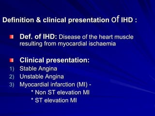
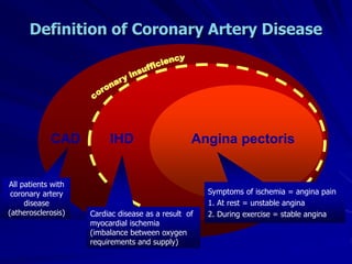
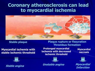
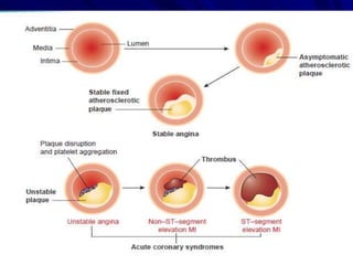
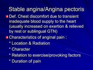
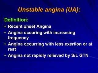
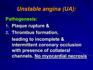
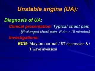
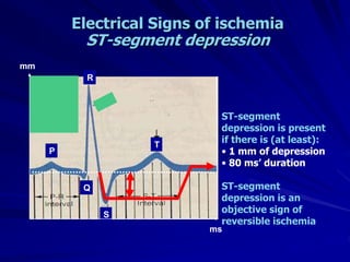
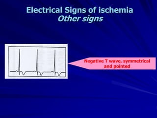
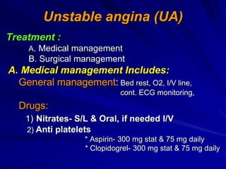
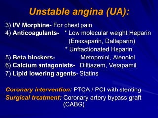
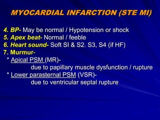
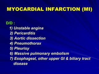
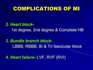
1) Ischaemic heart disease (IHD) results from a lack of oxygen supply to the heart muscle due to narrowed coronary arteries. It presents as stable angina, unstable angina, or myocardial infarction.
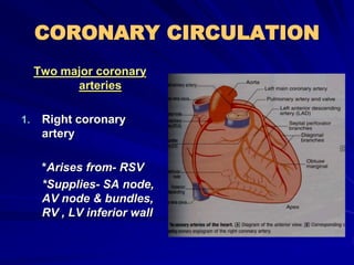
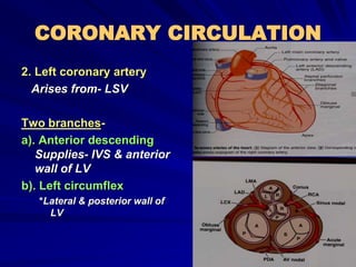
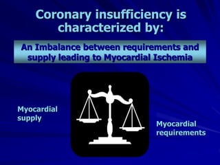
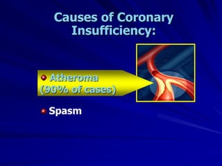
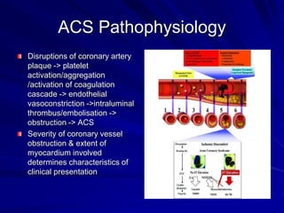
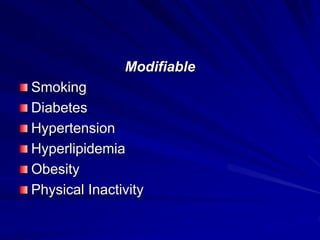
2) Coronary arteries supply oxygenated blood to the heart muscle. Blockages in these arteries due to atherosclerosis can cause angina or infarction depending on the severity and location of the blockage.
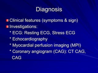
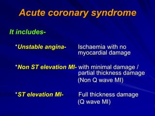
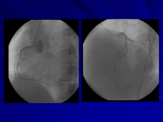
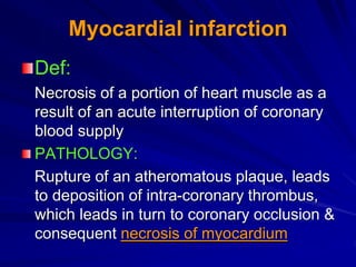
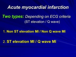
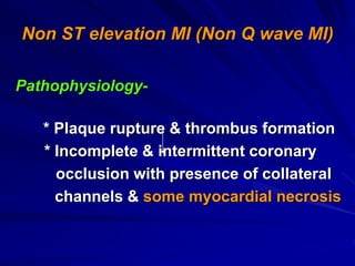
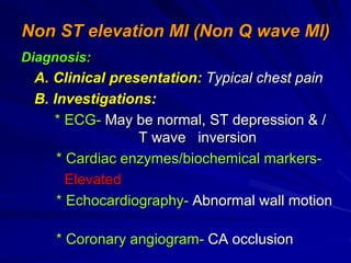
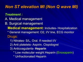
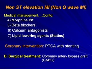
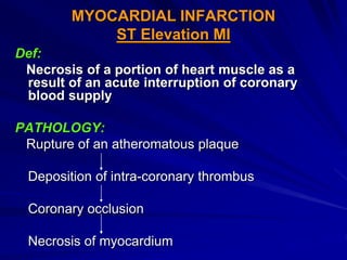
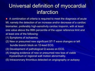
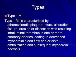
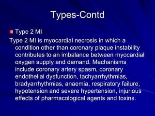
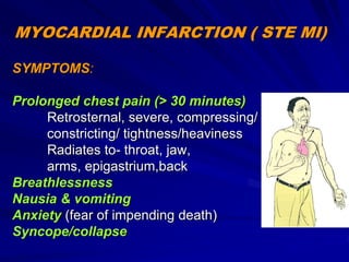
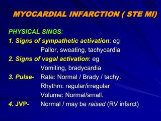
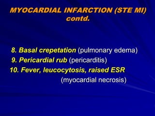
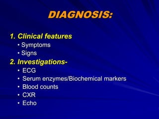
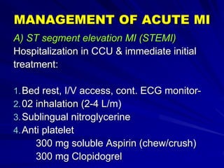
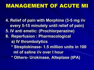
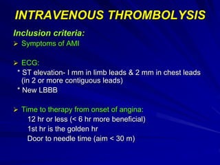
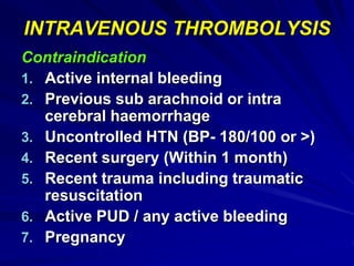
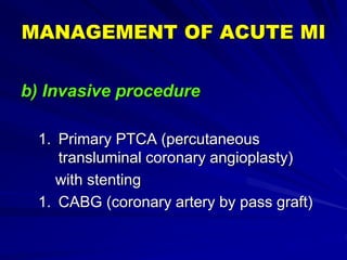
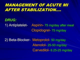
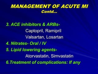
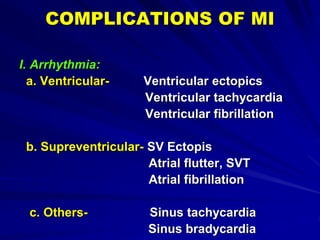
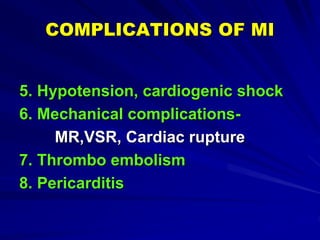
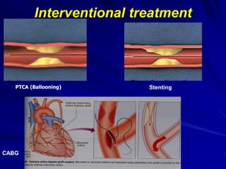
3) Myocardial infarction occurs when a blockage completely cuts off blood flow, causing death of heart muscle tissue. It is classified as non-ST elevation or ST-elevation MI depending on ECG findings. Treatment involves reperfusion through thrombolysis or angioplasty as well
Heart is a muscular organ. Pathology of heart indicates any disorder of heart, a properly functioning heart is important to sustain a live because it pumps blood out to whole body providing oxygen and nutrients. Here I've discussed on two common cardiovascular pathological condition.
Rasamanikya is a excellent preparation in the field of Rasashastra, it is used in various Kushtha Roga, Shwasa, Vicharchika, Bhagandara, Vatarakta, and Phiranga Roga. In this article Preparation& Comparative analytical profile for both Formulationon i.e Rasamanikya prepared by Kushmanda swarasa & Churnodhaka Shodita Haratala. The study aims to provide insights into the comparative efficacy and analytical aspects of these formulations for enhanced therapeutic outcomes.
263778731218 Abortion Clinic /Pills In Harare ,sisternakatoto
263778731218 Abortion Clinic /Pills In Harare ,ABORTION WOMEN’S CLINIC +27730423979 IN women clinic we believe that every woman should be able to make choices in her pregnancy. Our job is to provide compassionate care, safety,affordable and confidential services. That’s why we have won the trust from all generations of women all over the world. we use non surgical method(Abortion pills) to terminate…Dr.LISA +27730423979women Clinic is committed to providing the highest quality of obstetrical and gynecological care to women of all ages. Our dedicated staff aim to treat each patient and her health concerns with compassion and respect.Our dedicated group ABORTION WOMEN’S CLINIC +27730423979 IN women clinic we believe that every woman should be able to make choices in her pregnancy. Our job is to provide compassionate care, safety,affordable and confidential services. That’s why we have won the trust from all generations of women all over the world. we use non surgical method(Abortion pills) to terminate…Dr.LISA +27730423979women Clinic is committed to providing the highest quality of obstetrical and gynecological care to women of all ages. Our dedicated staff aim to treat each patient and her health concerns with compassion and respect.Our dedicated group of receptionists, nurses, and physicians have worked together as a teamof receptionists, nurses, and physicians have worked together as a team wwww.lisywomensclinic.co.za/
These simplified slides by Dr. Sidra Arshad present an overview of the non-respiratory functions of the respiratory tract.
Learning objectives:
1. Enlist the non-respiratory functions of the respiratory tract
2. Briefly explain how these functions are carried out
3. Discuss the significance of dead space
4. Differentiate between minute ventilation and alveolar ventilation
5. Describe the cough and sneeze reflexes
Study Resources:
1. Chapter 39, Guyton and Hall Textbook of Medical Physiology, 14th edition
2. Chapter 34, Ganong’s Review of Medical Physiology, 26th edition
3. Chapter 17, Human Physiology by Lauralee Sherwood, 9th edition
4. Non-respiratory functions of the lungs https://academic.oup.com/bjaed/article/13/3/98/278874
Basavarajeeyam is an important text for ayurvedic physician belonging to andhra pradehs. It is a popular compendium in various parts of our country as well as in andhra pradesh. The content of the text was presented in sanskrit and telugu language (Bilingual). One of the most famous book in ayurvedic pharmaceutics and therapeutics. This book contains 25 chapters called as prakaranas. Many rasaoushadis were explained, pioneer of dhatu druti, nadi pareeksha, mutra pareeksha etc. Belongs to the period of 15-16 century. New diseases like upadamsha, phiranga rogas are explained.
Integrating Ayurveda into Parkinson’s Management: A Holistic ApproachAyurveda ForAll
Explore the benefits of combining Ayurveda with conventional Parkinson's treatments. Learn how a holistic approach can manage symptoms, enhance well-being, and balance body energies. Discover the steps to safely integrate Ayurvedic practices into your Parkinson’s care plan, including expert guidance on diet, herbal remedies, and lifestyle modifications.
Basavarajeeyam is a Sreshta Sangraha grantha (Compiled book ), written by Neelkanta kotturu Basavaraja Virachita. It contains 25 Prakaranas, First 24 Chapters related to Rogas& 25th to Rasadravyas.
These lecture slides, by Dr Sidra Arshad, offer a quick overview of the physiological basis of a normal electrocardiogram.
Learning objectives:
1. Define an electrocardiogram (ECG) and electrocardiography
2. Describe how dipoles generated by the heart produce the waveforms of the ECG
3. Describe the components of a normal electrocardiogram of a typical bipolar lead (limb II)
4. Differentiate between intervals and segments
5. Enlist some common indications for obtaining an ECG
6. Describe the flow of current around the heart during the cardiac cycle
7. Discuss the placement and polarity of the leads of electrocardiograph
8. Describe the normal electrocardiograms recorded from the limb leads and explain the physiological basis of the different records that are obtained
9. Define mean electrical vector (axis) of the heart and give the normal range
10. Define the mean QRS vector
11. Describe the axes of leads (hexagonal reference system)
12. Comprehend the vectorial analysis of the normal ECG
13. Determine the mean electrical axis of the ventricular QRS and appreciate the mean axis deviation
14. Explain the concepts of current of injury, J point, and their significance
Study Resources:
1. Chapter 11, Guyton and Hall Textbook of Medical Physiology, 14th edition
2. Chapter 9, Human Physiology - From Cells to Systems, Lauralee Sherwood, 9th edition
3. Chapter 29, Ganong’s Review of Medical Physiology, 26th edition
4. Electrocardiogram, StatPearls - https://www.ncbi.nlm.nih.gov/books/NBK549803/
5. ECG in Medical Practice by ABM Abdullah, 4th edition
6. Chapter 3, Cardiology Explained, https://www.ncbi.nlm.nih.gov/books/NBK2214/
7. ECG Basics, http://www.nataliescasebook.com/tag/e-c-g-basics
Knee anatomy and clinical tests 2024.pdfvimalpl1234
This includes all relevant anatomy and clinical tests compiled from standard textbooks, Campbell,netter etc..It is comprehensive and best suited for orthopaedicians and orthopaedic residents.
Ozempic: Preoperative Management of Patients on GLP-1 Receptor Agonists Saeid Safari
Preoperative Management of Patients on GLP-1 Receptor Agonists like Ozempic and Semiglutide
ASA GUIDELINE
NYSORA Guideline
2 Case Reports of Gastric Ultrasound
Here is the updated list of Top Best Ayurvedic medicine for Gas and Indigestion and those are Gas-O-Go Syp for Dyspepsia | Lavizyme Syrup for Acidity | Yumzyme Hepatoprotective Capsules etc
micro teaching on communication m.sc nursing.pdfAnurag Sharma
Microteaching is a unique model of practice teaching. It is a viable instrument for the. desired change in the teaching behavior or the behavior potential which, in specified types of real. classroom situations, tends to facilitate the achievement of specified types of objectives.
1. Ischaemic Heart Disease (IHD)
Dr.A.T.M.Mahfuzul Hoque
M D (Cardiology)
Asst. Prof.
Department of Cardiology
Dhaka Medical College& Hospital
2. Definition & clinical presentation of IHD :
Def. of IHD: Disease of the heart muscle
resulting from myocardial ischaemia
Clinical presentation:
1) Stable Angina
2) Unstable Angina
3) Myocardial infarction (MI) -
* Non ST elevation MI
* ST elevation MI
3. CORONARY CIRCULATION
Two major coronary
arteries
1. Right coronary
artery
*Arises from- RSV
*Supplies- SA node,
AV node & bundles,
RV , LV inferior wall
4. CORONARY CIRCULATION
2. Left coronary artery
Arises from- LSV
Two branches-
a). Anterior descending
Supplies- IVS & anterior
wall of LV
b). Left circumflex
*Lateral & posterior wall of
LV
5. CAD IHD Angina pectoris
Definition of Coronary Artery Disease
All patients with
coronary artery
disease
(atherosclerosis) Cardiac disease as a result of
myocardial ischemia
(imbalance between oxygen
requirements and supply)
Symptoms of ischemia = angina pain
1. At rest = unstable angina
2. During exercise = stable angina
6. Coronary insufficiency is
characterized by:
Myocardial
requirements
Myocardial
supply
An Imbalance between requirements and
supply leading to Myocardial Ischemia
8. Stable plaque Plaque rupture or fissuration
Thrombus formation
Coronary atherosclerosis can
lead to myocardial ischemia
Myocardial ischemia with
Moderate/severe
exertion
Chronic Ischemic Disease
Myocardial
necrosis
Prolonged myocardial
ischemia with less
exertion/rest
Acute Ischaemic Events
9. Stable angina
Unstable angina Myocardial
Infarction
Stable plaque Plaque rupture or fissuration
Thrombus formation
Myocardial ischemia with
stable ischemic threshold
Myocardial
necrosis
Prolonged myocardial
ischemia with decreased
ischemic threshold
Coronary atherosclerosis can lead
to myocardial ischemia
10. ACS Pathophysiology
Disruptions of coronary artery
plaque -> platelet
activation/aggregation
/activation of coagulation
cascade -> endothelial
vasoconstriction ->intraluminal
thrombus/embolisation ->
obstruction -> ACS
Severity of coronary vessel
obstruction & extent of
myocardium involved
determines characteristics of
clinical presentation
11.
12. Presentation of IHD
Stable angina
Unstable angina
Acute myocardial infarction (Acute MI)
* Non ST elevation MI (NSTEMI)
* ST elevation MI (STEMI)
Acute coronary syndrome (ACS)
13. Stable angina/Angina pectoris
Def. Chest discomfort due to transient
inadequate blood supply to the heart
(usually increased on exertion & relieved
by rest or sublingual GTN)
Characteristics of anginal pain :
* Location & Radiation
* Character
* Relation to exercise/provoking factors
* Duration of pain
15. Acute coronary syndrome
It includes-
*Unstable angina- Ischaemia with no
myocardial damage
*Non ST elevation MI- with minimal damage /
partial thickness damage
(Non Q wave MI)
*ST elevation MI- Full thickness damage
(Q wave MI)
16. Unstable angina (UA):
Definition:
• Recent onset Angina
• Angina occuring with increasing
frequency
• Angina occurring with less exertion or at
rest
• Angina not rapidly relieved by S/L GTN
17. Unstable angina (UA):
Pathogenesis:
1. Plaque rupture &
2. Thrombus formation,
leading to incomplete &
intermittent coronary occlusion
with presence of collateral
channels. No myocardial necrosis
18. Unstable angina (UA):
Diagnosis of UA:
Clinical presentation: Typical chest pain
(Prolonged chest pain- Pain > 15 minutes)
Investigations:
ECG- May be normal / ST depression & /
T wave inversion
19. Electrical Signs of ischemia
ST-segment depression
P
Q
R
S
T
ST-segment
depression is present
if there is (at least):
• 1 mm of depression
• 80 ms’ duration
ST-segment
depression is an
objective sign of
reversible ischemia
mm
ms
20. Electrical Signs of ischemia
Other signs
Negative T wave, symmetrical
and pointed
28. Myocardial infarction
Def:
Necrosis of a portion of heart muscle as a
result of an acute interruption of coronary
blood supply
PATHOLOGY:
Rupture of an atheromatous plaque, leads
to deposition of intra-coronary thrombus,
which leads in turn to coronary occlusion &
consequent necrosis of myocardium
29. Two types: Depending on ECG criteria
(ST elevation / Q wave)
1. Non ST elevation MI / Non Q wave MI
2. ST elevation MI / Q wave MI
Acute myocardial infarction
30. Non ST elevation MI (Non Q wave MI)
Pathophysiology-
* Plaque rupture & thrombus formation
* Incomplete & intermittent coronary
occlusion with presence of collateral
channels & some myocardial necrosis
31. Non ST elevation MI (Non Q wave MI)
Diagnosis:
A. Clinical presentation: Typical chest pain
B. Investigations:
* ECG- May be normal, ST depression & /
T wave inversion
* Cardiac enzymes/biochemical markers-
Elevated
* Echocardiography- Abnormal wall motion
* Coronary angiogram- CA occlusion
32. Non ST elevation MI (Non Q wave MI)
Treatment :
A. Medical management
B. Surgical management
Medical management: Includes- Hospitalization
* General management: O2, I/V line, ECG monitor
Drugs:
1) Nitrates- S/L, Oral. If needed I/V
2) Anti platelets- Aspirin, Clopidogrel
3) Anticoagulants- Heparin
* Low molecular weight Heparin (Enoxaparin)
* Unfractionated Heparin
33. Non ST elevation MI (Non Q wave MI)
Medical management….Contd:
4) Morphine I/V
5) Beta blockers
6) Calcium antagonists
7) Lipid lowering agents (Statins)
Coronary intervention: PTCA with stenting
B. Surgical treatment: Coronary artery bypass graft
(CABG)
34. MYOCARDIAL INFARCTION
ST Elevation MI
Def:
Necrosis of a portion of heart muscle as a
result of an acute interruption of coronary
blood supply
PATHOLOGY:
Rupture of an atheromatous plaque
Deposition of intra-coronary thrombus
Coronary occlusion
Necrosis of myocardium
35. Universal definition of myocardial
infarction
A combination of criteria is required to meet the diagnosis of acute
MI, namely the detection of an increase and/or decrease of a cardiac
biomarker, preferably high-sensitivity cardiac troponin, with at least
one value above the 99th percentile of the upper reference limit and
at least one of the following:
(1) Symptoms of ischaemia.
(2) New or presumed new significant ST-T wave changes or left
bundle branch block on 12-lead ECG.
(3) Development of pathological Q waves on ECG.
(4) Imaging evidence of new or presumed new loss of viable
myocardium or regional wall motion abnormality.
(5) Intracoronary thrombus detected on angiography or autopsy
36. Types
Type 1 MI
Type 1 MI is characterized by
atherosclerotic plaque rupture, ulceration,
fissure, erosion or dissection with resulting
intraluminal thrombus in one or more
coronary arteries leading to decreased
myocardial blood flow and/or distal
embolization and subsequent myocardial
necrosis.
37. Types-Contd
Type 2 MI
Type 2 MI is myocardial necrosis in which a
condition other than coronary plaque instability
contributes to an imbalance between myocardial
oxygen supply and demand. Mechanisms
include coronary artery spasm, coronary
endothelial dysfunction, tachyarrhythmias,
bradyarrhythmias, anaemia, respiratory failure,
hypotension and severe hypertension, injurious
effects of pharmacological agents and toxins.
38. Risk factors
The presence of any risk factor is
associated with doubling the risk of an MI.
Non Modifiable
Age
Gender
Family history
46. INVESTIGATIONS
1.ECG:
* ST segment elevation
* Pathological Q wave
* T wave inversion
a) Infrior MI: changes in
II, III, aVF
b) Anterior MI: VI- V6
c) Anteroseptal MI: VI-
V4 d)
d) Anterolateral MI. I,
aVL & V4- V6
e) Posterior MI: VI & V2
f) RV infarction: V4 R
47. INVESTIGATIONS
2) SERUM ENZYMES/
Biochemical markers:
a. Troponin T, Troponin I
Advantage- Rapid release
Early diagnosis
b. Creatine Kinase (CK)-
heart, brain & skeletal
muscle ,
CK-MB - cadiospecific
Rises within 6 hrs of onset.
Reaches peak in 18-24 hrs
Becomes normal after 72
hrs
48. INVESTIGATIONS
c. SGOT/AST:
Rises 12 hrs.
Peak 24-36 hrs,
Normal 3-5 days
d. LDH:
Rises later after MI
Peak- after 24-48 hrs
Normal after 1-3
weeks
50. MANAGEMENT OF ACUTE MI
A) ST segment elevation MI (STEMI)
Hospitalization in CCU & immediate initial
treatment:
1.Bed rest, I/V access, cont. ECG monitor-
2.02 inhalation (2-4 L/m)
3.Sublingual nitroglycerine
4.Anti platelet
300 mg soluble Aspirin (chew/crush)
300 mg Clopidogrel
51. MANAGEMENT OF ACUTE MI
4. Relief of pain with Morphine (3-5 mg i/v
every 5-15 minutely until relief of pain)
5. IV anti emetic: (Prochlorperazine)
8. Reperfusion : Pharmacological
a) IV thrombolytics
* Streptokinase- 1.5 million units in 100
ml of saline i/v over I hour
* Others- Urokinase, Alteplase (tPA)
52. INTRAVENOUS THROMBOLYSIS
Inclusion criteria:
Symptoms of AMI
ECG:
* ST elevation- I mm in limb leads & 2 mm in chest leads
(in 2 or more contiguous leads)
* New LBBB
Time to therapy from onset of angina:
12 hr or less (< 6 hr more beneficial)
1st hr is the golden hr
Door to needle time (aim < 30 m)
53. INTRAVENOUS THROMBOLYSIS
Contraindication
1. Active internal bleeding
2. Previous sub arachnoid or intra
cerebral haemorrhage
3. Uncontrolled HTN (BP- 180/100 or >)
4. Recent surgery (Within 1 month)
5. Recent trauma including traumatic
resuscitation
6. Active PUD / any active bleeding
7. Pregnancy
54. MANAGEMENT OF ACUTE MI
b) Invasive procedure
1. Primary PTCA (percutaneous
transluminal coronary angioplasty)
with stenting
1. CABG (coronary artery by pass graft)
55. MANAGEMENT OF ACUTE MI
AFTER STABILIZATION….
DRUG:
1) Antiplatelet- Aspirin- 75 mg/day after meal
Clopidogrel- 75 mg/day
2) Beta-Blocker- Metoprolol- 50 mg/day
Atenolol- 25-50 mg/day
Carvedilol- 6.25-25 mg/day
56. MANAGEMENT OF ACUTE MI
Contd…
3. ACE inhibitors & ARBs-
Captopril, Ramipril
Valsartan, Losartan
4. Nitrates- Oral / IV
5. Lipid lowering agents-
Atorvastatin, Simvastatin
6.Treatment of complications: If any
57. COMPLICATIONS OF MI
I. Arrhythmia:
a. Ventricular- Ventricular ectopics
Ventricular tachycardia
Ventricular fibrillation
b. Supreventricular- SV Ectopis
Atrial flutter, SVT
Atrial fibrillation
c. Others- Sinus tachycardia
Sinus bradycardia
58. COMPLICATIONS OF MI
2. Heart block-
1st degree, 2nd degree & Complete HB
3. Bundle branch block-
LBBB, RBBB, Bi & Tri fascicular block
4. Heart failure- LVF, RVF (RVI)
59. COMPLICATIONS OF MI
5. Hypotension, cardiogenic shock
6. Mechanical complications-
MR,VSR, Cardiac rupture
7. Thrombo embolism
8. Pericarditis
60. COMPLICATIONS OF MI
Late complications-
1. Post MI angina, Re infarction
2. LV Aneurysm
3. Late arrhythmias
4. Thrombo embolism (systemic,
pulmonary)
5. Dressler’s syndrome
6. Heart failure
61. Secondary prevention:
1. Life style/ Risk factor modification
2. Drugs to be continued for indefinite
period
Aspirin/Clopidogrel
Beta-Blocker
Lipid lowering agent
ACE inhibitors
62. Future plan of management
For further evaluation-
CAG-To see coronary artery anatomy
* Accordingly 2 types of treatment
A) Medical management
* Drug treatment & /
* PTCA (Balloon angioplasty with stenting)
B) Surgical treatment- CABG