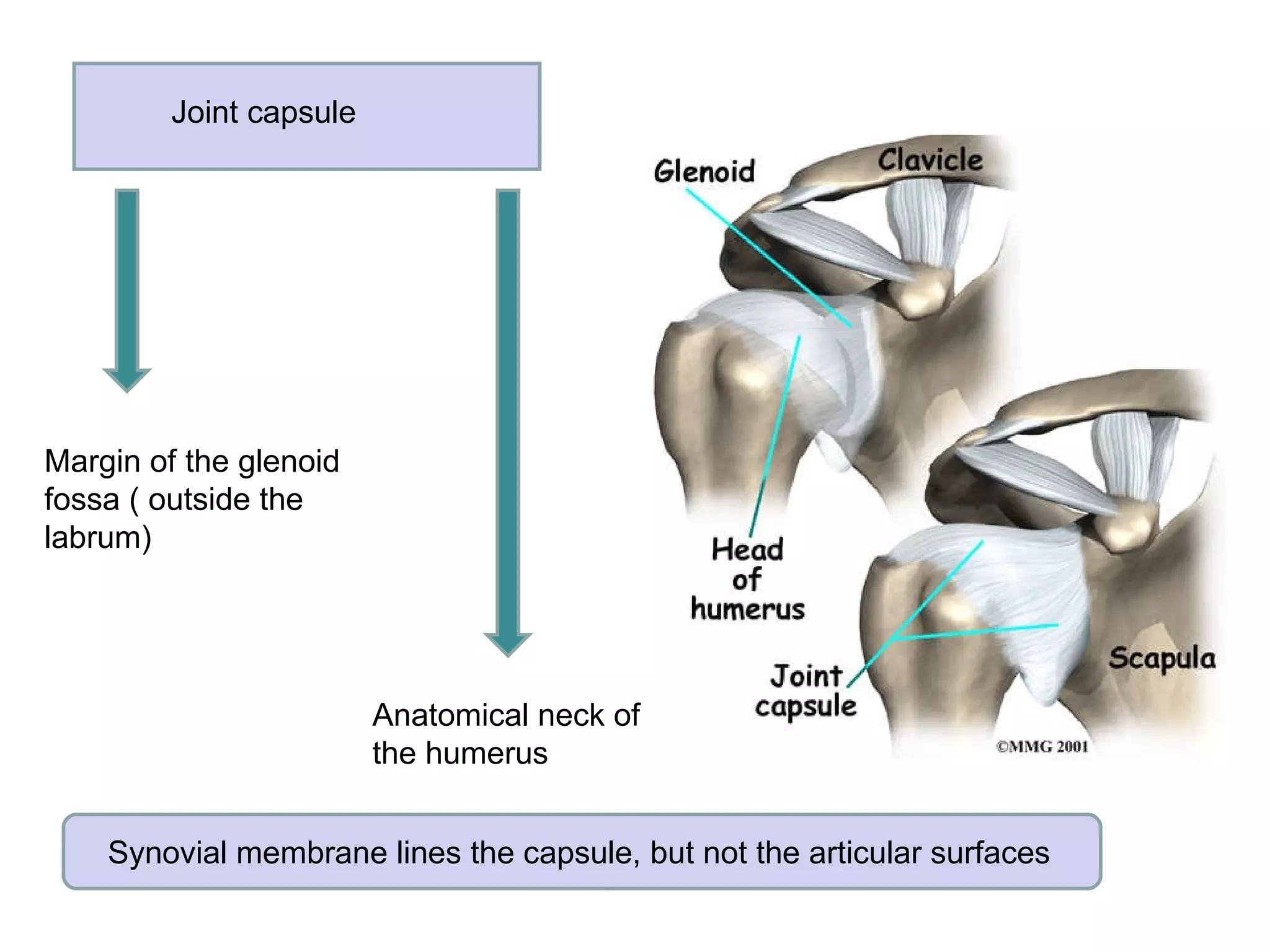
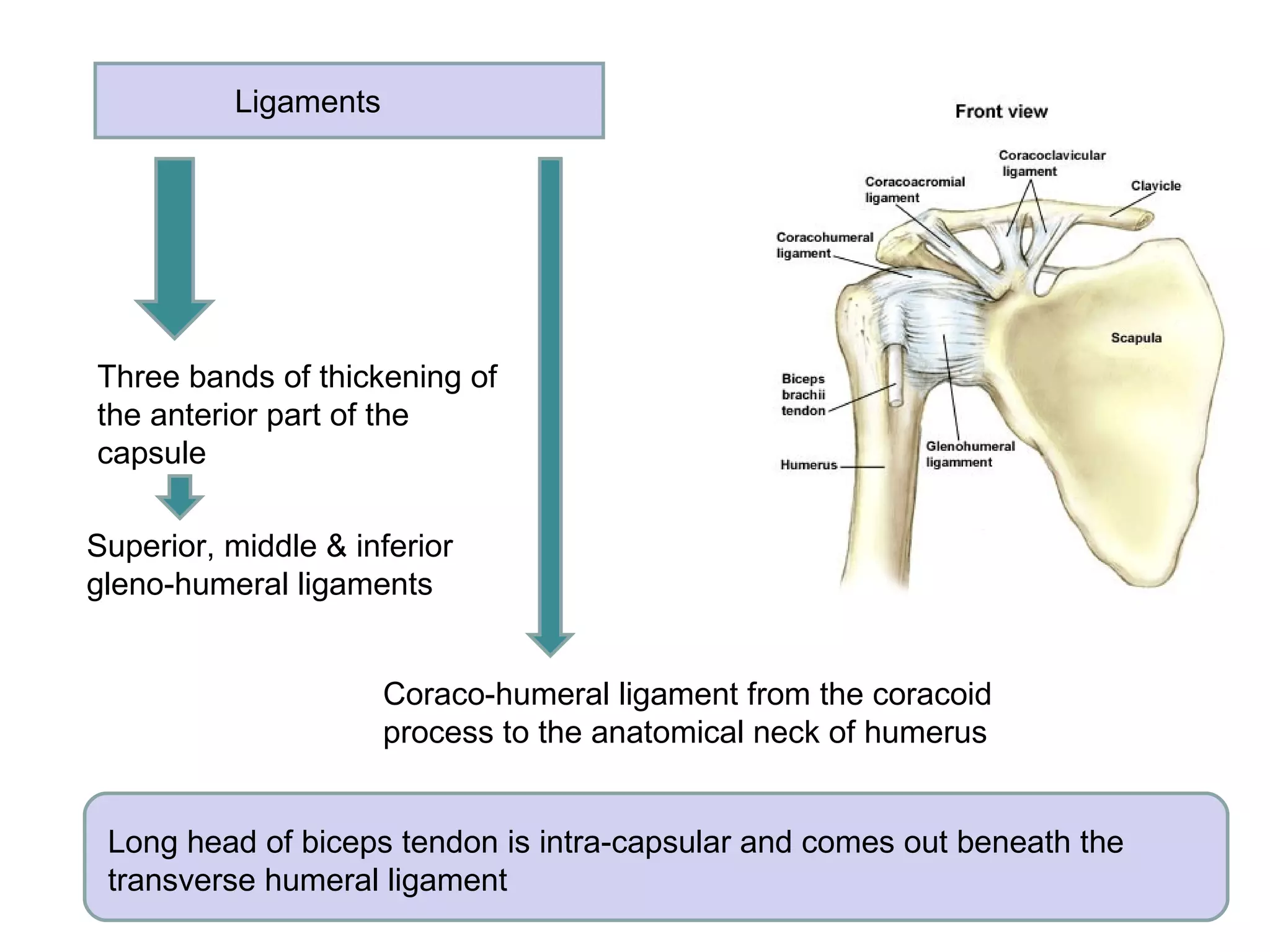
Joints of the  Limbs
Learning outcomes Describe the type of joint Describe the articular surfaces Describe the capsule and its attachment Describe the synovial membrane Describe the arrangement of ligaments ( extra-capsular & intra-capsular) Describe the movement and name the muscles producing movements Discuss the anatomical basis of common problems associated with the joint
Shoulder ( glenohumeral) joint Synovial joint Ball & socket Multi - axial Articular surfaces Glenoid fossa of scapula Head of the humerus Glenoid labrum ( fibro-cartilagenous rim) Increase the depth Only 1/3 of the humeral head is in contact with the glenoid fossa
Joint capsule Margin of the glenoid fossa ( outside the labrum) Anatomical neck of the humerus Synovial membrane lines the capsule, but not the articular surfaces
Ligaments Three bands of thickening of the anterior part of the capsule Superior, middle & inferior gleno-humeral ligaments Coraco-humeral ligament from the coracoid process to the anatomical neck of humerus Long head of biceps tendon is intra-capsular and comes out beneath the transverse humeral ligament
Musculo-tendinous rotator cuff Subscapularis Supraspinatus Infraspinatus Teres minor No rotator cuff Inferior dislocations are commoner
Dislocation of the shoulder Dislocated humeral head can press on  the axillary nerve Test for axillary nerve function in  dislocation of the shoulder
Lateral rotation Infraspinatus Teres minor Movement Muscles Flexion Pectoralis major Deltoid anterior fibers Extension Deltoid posterior fibers Teres major Abduction Suprspinatus Deltoid middle fibers Adduction Deltoid anterior & posterior fibers Pectoralis major Teres major Lattisimus dorsi Medial rotation Deltoid anterior fibers Teres major Lattisimus dorsi
Elbow joint Synovial joint Hinge joint uni-axial Articular surfaces Trochlea of humerus Capitulum of humerus Trochlear notch of ulna Upper surface of the head of radius Proximally capsule is attached to the lower end of humerus Distally to the trochlear notch of the ulna & the annular ligament of the head of radius
Ligaments Ulnar collateral ligament ( from medial epicondyle to trochlear notch) Radial collateral ligament ( from lateral epicondyle to annular ligament) Ulna nerve runs posterior to the ulnar collateral ligament and it is palpable at this location
Pronation & supination are  NOT  movements of the elbow joint Muscles Movements Flexion Biceps & brachialis Extension Triceps
Dislocation of the elbow Posterior dislocation commoner Ulnar nerve may be damaged in posterior dislocation
Wrist ( radio-carpal) joint Synovial joint ellipsoid Bi - axial Articular surfaces Distal articular  surface of radius Articular disc of  inferior radio-ulnar joint Scaphoid, lunate & triquetram
Joint capsule Peripheral margin of the radius and articular disc of the inferior radio-ulnar joint Peripheral margins of the scaphoid, lunate & triquetram Head of the ulnar is  NOT  a component of the wrist joint
Ligaments Palmar & dorsal radio-carpal ligaments are thickenings of the capsule Radial collateral ligament ( styloid process of radius to scaphoid and  trapezium Ulnar collateral ligament ( ulnar styloid process to triqutram &  pisiform)
Midcarpal joint Synovial joint Plane joints Articular surfaces Proximal: Scaphoid, lunate & triquetram Distal: trapezium, trapezoid, capitate & hamate
Movements of the wrist Movement Joint Muscle Flexion Midcarpal Flexor carpi ulnaris, Flexor carpi radialis Extension Radio-carpal Extensor carpi radialis longus & brevis, Extensor carpi ulnaris Adduction Radio-carpal Flexor carpi ulnaris & extensor carpi ulnaris Abduction Midcarpal Flexor carpi radialis & extensor carpi radialis
1 st  carpometacarpal joint Synovial joint Saddle Multi - axial Articular surfaces Trapezium Base of 1 st  metacarpal bone Movements Muscles Flexion Flexor pollicis brevis & longus Extension Extensor pollicis brevis & longus Abduction Abductor pollicis longus & brevis Adduction Adductor pollicis Opposition Opponens pollicis
Metacarpo - phalangeal joints Synovial joint Ellipsoid Articular surfaces Head of metacarpal Base of proximal phalanx 1 st  MP joint Flexion: Flexor pollicis longus Extension: Extensor pollicis longus & brevis Movements Muscles Flexion Lumbricals Extension Extensor digitorum Abduction Dorsal interossei Adduction Palmar interossei
Inter-phalangeal joints Synovial joint Hinge Uni-axial IP joint: Thumb Flexion: Flexor pollicis longus Extension: Extensor pollicis  longus Thumb: Inter-phalngeal joint (IP) Other fingers: Proxiaml & distal inter-phalangeal joints (PIP & DIP) Movements Joint Muscles Flexion PIP Flexor digitorum superficialis & profundus DIP Flexor digitorum profundus Extension PIP Lumbricals DIP Extensor digitorum
Hip joint Synovial joint Ball & socket Multi - axial Articular surfaces Lunate surface of the acetabulum Head of the femur acetabular labrum ( fibro-cartilagenous rim) Increase the depth Non articular area of the actebular fossa contains fat ( Haversian fat)
Joint capsule Margin of the actabular fossa  ( outside the labrum) Anteriorly: intertrochanteric line Posterirorly: middle of the neck Synovial membrane lines the capsule, but not the articular surfaces
Ligaments Ilio-femoral ligament Pubo-femoral ligament Ischio-femoral ligament Ilio-femoral ligament is the strongest  of the three ligaments and prevents anterior dislocation of the joint
Ligaments Ligament of the head of the femur  Blood vessels reach the head of the femur along ligament of the head of the femur & retinacular fibers From the inferior margin of the acetabulum to the fovea of the head of the femur Joint capsule is reflected along the neck of the femur as retinacular fibers
Movement Muscles Flexion Ilio-psoas Extension Gluteus maximus Adduction Adductor longus, brevis & magnus Abduction Gluteus mediaus & minimus Medial rotation Ilio-psoas, pectineus Lateral rotation Piriformis, obturator internus , obturator externus, gemmeli & quadratus femoris
Dislocation of the hip Posterior dislocation commoner Dislocated femoral head can fracture acetabular lip & damage the sciatic nerve Shenton’s line
Knee joint Synovial joint Modified hinge Articular surfaces Medial & lateral condyles of femur Medial & lateral condyles of tibia Articular surface of patella
Joint capsule Above the condyles of the femur Articular margins of the tibial condyles
Ligaments Tibial collateral Ligament Fibular collateral ligament Anterior cruciate ligament: prevents forward displacement of tibial condyles  Posterior cruciate ligament: prevents backward displacement of tibial condyles  Movements Muscles Flexion Semimembranosus, semitendinosus, biceps femoris Extension Quadreceps femoris
Ankle joint Synovial joint Modified hinge Uni-axial Articular surfaces Inferior surface of tibia Medial malleolus of tibia Lateral malleolus of  fibula Upper surface of body of the talus Medail surface of talus Lateral surface of talus Tibio-fibular mortise Trochlea tali
Ligaments Deltod ligament ( medial) Anterior & posterior talo-fibular ligaments Calcaneo-fibular ligament Movements Muscles Dorsi flexion Tibialis anterior, extensor digitorum longus, extensor hallucis longus Plantar flexion Gastrocnemius & soleus  ( tendo-calcaneus)
Mid-tarsal joints Talo-calcaneo-navicular joint Calcaneo-cuboid joint Subtalar joint Movements: Inversion ( Tibialis anterior & posterior Eversion ( Peroneus muscles) Metatarso phalangeal joints ( flexion, extension, abduction & adduction) Interphalangeal joints  ( flexion & extension)

Joints of the limbs

  • 1.
  • 2.
    Learning outcomes Describethe type of joint Describe the articular surfaces Describe the capsule and its attachment Describe the synovial membrane Describe the arrangement of ligaments ( extra-capsular & intra-capsular) Describe the movement and name the muscles producing movements Discuss the anatomical basis of common problems associated with the joint
  • 3.
    Shoulder ( glenohumeral)joint Synovial joint Ball & socket Multi - axial Articular surfaces Glenoid fossa of scapula Head of the humerus Glenoid labrum ( fibro-cartilagenous rim) Increase the depth Only 1/3 of the humeral head is in contact with the glenoid fossa
  • 4.
    Joint capsule Marginof the glenoid fossa ( outside the labrum) Anatomical neck of the humerus Synovial membrane lines the capsule, but not the articular surfaces
  • 5.
    Ligaments Three bandsof thickening of the anterior part of the capsule Superior, middle & inferior gleno-humeral ligaments Coraco-humeral ligament from the coracoid process to the anatomical neck of humerus Long head of biceps tendon is intra-capsular and comes out beneath the transverse humeral ligament
  • 6.
    Musculo-tendinous rotator cuffSubscapularis Supraspinatus Infraspinatus Teres minor No rotator cuff Inferior dislocations are commoner
  • 7.
    Dislocation of theshoulder Dislocated humeral head can press on the axillary nerve Test for axillary nerve function in dislocation of the shoulder
  • 8.
    Lateral rotation InfraspinatusTeres minor Movement Muscles Flexion Pectoralis major Deltoid anterior fibers Extension Deltoid posterior fibers Teres major Abduction Suprspinatus Deltoid middle fibers Adduction Deltoid anterior & posterior fibers Pectoralis major Teres major Lattisimus dorsi Medial rotation Deltoid anterior fibers Teres major Lattisimus dorsi
  • 9.
    Elbow joint Synovialjoint Hinge joint uni-axial Articular surfaces Trochlea of humerus Capitulum of humerus Trochlear notch of ulna Upper surface of the head of radius Proximally capsule is attached to the lower end of humerus Distally to the trochlear notch of the ulna & the annular ligament of the head of radius
  • 10.
    Ligaments Ulnar collateralligament ( from medial epicondyle to trochlear notch) Radial collateral ligament ( from lateral epicondyle to annular ligament) Ulna nerve runs posterior to the ulnar collateral ligament and it is palpable at this location
  • 11.
    Pronation & supinationare NOT movements of the elbow joint Muscles Movements Flexion Biceps & brachialis Extension Triceps
  • 12.
    Dislocation of theelbow Posterior dislocation commoner Ulnar nerve may be damaged in posterior dislocation
  • 13.
    Wrist ( radio-carpal)joint Synovial joint ellipsoid Bi - axial Articular surfaces Distal articular surface of radius Articular disc of inferior radio-ulnar joint Scaphoid, lunate & triquetram
  • 14.
    Joint capsule Peripheralmargin of the radius and articular disc of the inferior radio-ulnar joint Peripheral margins of the scaphoid, lunate & triquetram Head of the ulnar is NOT a component of the wrist joint
  • 15.
    Ligaments Palmar &dorsal radio-carpal ligaments are thickenings of the capsule Radial collateral ligament ( styloid process of radius to scaphoid and trapezium Ulnar collateral ligament ( ulnar styloid process to triqutram & pisiform)
  • 16.
    Midcarpal joint Synovialjoint Plane joints Articular surfaces Proximal: Scaphoid, lunate & triquetram Distal: trapezium, trapezoid, capitate & hamate
  • 17.
    Movements of thewrist Movement Joint Muscle Flexion Midcarpal Flexor carpi ulnaris, Flexor carpi radialis Extension Radio-carpal Extensor carpi radialis longus & brevis, Extensor carpi ulnaris Adduction Radio-carpal Flexor carpi ulnaris & extensor carpi ulnaris Abduction Midcarpal Flexor carpi radialis & extensor carpi radialis
  • 18.
    1 st carpometacarpal joint Synovial joint Saddle Multi - axial Articular surfaces Trapezium Base of 1 st metacarpal bone Movements Muscles Flexion Flexor pollicis brevis & longus Extension Extensor pollicis brevis & longus Abduction Abductor pollicis longus & brevis Adduction Adductor pollicis Opposition Opponens pollicis
  • 19.
    Metacarpo - phalangealjoints Synovial joint Ellipsoid Articular surfaces Head of metacarpal Base of proximal phalanx 1 st MP joint Flexion: Flexor pollicis longus Extension: Extensor pollicis longus & brevis Movements Muscles Flexion Lumbricals Extension Extensor digitorum Abduction Dorsal interossei Adduction Palmar interossei
  • 20.
    Inter-phalangeal joints Synovialjoint Hinge Uni-axial IP joint: Thumb Flexion: Flexor pollicis longus Extension: Extensor pollicis longus Thumb: Inter-phalngeal joint (IP) Other fingers: Proxiaml & distal inter-phalangeal joints (PIP & DIP) Movements Joint Muscles Flexion PIP Flexor digitorum superficialis & profundus DIP Flexor digitorum profundus Extension PIP Lumbricals DIP Extensor digitorum
  • 21.
    Hip joint Synovialjoint Ball & socket Multi - axial Articular surfaces Lunate surface of the acetabulum Head of the femur acetabular labrum ( fibro-cartilagenous rim) Increase the depth Non articular area of the actebular fossa contains fat ( Haversian fat)
  • 22.
    Joint capsule Marginof the actabular fossa ( outside the labrum) Anteriorly: intertrochanteric line Posterirorly: middle of the neck Synovial membrane lines the capsule, but not the articular surfaces
  • 23.
    Ligaments Ilio-femoral ligamentPubo-femoral ligament Ischio-femoral ligament Ilio-femoral ligament is the strongest of the three ligaments and prevents anterior dislocation of the joint
  • 24.
    Ligaments Ligament ofthe head of the femur Blood vessels reach the head of the femur along ligament of the head of the femur & retinacular fibers From the inferior margin of the acetabulum to the fovea of the head of the femur Joint capsule is reflected along the neck of the femur as retinacular fibers
  • 25.
    Movement Muscles FlexionIlio-psoas Extension Gluteus maximus Adduction Adductor longus, brevis & magnus Abduction Gluteus mediaus & minimus Medial rotation Ilio-psoas, pectineus Lateral rotation Piriformis, obturator internus , obturator externus, gemmeli & quadratus femoris
  • 26.
    Dislocation of thehip Posterior dislocation commoner Dislocated femoral head can fracture acetabular lip & damage the sciatic nerve Shenton’s line
  • 27.
    Knee joint Synovialjoint Modified hinge Articular surfaces Medial & lateral condyles of femur Medial & lateral condyles of tibia Articular surface of patella
  • 28.
    Joint capsule Abovethe condyles of the femur Articular margins of the tibial condyles
  • 29.
    Ligaments Tibial collateralLigament Fibular collateral ligament Anterior cruciate ligament: prevents forward displacement of tibial condyles Posterior cruciate ligament: prevents backward displacement of tibial condyles Movements Muscles Flexion Semimembranosus, semitendinosus, biceps femoris Extension Quadreceps femoris
  • 30.
    Ankle joint Synovialjoint Modified hinge Uni-axial Articular surfaces Inferior surface of tibia Medial malleolus of tibia Lateral malleolus of fibula Upper surface of body of the talus Medail surface of talus Lateral surface of talus Tibio-fibular mortise Trochlea tali
  • 31.
    Ligaments Deltod ligament( medial) Anterior & posterior talo-fibular ligaments Calcaneo-fibular ligament Movements Muscles Dorsi flexion Tibialis anterior, extensor digitorum longus, extensor hallucis longus Plantar flexion Gastrocnemius & soleus ( tendo-calcaneus)
  • 32.
    Mid-tarsal joints Talo-calcaneo-navicularjoint Calcaneo-cuboid joint Subtalar joint Movements: Inversion ( Tibialis anterior & posterior Eversion ( Peroneus muscles) Metatarso phalangeal joints ( flexion, extension, abduction & adduction) Interphalangeal joints ( flexion & extension)