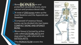
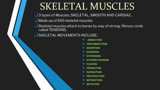
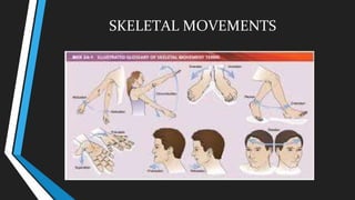
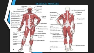
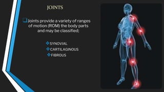
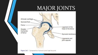
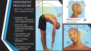
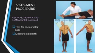
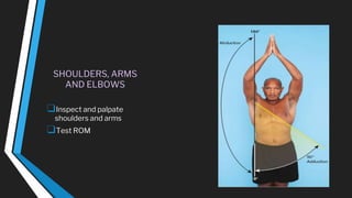
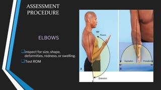
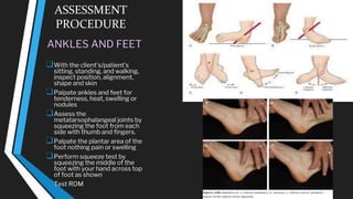
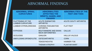
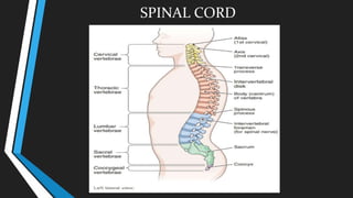
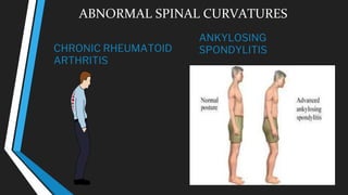
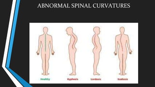
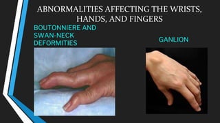
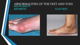
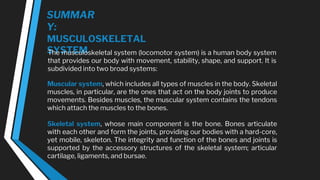
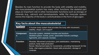
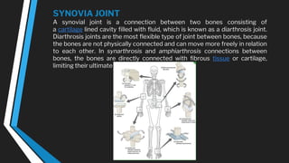
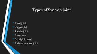
ASSESSING
MUSCULOSKELETA
L SYSTEM
STUCTURE AND FUNCTION
BONES
MUSCLES
JOINTS
BONES
❑ Bones provide structure and
protection, severe as levers, store
calcium and produce blood cells.
❑A total of 206 bones make up the
Axial Skeleton and the Appendicular
Skeleton.
❑Composed of osseous tissue,
bones can be divided into two types:
COMPACT BONE AND SPONGY
BONE.
❑Bone tissue is formed by active
cells called OSTEOBLASTS and
degraded by cells referred to as
OSTEOLASTS.
❑The PERIOSTEUM covers the
bones.
SKELETAL MUSCLES
❑3 types of Muscles; SKELETAL, SMOOTH AND CARDIAC.
❑Made up of 650 skeletal muscles.
❑Skeletal muscles attach to bones by way of strong, fibrous cords
called TENDONS.
❑SKELETAL MOVEMENTS INCLUDE;
❖ ABDUCTION
❖ CIRCUMDUCTION
❖ INVERTION
❖ EVERSION
❖ EXTENSION
❖ HYPEREXTENSION
❖ FLEXION
❖ PRONATION
❖ SUPINATION
❖ PROTRACTION
❖ RETRACTION
❖ ROTATION
SKELETAL MOVEMENTS
SKELETAL MUSCLES
JOINTS
❑Joints provide a variety of ranges
of motion (ROM) the body parts
and may be classified;
❖SYNOVIAL
❖CARTILAGINOUS
❖FIBROUS
MAJOR JOINTS
MAJOR JOINTS
TEMPOROMANDIBU
LAR ELBOW
MAJOR JOINTS
STERNOCLAVICULAR SHOULDER
WRIST, FINGERS, THUMB
MAJOR JOINTS
HIP VERTEBRAE
MAJOR JOINTS
MAJOR JOINTS
KNEE ANKLE AND FOOT
TECHNIQUES OF EXAMINATION
Important Areas of Examination for Each of the Major
Joints;
❑ Inspection for joint symmetry, alignment, bony deformities.
❑Inspection and palpation of surrounding tissues for skin changes, nodules,
muscle atrophy, crepitus.
❑Range of motion and maneuvers to test joint function and stability, integrity of
ligaments,
tendons, bursae, especially if pain or trauma.
❑Assessment of inflammation or arthritis, especially swelling, warmth,
tenderness, redness.
Common or Concern Symptoms
o Low back pain
o Neck pain
o Monoarticular or polyarticular joint pain
o Inflammatory or infectious joint pain
o Joint pain with systemic features
o Joint pain with symptoms from other organ
system
Remember the following clues To examination
⮚Use inspection and palpation to assess the surroundings tissues, noting skin
changes, subcutaneous nodules and muscle atrophy.
NOTE: any crepitus and audible and/or palpable crunching during movement of
tendons or ligaments over bone. This may occur in normal joints but is more
significant when associated with symptoms or signs.
⮚Testing range of motion and maneuvers (described for each joint) may demonstrate
limitations in range of motion or increased mobility and joint instability from excess
mobility of joint ligaments, called ligamentous laxity.
⮚Testing muscle strength may aid in the assessment of joint function.
Be especially alert to signs of inflammation and arthritis.
⮚ Swelling
⮚Warmth
⮚Tenderness
⮚ Redness
NURSING ASSESSMENT
COLLETING SUBJECTIVE DATA
❑Assessment of the musculoskeletal
system helps to evaluate the clients
level of functioning with activities of
daily living (ADLS).
❑This system affects the entire
body, from head to toe, and greatly
influences what physical activities a
client can and cannot do,
❑The neurologic system is
responsible for coordinating the
functions of the skeleton and
muscles.
COLLETING OBJECTIVE DATA
❑Physical assessment of the
musculoskeletal system provides
data regarding the clients posture,
gait, bone structure, muscle
strength, and joint mobility, as well
as the client ability to perform
ADLs.
❑Preparing the client.
EQUIPMENT
❑TAPE MEASURE
❑GONIOMETER (OPTIONAL)
❑SKIN MARKING PEN (OPTIONAL)
PHYSICAL ASSESSMENT
❑Observe gait and posture.
❑Inspect joints, muscles, and
extremities for size, symmetry and
color.
❑Palpate joints, muscles, and
extremities for tenderness, edema,
heat, nodules, or crepitus.
❑Test muscle strength and ROM of
joints.
❑Compare bilateral findings of joints
and muscles.
❑Perform special tests for carpal
tunnel syndrome.
❑Perform the bulge ballottement and
McMurray knee test.
WHAT IS COMPOSED OF UPPER AND LOWER
EXTREMITIES?
• Appendicular bones: the 126 appendicular bones are
made up of 64 bones in the upper extremities (the arms,
wrists, and hand) and 62 bones in the lower extremities
(the legs, ankles, and feet).
INSPECTION
Observe any lack of symmetry and any
evidence of trauma or disease.
•Look for muscle wasting;
•Inspect the joint contour (shape) and
observe any evidence of swelling, deformity
or inflammation.
PALPATION
Examine the ankle for discoloration and
swelling and palpate for tenderness,
swelling, effusion, and crepitus on range of
motion. Ask the patient to dorsiflex the
ankles (this should be possible to
approximately 20 degrees) and to plantar-
fex the ankles (this should be possible to
approximately 45 degrees)
GENERAL ROUTINE SCREENING VERSUS
FOCUSED SPECIALTY ASSESSMENT FOR THE
MUSCULOSKELETAL SYSTEM
GENERAL ROUTINE
SCREENING
❑OBSERVE POSTURE
AND GAIT.
❑INSPECT THE
FOLLOWING FOR
SYMMETRY, COLOR AND
MOBILITY;
❑PALAPATE THE
FOLLOWING FOR
TENDERNESS, HEAT,
FOCUSED SPECIALTY
ASSESSMENT
❑MEASURE THE ROM
WITH A GONIOMETER.
❑PALPATE THE
ANATOMIC SNUFFBOX.
ASSESSMENT
PROCEDURE
INSPECTION
❑OBSERVE
POSTURE
❑OBSERVE GAIT
ASSESSMENT
PROCEDURE
TEMPOROMANDIBUL
AR JOINT (TMJ)
❑INSPECT AND PALPATE TMJ
Move the jaw side to side
Protrude and retract
❑TEST ROM
ASSESSMENT
PROCEDURE
STERNOCLAVICULAR
JOINT
❑Inspect the location in
midline, color, swelling,
and masses.
❑Palpate for tenderness or
pain
ASSESSMENT
PROCEDURE
CERVICAL, THORACIC
AND LUMBAR SPINE
❑OBSERVE THE
CERVICAL, THORACIC
AND LUMBAR CURVES
❑PALPATE THE
SPINOUS PROCESSES
AND PARAVERTEBRAL
MUSCLES
❑TEST ROM OF THE
CERVICAL SPINE
❑TEST LATERAL
BENDING
❑EVALUATE ROTATION
❑TEST ROM OF THE
LUMBAR SPINE
ASSESSMENT
PROCEDURE
CERVICAL, THORACIC AND
LUMBAR SPINE (continued)
❑Test for back and leg
pain
❑Measure leg length
SHOULDERS, ARMS
AND ELBOWS
❑Inspect and palpate
shoulders and arms
❑Test ROM
ASSESSMENT
PROCEDURE
ELBOWS
❑inspect for size, shape,
deformities, redness, or swelling.
❑Test ROM
ASSESSMENT
PROCEDURE
WRISTS
❑Inspect wrists size, shape,
symmetry, color and swelling
❑Perform the squeeze test
❑Palpate the anatomic
snuffbox no tenderness
palpated in anatomic snuffbox
❑Test ROM
❑Test for carpal tunnel
syndrome (CTS)
❑Perform Phalen test
❑Perform test for Tinel
❑Observe for the flick signal
❑Test for thumb weakness
ASSESSMENT
PROCEDURE
HANDS AND
FINGERS
❑ Inspect size, shape
symmetry, swelling and
color
❑ Test ROM
ASSESSMENT
PROCEDURE
HIPS
❑With the client standing,
inspect symmetry and
shape of the hips.
❑Observe for convex
thoracic curve and
concave lumbar curve.
❑Palpate for stability,
tenderness and crepitus.
❑Test ROM
ASSESSMENT
PROCEDURE
KNEES
❑ with the client supine then
sitting with knees dangling,
inspect for the size, shape,
symmetry, swelling,
deformities, and alignment
❑ Observe for quadriceps muscle
atrophy
❑ Palpate for tenderness,
warmth, consistency and
nodules.
❑ Perform the bulge test if
swelling is present
❑ Perform the ballottment test
❑ Palpate the tibiofemoral space
❑ Test ROM
❑ Test for pain and injury
ASSESSMENT
PROCEDURE
ANKLES AND FEET
❑With the client’s/patient’s
sitting, standing, and walking,
inspect position, alignment,
shape and skin
❑Palpate ankles and feet for
tenderness, heat, swelling or
nodules
❑Assess the
metatarsophalangeal joints by
squeezing the foot from each
side with thumb and fingers.
❑Palpate the plantar area of the
foot nothing pain or swelling
❑Perform squeeze test by
squeezing the middle of the
foot with your hand across top
of foot as shown
❑Test ROM
ABNORMAL FINDINGS
ABNORMAL SPINAL
CURVATURES
ABNORMALITIES
AFFECTING THE
WRISTS, HANDS, AND
FINGERS
ABNORMALITIES OF THE
FEET AND TOES
FLATTENING OF THE
LUMBAR CURVATURE
ACUTE RHEMATOID
ARTHEITIS
ACUTE GOUTY ARTHRITIS
LUMBAR HYPERLORDOSIS CHRONIC RHEUMATOID
ARTHRITIS
FLAT FEET
KYPHOSIS BOUTONNIERE AND SWAN-
NECK DEFORMITIES
CALLUS
SCOLIOSIS GANLION HALLUX VALGUS
ANKYLOSING SPONDYLITIS OSTEOARTHRITIS CORN
TENOSYNOVITIS HAMMER TOE
THENAR ATROPHY PLANTAR WART
SPINAL CORD
ABNORMAL SPINAL CURVATURES
CHRONIC RHEUMATOID
ARTHRITIS
ANKYLOSING
SPONDYLITIS
ABNORMAL SPINAL CURVATURES
ABNORMALITIES AFFECTING THE WRISTS,
HANDS, AND FINGERS
ACUTE RHEMATOID
ARTHEITIS
CHRONIC RHEUMATOID
ARTHRITIS
ABNORMALITIES AFFECTING THE WRISTS,
HANDS, AND FINGERS
BOUTONNIERE AND
SWAN-NECK
DEFORMITIES
GANLION
ABNORMALITIES AFFECTING THE WRISTS,
HANDS, AND FINGERS
OSTEOARTHRITIS TENOSYNOVITIS
ABNORMALITIES AFFECTING THE WRISTS, HANDS, AND
FINGERS
THENAR ATROPHY
ABNORMALITIES OF THE FEET AND TOES
ACUTE GOUTY
ARTHRITIS FLAT FEET
ABNORMALITIES OF THE FEET AND TOES
CALLUS HALLUX VALGUS
ABNORMALITIES OF THE FEET AND TOES
CORN HAMMER TOE
ABNORMALITIES OF THE FEET AND TOES
PLANTAR WART
SUMMAR
Y:
MUSCULOSKELETAL
SYSTEM
The musculoskeletal system (locomotor system) is a human body system
that provides our body with movement, stability, shape, and support. It is
subdivided into two broad systems:
Muscular system, which includes all types of muscles in the body. Skeletal
muscles, in particular, are the ones that act on the body joints to produce
movements. Besides muscles, the muscular system contains the tendons
which attach the muscles to the bones.
Skeletal system, whose main component is the bone. Bones articulate
with each other and form the joints, providing our bodies with a hard-core,
yet mobile, skeleton. The integrity and function of the bones and joints is
supported by the accessory structures of the skeletal system; articular
cartilage, ligaments, and bursae.
Besides its main function to provide the body with stability and mobility,
the musculoskeletal system has many other functions; the skeletal part
plays an important role in other homeostatic functions such as storage of
minerals (e.g., calcium) and hematopoiesis, while the muscular system
stores the majority of the body's carbohydrates in the form of glycogen.
Definition A human body system that provides the body with movement,
stability, shape, and support
Component
s
Muscular system: skeletal muscles and tendons
Skeletal system: bones, joints; associated tissues (cartilage,
ligaments, joint capsule, bursae)
Function Muscles: Movement production, joint stabilization, maintaining
posture, body heat production
Bones: Mechanical basis for movements, providing framework for the
body, vital organs protection, blood cells production, storage of
minerals
Key facts about the musculoskeletal
system
NURSING HEALTH
ASSESSMENT
The musculoskeletal system consists of the muscles, bones, cartilage, and
joints. This system provides the body with support and movement. Also,
the musculoskeletal system protects major organs, produces red blood
cells and store important minerals such as calcium and phosphorus.
A properly functioning musculoskeletal system is important for a patient
to perform activities of daily living (ADLs). The techniques for the
assessment of the musculoskeletal system are inspection, palpation, and
observing the range of motion of the joints.
Perform Inspection of the Musculoskeletal System.
A nursing health assessment of the musculoskeletal system involves inspection
of the joints. Use inspection to assess the joints for symmetry. A problem in one
joint can mean trauma. A problem in more than one joint can mean a systemic
condition. Note the patient’s movements when performing the range of motion
maneuvers. The movements should be smooth.
•Check for joint deformities, muscle atrophy or abnormal positioning of the
limb.
•Assess for immobility in all joints.
•Inspect the surrounding tissue and muscle for swelling,
•Inspect the area for redness.
•Assess the joint area for any skin abnormalities. (Different color, or protruding
bony prominences.)
•Listen for an audible crunching sound resonating from the joint. This sound
indicates crepitus. Crepitus can also be palpated.
•Observe the patients posture.
•Note the position of the patients head and neck.
•Assess the patient’s gait. A waddling gait could indicate a hip problem.
•Note how the patient bears weight on each side as they walk.
•Note the swing of the leg that is not bearing the weight as the patient walks.
•Assess the knee as the patient walks. The knee should be extended when the
heel strikes the ground and flexed when the leg is swinging.
•Note how far the patient’s feet are apart while standing. The normal width is
between 2-4 inches.
Perform Palpation of the Musculoskeletal System.
Also, a nursing health assessment of the musculoskeletal system involves
palpation of the joints. Palpate the joints and assess the temperature of the
skin and the muscles. Palpate for warmth, tenderness, swelling or masses. If
pain or tenderness are noted, further assess to specify the joint or structure
involved. If there is any pain proceed carefully.
•Use the back of the hand to palpate for warmth.
•Compare the affected area with an unaffected area.
•Palpate the bony landmarks of each joint.
•Assess each area for pain.
•Palpate each area for swelling especially in the synovial joint area.
SYNOVIA JOINT
A synovial joint is a connection between two bones consisting of
a cartilage lined cavity filled with fluid, which is known as a diarthrosis joint.
Diarthrosis joints are the most flexible type of joint between bones, because
the bones are not physically connected and can move more freely in relation
to each other. In synarthrosis and amphiarthrosis connections between
bones, the bones are directly connected with fibrous tissue or cartilage,
limiting their ultimate range of motion.
Types of Synovia joint
• Pivot joint
• Hinge joint
• Saddle joint
• Plane joint
• Condyloid joint
• Ball-and-sacket joint
ASSESSING-MUSCULOSKELETAL-SYSTEM.pptx

ASSESSING-MUSCULOSKELETAL-SYSTEM.pptx

  • 1.
  • 2.
  • 3.
    BONES ❑ Bones providestructure and protection, severe as levers, store calcium and produce blood cells. ❑A total of 206 bones make up the Axial Skeleton and the Appendicular Skeleton. ❑Composed of osseous tissue, bones can be divided into two types: COMPACT BONE AND SPONGY BONE. ❑Bone tissue is formed by active cells called OSTEOBLASTS and degraded by cells referred to as OSTEOLASTS. ❑The PERIOSTEUM covers the bones.
  • 4.
    SKELETAL MUSCLES ❑3 typesof Muscles; SKELETAL, SMOOTH AND CARDIAC. ❑Made up of 650 skeletal muscles. ❑Skeletal muscles attach to bones by way of strong, fibrous cords called TENDONS. ❑SKELETAL MOVEMENTS INCLUDE; ❖ ABDUCTION ❖ CIRCUMDUCTION ❖ INVERTION ❖ EVERSION ❖ EXTENSION ❖ HYPEREXTENSION ❖ FLEXION ❖ PRONATION ❖ SUPINATION ❖ PROTRACTION ❖ RETRACTION ❖ ROTATION
  • 5.
  • 6.
  • 7.
    JOINTS ❑Joints provide avariety of ranges of motion (ROM) the body parts and may be classified; ❖SYNOVIAL ❖CARTILAGINOUS ❖FIBROUS
  • 8.
  • 9.
  • 10.
  • 11.
  • 12.
  • 13.
  • 14.
    TECHNIQUES OF EXAMINATION ImportantAreas of Examination for Each of the Major Joints; ❑ Inspection for joint symmetry, alignment, bony deformities. ❑Inspection and palpation of surrounding tissues for skin changes, nodules, muscle atrophy, crepitus. ❑Range of motion and maneuvers to test joint function and stability, integrity of ligaments, tendons, bursae, especially if pain or trauma. ❑Assessment of inflammation or arthritis, especially swelling, warmth, tenderness, redness.
  • 15.
    Common or ConcernSymptoms o Low back pain o Neck pain o Monoarticular or polyarticular joint pain o Inflammatory or infectious joint pain o Joint pain with systemic features o Joint pain with symptoms from other organ system
  • 16.
    Remember the followingclues To examination ⮚Use inspection and palpation to assess the surroundings tissues, noting skin changes, subcutaneous nodules and muscle atrophy. NOTE: any crepitus and audible and/or palpable crunching during movement of tendons or ligaments over bone. This may occur in normal joints but is more significant when associated with symptoms or signs. ⮚Testing range of motion and maneuvers (described for each joint) may demonstrate limitations in range of motion or increased mobility and joint instability from excess mobility of joint ligaments, called ligamentous laxity. ⮚Testing muscle strength may aid in the assessment of joint function. Be especially alert to signs of inflammation and arthritis. ⮚ Swelling ⮚Warmth ⮚Tenderness ⮚ Redness
  • 17.
    NURSING ASSESSMENT COLLETING SUBJECTIVEDATA ❑Assessment of the musculoskeletal system helps to evaluate the clients level of functioning with activities of daily living (ADLS). ❑This system affects the entire body, from head to toe, and greatly influences what physical activities a client can and cannot do, ❑The neurologic system is responsible for coordinating the functions of the skeleton and muscles. COLLETING OBJECTIVE DATA ❑Physical assessment of the musculoskeletal system provides data regarding the clients posture, gait, bone structure, muscle strength, and joint mobility, as well as the client ability to perform ADLs. ❑Preparing the client.
  • 18.
    EQUIPMENT ❑TAPE MEASURE ❑GONIOMETER (OPTIONAL) ❑SKINMARKING PEN (OPTIONAL) PHYSICAL ASSESSMENT ❑Observe gait and posture. ❑Inspect joints, muscles, and extremities for size, symmetry and color. ❑Palpate joints, muscles, and extremities for tenderness, edema, heat, nodules, or crepitus. ❑Test muscle strength and ROM of joints. ❑Compare bilateral findings of joints and muscles. ❑Perform special tests for carpal tunnel syndrome. ❑Perform the bulge ballottement and McMurray knee test.
  • 19.
    WHAT IS COMPOSEDOF UPPER AND LOWER EXTREMITIES? • Appendicular bones: the 126 appendicular bones are made up of 64 bones in the upper extremities (the arms, wrists, and hand) and 62 bones in the lower extremities (the legs, ankles, and feet).
  • 20.
    INSPECTION Observe any lackof symmetry and any evidence of trauma or disease. •Look for muscle wasting; •Inspect the joint contour (shape) and observe any evidence of swelling, deformity or inflammation. PALPATION Examine the ankle for discoloration and swelling and palpate for tenderness, swelling, effusion, and crepitus on range of motion. Ask the patient to dorsiflex the ankles (this should be possible to approximately 20 degrees) and to plantar- fex the ankles (this should be possible to approximately 45 degrees)
  • 21.
    GENERAL ROUTINE SCREENINGVERSUS FOCUSED SPECIALTY ASSESSMENT FOR THE MUSCULOSKELETAL SYSTEM GENERAL ROUTINE SCREENING ❑OBSERVE POSTURE AND GAIT. ❑INSPECT THE FOLLOWING FOR SYMMETRY, COLOR AND MOBILITY; ❑PALAPATE THE FOLLOWING FOR TENDERNESS, HEAT, FOCUSED SPECIALTY ASSESSMENT ❑MEASURE THE ROM WITH A GONIOMETER. ❑PALPATE THE ANATOMIC SNUFFBOX.
  • 22.
  • 23.
    ASSESSMENT PROCEDURE TEMPOROMANDIBUL AR JOINT (TMJ) ❑INSPECTAND PALPATE TMJ Move the jaw side to side Protrude and retract ❑TEST ROM
  • 24.
    ASSESSMENT PROCEDURE STERNOCLAVICULAR JOINT ❑Inspect the locationin midline, color, swelling, and masses. ❑Palpate for tenderness or pain
  • 25.
    ASSESSMENT PROCEDURE CERVICAL, THORACIC AND LUMBARSPINE ❑OBSERVE THE CERVICAL, THORACIC AND LUMBAR CURVES ❑PALPATE THE SPINOUS PROCESSES AND PARAVERTEBRAL MUSCLES ❑TEST ROM OF THE CERVICAL SPINE ❑TEST LATERAL BENDING ❑EVALUATE ROTATION ❑TEST ROM OF THE LUMBAR SPINE
  • 26.
    ASSESSMENT PROCEDURE CERVICAL, THORACIC AND LUMBARSPINE (continued) ❑Test for back and leg pain ❑Measure leg length
  • 27.
    SHOULDERS, ARMS AND ELBOWS ❑Inspectand palpate shoulders and arms ❑Test ROM
  • 28.
    ASSESSMENT PROCEDURE ELBOWS ❑inspect for size,shape, deformities, redness, or swelling. ❑Test ROM
  • 29.
    ASSESSMENT PROCEDURE WRISTS ❑Inspect wrists size,shape, symmetry, color and swelling ❑Perform the squeeze test ❑Palpate the anatomic snuffbox no tenderness palpated in anatomic snuffbox ❑Test ROM ❑Test for carpal tunnel syndrome (CTS) ❑Perform Phalen test ❑Perform test for Tinel ❑Observe for the flick signal ❑Test for thumb weakness
  • 30.
    ASSESSMENT PROCEDURE HANDS AND FINGERS ❑ Inspectsize, shape symmetry, swelling and color ❑ Test ROM
  • 31.
    ASSESSMENT PROCEDURE HIPS ❑With the clientstanding, inspect symmetry and shape of the hips. ❑Observe for convex thoracic curve and concave lumbar curve. ❑Palpate for stability, tenderness and crepitus. ❑Test ROM
  • 32.
    ASSESSMENT PROCEDURE KNEES ❑ with theclient supine then sitting with knees dangling, inspect for the size, shape, symmetry, swelling, deformities, and alignment ❑ Observe for quadriceps muscle atrophy ❑ Palpate for tenderness, warmth, consistency and nodules. ❑ Perform the bulge test if swelling is present ❑ Perform the ballottment test ❑ Palpate the tibiofemoral space ❑ Test ROM ❑ Test for pain and injury
  • 33.
    ASSESSMENT PROCEDURE ANKLES AND FEET ❑Withthe client’s/patient’s sitting, standing, and walking, inspect position, alignment, shape and skin ❑Palpate ankles and feet for tenderness, heat, swelling or nodules ❑Assess the metatarsophalangeal joints by squeezing the foot from each side with thumb and fingers. ❑Palpate the plantar area of the foot nothing pain or swelling ❑Perform squeeze test by squeezing the middle of the foot with your hand across top of foot as shown ❑Test ROM
  • 34.
    ABNORMAL FINDINGS ABNORMAL SPINAL CURVATURES ABNORMALITIES AFFECTINGTHE WRISTS, HANDS, AND FINGERS ABNORMALITIES OF THE FEET AND TOES FLATTENING OF THE LUMBAR CURVATURE ACUTE RHEMATOID ARTHEITIS ACUTE GOUTY ARTHRITIS LUMBAR HYPERLORDOSIS CHRONIC RHEUMATOID ARTHRITIS FLAT FEET KYPHOSIS BOUTONNIERE AND SWAN- NECK DEFORMITIES CALLUS SCOLIOSIS GANLION HALLUX VALGUS ANKYLOSING SPONDYLITIS OSTEOARTHRITIS CORN TENOSYNOVITIS HAMMER TOE THENAR ATROPHY PLANTAR WART
  • 35.
  • 36.
    ABNORMAL SPINAL CURVATURES CHRONICRHEUMATOID ARTHRITIS ANKYLOSING SPONDYLITIS
  • 37.
  • 38.
    ABNORMALITIES AFFECTING THEWRISTS, HANDS, AND FINGERS ACUTE RHEMATOID ARTHEITIS CHRONIC RHEUMATOID ARTHRITIS
  • 39.
    ABNORMALITIES AFFECTING THEWRISTS, HANDS, AND FINGERS BOUTONNIERE AND SWAN-NECK DEFORMITIES GANLION
  • 40.
    ABNORMALITIES AFFECTING THEWRISTS, HANDS, AND FINGERS OSTEOARTHRITIS TENOSYNOVITIS
  • 41.
    ABNORMALITIES AFFECTING THEWRISTS, HANDS, AND FINGERS THENAR ATROPHY
  • 42.
    ABNORMALITIES OF THEFEET AND TOES ACUTE GOUTY ARTHRITIS FLAT FEET
  • 43.
    ABNORMALITIES OF THEFEET AND TOES CALLUS HALLUX VALGUS
  • 44.
    ABNORMALITIES OF THEFEET AND TOES CORN HAMMER TOE
  • 45.
    ABNORMALITIES OF THEFEET AND TOES PLANTAR WART
  • 46.
    SUMMAR Y: MUSCULOSKELETAL SYSTEM The musculoskeletal system(locomotor system) is a human body system that provides our body with movement, stability, shape, and support. It is subdivided into two broad systems: Muscular system, which includes all types of muscles in the body. Skeletal muscles, in particular, are the ones that act on the body joints to produce movements. Besides muscles, the muscular system contains the tendons which attach the muscles to the bones. Skeletal system, whose main component is the bone. Bones articulate with each other and form the joints, providing our bodies with a hard-core, yet mobile, skeleton. The integrity and function of the bones and joints is supported by the accessory structures of the skeletal system; articular cartilage, ligaments, and bursae.
  • 47.
    Besides its mainfunction to provide the body with stability and mobility, the musculoskeletal system has many other functions; the skeletal part plays an important role in other homeostatic functions such as storage of minerals (e.g., calcium) and hematopoiesis, while the muscular system stores the majority of the body's carbohydrates in the form of glycogen. Definition A human body system that provides the body with movement, stability, shape, and support Component s Muscular system: skeletal muscles and tendons Skeletal system: bones, joints; associated tissues (cartilage, ligaments, joint capsule, bursae) Function Muscles: Movement production, joint stabilization, maintaining posture, body heat production Bones: Mechanical basis for movements, providing framework for the body, vital organs protection, blood cells production, storage of minerals Key facts about the musculoskeletal system
  • 48.
    NURSING HEALTH ASSESSMENT The musculoskeletalsystem consists of the muscles, bones, cartilage, and joints. This system provides the body with support and movement. Also, the musculoskeletal system protects major organs, produces red blood cells and store important minerals such as calcium and phosphorus. A properly functioning musculoskeletal system is important for a patient to perform activities of daily living (ADLs). The techniques for the assessment of the musculoskeletal system are inspection, palpation, and observing the range of motion of the joints.
  • 49.
    Perform Inspection ofthe Musculoskeletal System. A nursing health assessment of the musculoskeletal system involves inspection of the joints. Use inspection to assess the joints for symmetry. A problem in one joint can mean trauma. A problem in more than one joint can mean a systemic condition. Note the patient’s movements when performing the range of motion maneuvers. The movements should be smooth. •Check for joint deformities, muscle atrophy or abnormal positioning of the limb. •Assess for immobility in all joints. •Inspect the surrounding tissue and muscle for swelling, •Inspect the area for redness. •Assess the joint area for any skin abnormalities. (Different color, or protruding bony prominences.) •Listen for an audible crunching sound resonating from the joint. This sound indicates crepitus. Crepitus can also be palpated. •Observe the patients posture. •Note the position of the patients head and neck. •Assess the patient’s gait. A waddling gait could indicate a hip problem. •Note how the patient bears weight on each side as they walk.
  • 50.
    •Note the swingof the leg that is not bearing the weight as the patient walks. •Assess the knee as the patient walks. The knee should be extended when the heel strikes the ground and flexed when the leg is swinging. •Note how far the patient’s feet are apart while standing. The normal width is between 2-4 inches. Perform Palpation of the Musculoskeletal System. Also, a nursing health assessment of the musculoskeletal system involves palpation of the joints. Palpate the joints and assess the temperature of the skin and the muscles. Palpate for warmth, tenderness, swelling or masses. If pain or tenderness are noted, further assess to specify the joint or structure involved. If there is any pain proceed carefully. •Use the back of the hand to palpate for warmth. •Compare the affected area with an unaffected area. •Palpate the bony landmarks of each joint. •Assess each area for pain. •Palpate each area for swelling especially in the synovial joint area.
  • 51.
    SYNOVIA JOINT A synovialjoint is a connection between two bones consisting of a cartilage lined cavity filled with fluid, which is known as a diarthrosis joint. Diarthrosis joints are the most flexible type of joint between bones, because the bones are not physically connected and can move more freely in relation to each other. In synarthrosis and amphiarthrosis connections between bones, the bones are directly connected with fibrous tissue or cartilage, limiting their ultimate range of motion.
  • 52.
    Types of Synoviajoint • Pivot joint • Hinge joint • Saddle joint • Plane joint • Condyloid joint • Ball-and-sacket joint