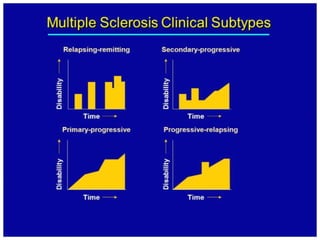
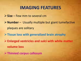
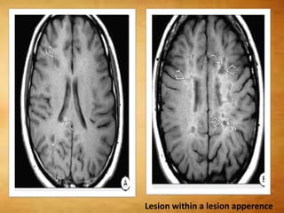
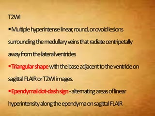
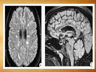
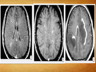
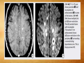
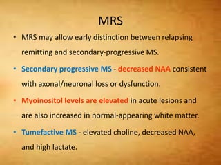
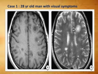
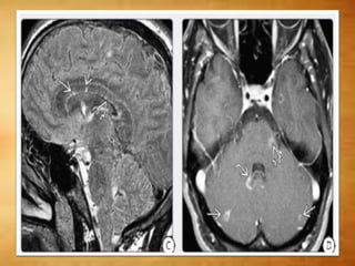
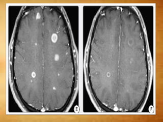
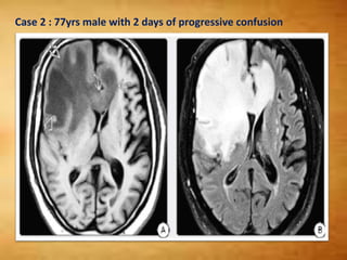
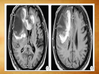
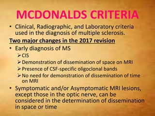
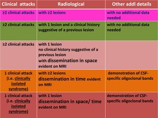
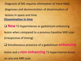
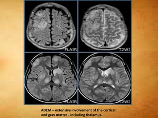
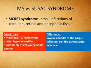
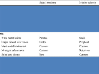
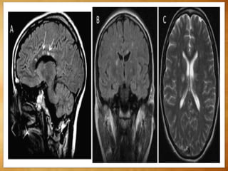
Multiple Sclerosis- Dr.N.Suriyaprakash
JR , Dept of RD , GKMCH
MULTIPLE SCLEROSIS
• MS is a progressive neurodegenerative
disorder characterized histopathologically by
multiple inflammatory demyelinating foci
called "plaques."
ETIOLOGY
• Multifactorial disease
• Autoimmune-Mediated Demyelination
• Environmental Factors - EBVexposure, chemicals,
smoking, diet, and geographic variability all
contribute to MS risk
• MS is a partially heritable autoimmune disease
LOCATION
• Supratentorial (90%), infratentorial (10%)
(higher in children)
• Deep cerebral/periventricular white matter
• Predilection for callososeptal interface
• Perivenular extension (Dawson fingers)
SIZE AND NUMBER
• Multiple > solitary
• Mostly small (5-10 mm)
Giant "tumefactive" plaques can be several cms
○ 30% of "tumefactive" MS lesions solitary
PATHOLOGY
Gross Pathology
• Active: Yellow-tan, ill-defined margins ± edema
• Chronic: Grayish, flat. Longstanding lesions are
depressed, excavated
Microscopic Features
• Plaques characterized by
○ Relatively sharp borders
○ Macrophages
○ Perivascular chronic inflammation
○ Reactive astrocytes
• Chronic: inflammation around borders
Longstanding plaques characterized
CLINICAL ISSUES
• Most common chronic nontraumatic neurologic
disease among young and middle-aged people.
• Risk of MS is increased 15-35 times in first-order
relatives of patients with clinically definite MS
• Overall F:M ratio is 1.77:1.00
PRESENTATION
• Intermittent neurologic disturbances followed
by progressive accumulation of disabilities.
• First attack of MS (most commonly optic
neuritis, transverse myelitis, or a brainstem
syndrome) is known as a CLINICALLY ISOLATED
SYNDROME
Radiologicall
y isolated
syndrome
(RIS)
Clinically
isolated
syndrome
(CIS)
Relapsing-
remitting
MS (RR-MS)
Relapsing
progressive
MS (RP-MS)
Secondary-
progressive
MS (SP-MS)
Primary-progressive MS (PP-MS)
CLINICAL SUBTYPES
Radiologically Isolated Syndrome
• Mildest of the demyelinating disease spectrum.
• RIS refers to MR findings of spatial dissemination
of T2/FLAIR lesions suggestive of MS in persons
with no history of neurologic symptoms and with
a normal neurologic examination.
• Half of the patients with RIS are initially
imaged because of headache, and the white
matter lesions are considered "incidental"
findings.
• When a clinical attack occurs in these patients,
a diagnosis of MS can be made.
• MS should not be diagnosed solely on the
basis of MR findings
Clinically Isolated Syndrome
• CIS refers to a first episode of neurologic
symptoms that lasts at least 24 hours and is
caused by inflammation or demyelination in the
CNS.
CIS can be monofocal or multifocal.
• Monofocal CIS - a single neurologic sign or
symptom (e.g., optic neuritis) is caused by a
single lesion.
• Multifocal CIS - more than one sign or symptom
caused by lesions in more than one location.
IMAGING FEATURES
Size – Few mm to several cm
Number – Usually multiple but giant tumefactive
plaques are solitary
Tissue loss with generalized brain atrophy
Enlarged ventricles and sulci with white matter
volume loss
Thinned corpus callosum
CT FINDINGS
• Early stage – NECT – Normal
• Solitary or multiple ill-defined white matter
hypodensities
• CECT - mild to moderate punctate, patchy, or
ring enhancement.
MRI FINDINGS
• MR is the procedure of choice for both initial
evaluation and treatment follow-up.
• Most recent revised McDonald criteria for MS
diagnosis rely on MR to demonstrate
dissemination in both space and time.
T1WI
 Hypo- or isointense - correlates with axonal
destruction.
Beveled or "lesion-within-a-lesion" appearance -
faint, poorly delineated peripheral rim of mild
hyperintensity (lipid peroxidation and macrophage
infiltration ) surrounds sharply delineated
hypointense black holes.
The corpus callosum becomes progressively thinner
and is best delineated on sagittal T1WI.
Lesion within a lesion apperence
T2WI
Multiplehyperintenselinear,round,orovoidlesions
surroundingthemedullaryveinsthatradiatecentripetally
awayfromthelateralventricles
Triangularshapewiththebaseadjacenttotheventricleon
sagittalFLAIRorT2WIimages.
Ependymaldot-dashsign-alternatingareasoflinear
hyperintensityalongtheependymaonsagittalFLAIR
T1 C+
 Active demyelination - transient enhancement
- Punctate, nodular, linear, and rim patterns are
seen
 Large tumefactive lesions – horseshoe
enhancement- open nonenhancing segment
facing the cortex
 Cortical demyelination - Leptomeningeal
enhancement
 Steroid administration significantly reduces
lesion enhancement
DWI
• Majority of acute plaques - normal or increased
diffusivity.
• Occasionally acute MS plaques can demonstrate
restriction on DWI - atypical - should not be
considered a reliable biomarker of plaque activity
MRS
• MRS may allow early distinction between relapsing
remitting and secondary-progressive MS.
• Secondary progressive MS - decreased NAA consistent
with axonal/neuronal loss or dysfunction.
• Myoinositol levels are elevated in acute lesions and
are also increased in normal-appearing white matter.
• Tumefactive MS - elevated choline, decreased NAA,
and high lactate.
Case 1 : 28 yr old man with visual symptoms
Case 2 : 77yrs male with 2 days of progressive confusion
MCDONALDS CRITERIA
• Clinical, Radiographic, and Laboratory criteria
used in the diagnosis of multiple sclerosis.
Two major changes in the 2017 revision
• Early diagnosis of MS
CIS
Demonstration of dissemination of space on MRI
Presence of CSF-specific oligoclonal bands
No need for demonstration of dissemination of time
on MRI
• Symptomatic and/or Asymptomatic MRI lesions,
except those in the optic nerve, can be
considered in the determination of dissemination
in space or time
Clinical attacks Radiological Other addl details
≥2 clinical attacks with ≥2 lesions with no additional data
needed
≥2 clinical attacks with 1 lesion and a clinical history
suggestive of a previous lesion
with no additional data
needed
≥2 clinical attacks with 1 lesion
no clinical history suggestive of a
previous lesion
with dissemination in space
evident on MRI
1 clinical attack
(i.e. clinically
isolated
syndrome)
with ≥2 lesions
dissemination in time evident
on MRI
demonstration of CSF-
specific oligoclonal bands
1 clinical attack
(i.e. clinically
isolated
syndrome)
with 1 lesion
dissemination in space/ time
evident on MRI
demonstration of CSF-
specific oligoclonal bands
Diagnosis of MS requires elimination of more likely
diagnoses and demonstration of dissemination of
lesions in space and time
Dissemination in time
 New T2-hyperintense or gadolinium-enhancing
lesion when compared to a previous baseline MRI scan
(irrespective of timing)
 Simultaneous presence of a gadolinium-enhancing
lesion and a non-enhancing T2-hyperintense lesion
on any one MRI scan
Dissemination in space
≥1 T2-hyperintense lesions (≥3 mm in long axis),
symptomatic and/or asymptomatic, that are characteristic
of MS in two or more of the four following locations 5:
Periventricular (≥1 lesion. Age >50yrs it is advised to
seek a higher number of lesions)
Cortical or juxtacortical (≥1 lesion)
Infratentorial (≥1 lesion)
Spinal cord (≥1 lesion)
DIFFERENTIAL DIAGNOSIS
Multifocal T2/FLAIR Hyperintensities
• ADEM
• Susac syndrome
• Lyme disease
• Vasculitis
Mass-Like ("Tumefactive") Lesion
• Neoplasm
oGlioblastoma multiforme
o Metastasis
• PML/PML-IRIS
○ HIV/AIDS
○ Natalizumab-treated MS
MS vs ADEM
• ADEM typically following arecent (1-2 weeks
prior) viral infection or vaccination.
• Slight male predominance
• Symptoms are more systemic
• Involvement of the callososeptal interface is
unusual . basal ganglia is often involved .
• MTR and diffusivity may help distinguish
ADEM from multiple sclerosis. In multiple
sclerosis both measurements are significantly
decreased.
ADEM – extensive involvement of the cortical
and gray matter - including thalamus.
MS vs SUSAC SYNDROME
• SICRET syndrome - small infarctions of
cochlear , retinal and encephalic tissue
Similarities
• Multifocal T2/FLAIR white
matter hyperintensities
• Commonly affect young adult
women
Differences
Involves middle of the corpus
callosum, not the callososeptal
interface.
• Vasculitis - Preferentially involves the basal
ganglia and spares the callososeptal interface.
• Lyme disease - Cranial nerve enhancement is
more common in LD than in MS.
MULTIPLE SCLEROSIS VARIANTS
Major MS variants are
(1) Marburg disease (MD)
(2) Schilder disease (SD)
(3) Balo concentric sclerosis (BCS).
MARBURG DISEASE
• Rare acute fulminate MS variant
○ Rapid neurologic deterioration
○ Monophasic, relentless progression
○ Death usually within 1 year
• Usually young adults
Pathology and Imaging
• Multifocal > solitary disease
○ Characterized by coalescent white matter plaques
○ Brain (including posterior fossa), spinal cord
lesions . Lesions characterized by massive
inflammation and necrosis.
MARBURG DISEASE
Imaging shows diffusely disseminated disease
o Large cavitating lesions
○ Incomplete enhancing rim
○ Multiple other patchy enhancing foci
26/M with short H/O visual disturbance and upper extremity weakness
SCHILDER DISEASE
• Myelinoclastic diffuse sclerosis
○ Rare acute/subacute demyelinating disorder
○ Lesions may resolve; 15% progress to MS
• Young adults , female preponderance
• Clinical features atypical for MS, ADEM
○ CSF normal
○ No history of fever, flu, vaccination
• Solitary > multifocal lesions
• Lesions look like tumefactive MS
• Differential diagnosis: Neoplasm and Abscess
BALO CONCENTRIC SCLEROSIS
• Concentric rings of demyelination/myelin
preservation
○ Resemble tree trunk or onion bulb
○ Solitary > multifocal
• Acute onset and rapid clinical deterioration
• "Whirlpool" hyperintense concentric rings on
T2WI
○ Minimal mass effect, edema
• Actively demyelinating layers enhance
Multiple sclerosis
Multiple sclerosis
Multiple sclerosis

Multiple sclerosis

  • 1.
  • 2.
    MULTIPLE SCLEROSIS • MSis a progressive neurodegenerative disorder characterized histopathologically by multiple inflammatory demyelinating foci called "plaques."
  • 3.
    ETIOLOGY • Multifactorial disease •Autoimmune-Mediated Demyelination • Environmental Factors - EBVexposure, chemicals, smoking, diet, and geographic variability all contribute to MS risk • MS is a partially heritable autoimmune disease
  • 4.
    LOCATION • Supratentorial (90%),infratentorial (10%) (higher in children) • Deep cerebral/periventricular white matter • Predilection for callososeptal interface • Perivenular extension (Dawson fingers)
  • 6.
    SIZE AND NUMBER •Multiple > solitary • Mostly small (5-10 mm) Giant "tumefactive" plaques can be several cms ○ 30% of "tumefactive" MS lesions solitary
  • 7.
    PATHOLOGY Gross Pathology • Active:Yellow-tan, ill-defined margins ± edema • Chronic: Grayish, flat. Longstanding lesions are depressed, excavated Microscopic Features • Plaques characterized by ○ Relatively sharp borders ○ Macrophages ○ Perivascular chronic inflammation ○ Reactive astrocytes • Chronic: inflammation around borders Longstanding plaques characterized
  • 9.
    CLINICAL ISSUES • Mostcommon chronic nontraumatic neurologic disease among young and middle-aged people. • Risk of MS is increased 15-35 times in first-order relatives of patients with clinically definite MS • Overall F:M ratio is 1.77:1.00
  • 10.
    PRESENTATION • Intermittent neurologicdisturbances followed by progressive accumulation of disabilities. • First attack of MS (most commonly optic neuritis, transverse myelitis, or a brainstem syndrome) is known as a CLINICALLY ISOLATED SYNDROME
  • 11.
    Radiologicall y isolated syndrome (RIS) Clinically isolated syndrome (CIS) Relapsing- remitting MS (RR-MS) Relapsing progressive MS(RP-MS) Secondary- progressive MS (SP-MS) Primary-progressive MS (PP-MS) CLINICAL SUBTYPES
  • 12.
    Radiologically Isolated Syndrome •Mildest of the demyelinating disease spectrum. • RIS refers to MR findings of spatial dissemination of T2/FLAIR lesions suggestive of MS in persons with no history of neurologic symptoms and with a normal neurologic examination.
  • 13.
    • Half ofthe patients with RIS are initially imaged because of headache, and the white matter lesions are considered "incidental" findings. • When a clinical attack occurs in these patients, a diagnosis of MS can be made. • MS should not be diagnosed solely on the basis of MR findings
  • 14.
    Clinically Isolated Syndrome •CIS refers to a first episode of neurologic symptoms that lasts at least 24 hours and is caused by inflammation or demyelination in the CNS. CIS can be monofocal or multifocal. • Monofocal CIS - a single neurologic sign or symptom (e.g., optic neuritis) is caused by a single lesion. • Multifocal CIS - more than one sign or symptom caused by lesions in more than one location.
  • 16.
    IMAGING FEATURES Size –Few mm to several cm Number – Usually multiple but giant tumefactive plaques are solitary Tissue loss with generalized brain atrophy Enlarged ventricles and sulci with white matter volume loss Thinned corpus callosum
  • 17.
    CT FINDINGS • Earlystage – NECT – Normal • Solitary or multiple ill-defined white matter hypodensities • CECT - mild to moderate punctate, patchy, or ring enhancement.
  • 18.
    MRI FINDINGS • MRis the procedure of choice for both initial evaluation and treatment follow-up. • Most recent revised McDonald criteria for MS diagnosis rely on MR to demonstrate dissemination in both space and time.
  • 19.
    T1WI  Hypo- orisointense - correlates with axonal destruction. Beveled or "lesion-within-a-lesion" appearance - faint, poorly delineated peripheral rim of mild hyperintensity (lipid peroxidation and macrophage infiltration ) surrounds sharply delineated hypointense black holes. The corpus callosum becomes progressively thinner and is best delineated on sagittal T1WI.
  • 20.
    Lesion within alesion apperence
  • 21.
  • 23.
    T1 C+  Activedemyelination - transient enhancement - Punctate, nodular, linear, and rim patterns are seen  Large tumefactive lesions – horseshoe enhancement- open nonenhancing segment facing the cortex  Cortical demyelination - Leptomeningeal enhancement  Steroid administration significantly reduces lesion enhancement
  • 25.
    DWI • Majority ofacute plaques - normal or increased diffusivity. • Occasionally acute MS plaques can demonstrate restriction on DWI - atypical - should not be considered a reliable biomarker of plaque activity
  • 27.
    MRS • MRS mayallow early distinction between relapsing remitting and secondary-progressive MS. • Secondary progressive MS - decreased NAA consistent with axonal/neuronal loss or dysfunction. • Myoinositol levels are elevated in acute lesions and are also increased in normal-appearing white matter. • Tumefactive MS - elevated choline, decreased NAA, and high lactate.
  • 28.
    Case 1 :28 yr old man with visual symptoms
  • 31.
    Case 2 :77yrs male with 2 days of progressive confusion
  • 33.
    MCDONALDS CRITERIA • Clinical,Radiographic, and Laboratory criteria used in the diagnosis of multiple sclerosis. Two major changes in the 2017 revision • Early diagnosis of MS CIS Demonstration of dissemination of space on MRI Presence of CSF-specific oligoclonal bands No need for demonstration of dissemination of time on MRI • Symptomatic and/or Asymptomatic MRI lesions, except those in the optic nerve, can be considered in the determination of dissemination in space or time
  • 34.
    Clinical attacks RadiologicalOther addl details ≥2 clinical attacks with ≥2 lesions with no additional data needed ≥2 clinical attacks with 1 lesion and a clinical history suggestive of a previous lesion with no additional data needed ≥2 clinical attacks with 1 lesion no clinical history suggestive of a previous lesion with dissemination in space evident on MRI 1 clinical attack (i.e. clinically isolated syndrome) with ≥2 lesions dissemination in time evident on MRI demonstration of CSF- specific oligoclonal bands 1 clinical attack (i.e. clinically isolated syndrome) with 1 lesion dissemination in space/ time evident on MRI demonstration of CSF- specific oligoclonal bands
  • 35.
    Diagnosis of MSrequires elimination of more likely diagnoses and demonstration of dissemination of lesions in space and time Dissemination in time  New T2-hyperintense or gadolinium-enhancing lesion when compared to a previous baseline MRI scan (irrespective of timing)  Simultaneous presence of a gadolinium-enhancing lesion and a non-enhancing T2-hyperintense lesion on any one MRI scan
  • 36.
    Dissemination in space ≥1T2-hyperintense lesions (≥3 mm in long axis), symptomatic and/or asymptomatic, that are characteristic of MS in two or more of the four following locations 5: Periventricular (≥1 lesion. Age >50yrs it is advised to seek a higher number of lesions) Cortical or juxtacortical (≥1 lesion) Infratentorial (≥1 lesion) Spinal cord (≥1 lesion)
  • 37.
    DIFFERENTIAL DIAGNOSIS Multifocal T2/FLAIRHyperintensities • ADEM • Susac syndrome • Lyme disease • Vasculitis Mass-Like ("Tumefactive") Lesion • Neoplasm oGlioblastoma multiforme o Metastasis • PML/PML-IRIS ○ HIV/AIDS ○ Natalizumab-treated MS
  • 38.
    MS vs ADEM •ADEM typically following arecent (1-2 weeks prior) viral infection or vaccination. • Slight male predominance • Symptoms are more systemic • Involvement of the callososeptal interface is unusual . basal ganglia is often involved . • MTR and diffusivity may help distinguish ADEM from multiple sclerosis. In multiple sclerosis both measurements are significantly decreased.
  • 40.
    ADEM – extensiveinvolvement of the cortical and gray matter - including thalamus.
  • 41.
    MS vs SUSACSYNDROME • SICRET syndrome - small infarctions of cochlear , retinal and encephalic tissue Similarities • Multifocal T2/FLAIR white matter hyperintensities • Commonly affect young adult women Differences Involves middle of the corpus callosum, not the callososeptal interface.
  • 44.
    • Vasculitis -Preferentially involves the basal ganglia and spares the callososeptal interface. • Lyme disease - Cranial nerve enhancement is more common in LD than in MS.
  • 45.
    MULTIPLE SCLEROSIS VARIANTS MajorMS variants are (1) Marburg disease (MD) (2) Schilder disease (SD) (3) Balo concentric sclerosis (BCS).
  • 46.
    MARBURG DISEASE • Rareacute fulminate MS variant ○ Rapid neurologic deterioration ○ Monophasic, relentless progression ○ Death usually within 1 year • Usually young adults Pathology and Imaging • Multifocal > solitary disease ○ Characterized by coalescent white matter plaques ○ Brain (including posterior fossa), spinal cord lesions . Lesions characterized by massive inflammation and necrosis.
  • 47.
    MARBURG DISEASE Imaging showsdiffusely disseminated disease o Large cavitating lesions ○ Incomplete enhancing rim ○ Multiple other patchy enhancing foci
  • 48.
    26/M with shortH/O visual disturbance and upper extremity weakness
  • 50.
    SCHILDER DISEASE • Myelinoclasticdiffuse sclerosis ○ Rare acute/subacute demyelinating disorder ○ Lesions may resolve; 15% progress to MS • Young adults , female preponderance • Clinical features atypical for MS, ADEM ○ CSF normal ○ No history of fever, flu, vaccination • Solitary > multifocal lesions • Lesions look like tumefactive MS • Differential diagnosis: Neoplasm and Abscess
  • 51.
    BALO CONCENTRIC SCLEROSIS •Concentric rings of demyelination/myelin preservation ○ Resemble tree trunk or onion bulb ○ Solitary > multifocal • Acute onset and rapid clinical deterioration • "Whirlpool" hyperintense concentric rings on T2WI ○ Minimal mass effect, edema • Actively demyelinating layers enhance